. 2018 Oct 11;13(10):e0204909.
doi: 10.1371/journal.pone.0204909. eCollection 2018.

Mice lacking RAP1 show early onset and higher rates of DEN-induced hepatocellular carcinomas in female mice

Affiliations

Mice lacking RAP1 show early onset and higher rates of DEN-induced hepatocellular carcinomas in female mice

Iole Ferrara-Romeo et al. PLoS One. .

Abstract

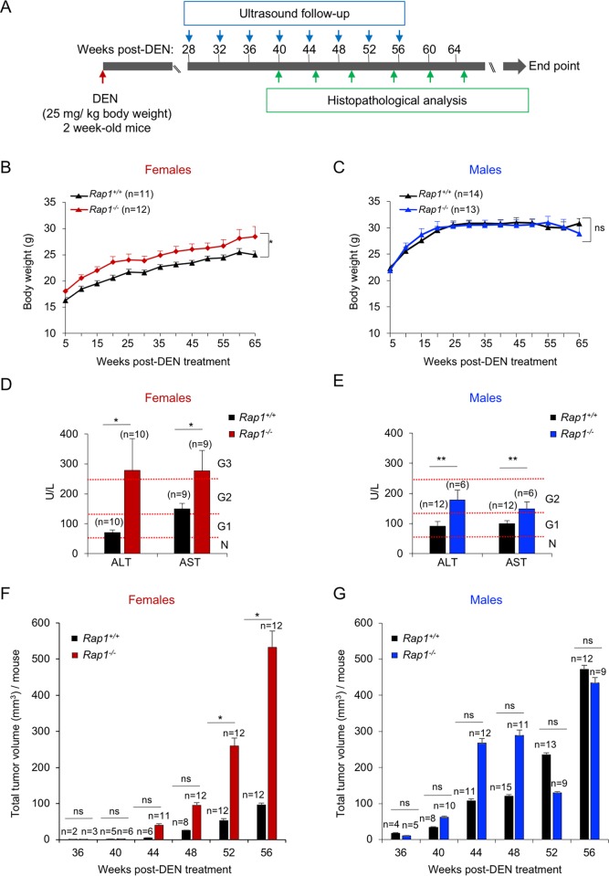
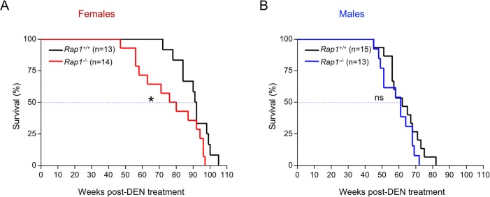
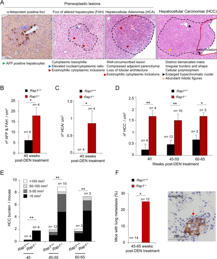
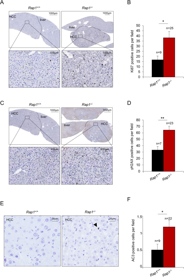
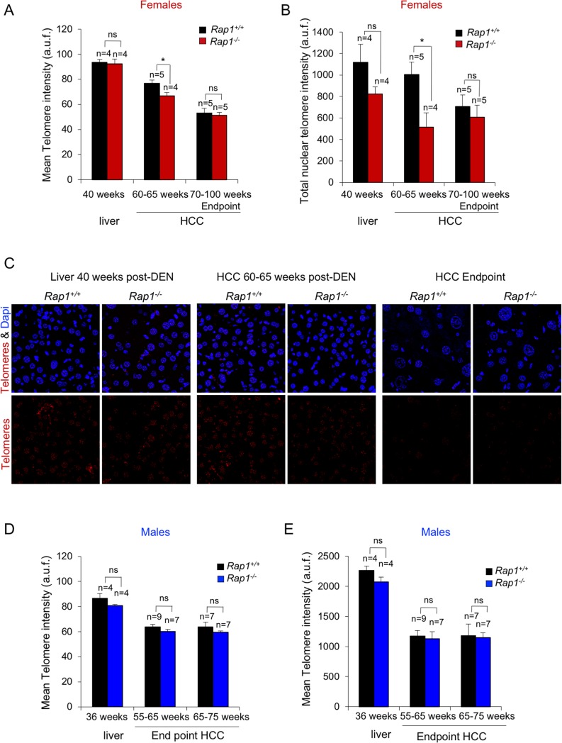
RAP1, a component of the telomere-protective shelterin complex, has been shown to have both telomeric and non-telomeric roles. In the liver, RAP1 is involved in the regulation of metabolic transcriptional programs. RAP1-deficient mice develop obesity and hepatic steatosis, these phenotypes being more severe in females than in males. As hepatic steatosis and obesity have been related to increased liver cancer in mice and humans, we set out to address whether RAP1 deficiency resulted in increased liver cancer upon chemical liver carcinogenesis. We found that Rap1-/- females were more susceptible to DEN-induced liver damage and hepatocellular carcinoma (HCC). DEN-treated Rap1-/- female livers showed an earlier onset of both premalignant and malignant liver lesions, which were characterized by increased abundance of γH2AX-positive cells, increased proliferation and shorter telomeres. These findings highlight an important role for RAP1 in protection from liver damage and liver cancer.


Conflict of interest statement

The authors have declared that no competing interests exist.

Figures

Fig 1
Fig 1. RAP1 deficiency leads to a higher susceptibility to DEN-induced liver damage and HCC development.
(A) Two-week-old Rap1+/+ and Rap1-/- female and male mice were intraperitoneally injected with DEN (25mg/kg body). Liver lesions were longitudinally monitored by ultrasound analysis from 28 weeks onwards after DEN treatment every fourth week. A group of Rap1+/+ and Rap1-/- females were sacrificed at 40-, 45-, 55-, 60- and 65 weeks post-DEN for histopathological analysis. The rest of the mice were sacrificed at humane endpoint. (B-C) Body weight gain of Rap1+/+ and Rap1-/- females (B) and males (C) from the fifth week post-DEN onward. (D-E) Plasma levels analysis of alanine (ALT) and aspartate (AST) aminotransferases in Rap1+/+ and Rap1-/- females at 50–60 weeks post-DEN (D) and males at 50–55 weeks post-DEN (E). Females at 50- and 60- weeks post-DEN and males at 50- and 55- weeks post-DEN were grouped since no significant differences were observed between these two ages. For ALT analysis, 5 Rap1+/+ and 6 Rap1-/- females at 50 weeks post-DEN and 5 Rap1+/+ and 4 Rap1-/- females at 60 weeks post-DEN were analyzed. For AST analysis, 4 Rap1+/+ and 5 Rap1-/- females at 50 weeks post-DEN and 5 Rap1+/+ and 4 Rap1-/- females at 60 weeks post-DEN were analyzed. For ALT and AST analysis in males, 4 Rap1+/+ and 2 Rap1-/- mice at 50 weeks post-DEN and 8 Rap1+/+ and 2 Rap1-/- mice at 55 weeks post-DEN were analyzed. Hepatotoxicity grades are shown to the right. N, normal; G1, grade 1; G2, grade 2 and G3, grade 3. G1 hepatotoxicity was defined as a serum ALT level of 51–125 U/L, G2 as a serum ALT level of 126–250 U/L and G3 as a serum ALT level of 251–500 U/L. (F-G) Quantification of total volume of hepatic lesion by ultrasound between 36- and 56-weeks post-DEN in Rap1+/+ and Rap1-/- females (F) and males (G). Values and error bars represent the mean and SE, respectively. N, number of mice. Statistical significance was determined by Student’s t test. *p<0.05, **p<0.01, ***p<0.001; ns, not significant.
Fig 2
Fig 2. RAP1 deficiency leads to a reduced lifespan of DEN-treated female mice.
(A-B) Kaplan-Meier survival curves of Rap1+/+ and Rap1-/- female (A) and male (B) mice. Statistical significance was determined by the log rank test. *, p ≤ 0.05.); ns, not significant.
Fig 3
Fig 3. Earlier onset of premalignant and malignant hepatic lesions in DEN-treated Rap1-/- females.
(A) Representative light microscopy images of α-fetoprotein (AFP) and hematoxylin-eosin (H&E) liver sections showing the different types of lesions induced by DEN. Focus of AFP-positive hepatocytes, focus of altered hepatocytes (FAH), hepatocellular adenoma (HCA) and hepatocellular carcinoma (HCC) are shown from left to right. For a detailed histological description see Material and Methods. (B-C) Quantification of FAHs and AFP-positive foci (B) and HCAs (C) in Rap1+/+ and Rap1-/- livers of female mice at 40 weeks after DEN injection. (D) Quantification of total number of HCC in Rap1+/+ and Rap1-/- livers of female mice at 40, 45–55 and 60–65 weeks post-DEN. (E) Quantification of the number of HCC of different size in the mice described in (D). (F) Incidence of lung metastasis in Rap1+/+ and Rap1-/- female mice between 45 and 65 weeks post-DEN. A representative light microscopy image of hepatocyte antigen-stained lung sections is shown to the right. Values and error bars represent the mean and SE, respectively. N, number of mice. Statistical significance was determined by Student’s t test.
Fig 4
Fig 4. Increased proliferation, DNA damage and apoptosis in HCC lacking RAP1 in females.
(A-F) Representative images and quantification of Ki67 (A, B) γH2AX (C-D) and AC3-positive cells (E-F) in Rap1+/+ and Rap1-/- tumor sections of female mice at 50–55 weeks post-DEN treatment. Tumors from 6 Rap1+/+ and 6 Rap1-/- females were analyzed. Three females from each cohort were at 50- and three at 55- weeks post-DEN. Mice of 50 and 55 weeks post-DEN were grouped since no significant differences were observed between these two ages. The size of the analyzed tumors ranged from 5 to 20 mm2. Values and error bars represent the mean and SE, respectively. N, number of tumors. Statistical significance was determined by Student’s t test. *p<0.05, **p<0.01, ***p<0.001.
Fig 5
Fig 5. RAP1 deficiency does not affect telomere length in full blown HCC.
(A-B) Mean telomere fluorescence (A) and total nuclear telomere fluorescence (B) in liver sections at 40 weeks post-DEN, 60–65 weeks post-DEN and at the endpoint at 70–100 weeks post-DEN. At 40 weeks the telomere intensity was determined in healthy hepatic tissue. At 60–65 weeks post-DEN before humane endpoint and at the humane endpoint, telomere intensity was determined in HCC. At 60–65 weeks post-DEN, the tumors analyzed presented an area between 10–30 mm2. At the endpoint, the analysis was performed in tumors with an area between 50–500 mm2. (C) Representative Q-fish images showing the telomere FISH (red) in Rap1+/+ and Rap1-/- liver and tumor sections of female mice at different time points. (D-E) Mean telomere fluorescence (D) and total nuclear telomere fluorescence (E) in liver sections at 36 weeks post-DEN and at the humane endpoint of male mice that died of liver tumors at either 55–65 or 65–75 weeks post DEN. At 36 weeks the telomere intensity was determined in healthy hepatic tissue. At the humane endpoint, telomere intensity was determined in HCC. In male mice at humane end points ranging from 55–65 or from 65–75 weeks post-DEN, the tumors analyzed presented an area between 50–350 mm2 and 100–500 mm2, respectively. a.u.f., arbitrary units of fluorescence. Values and error bars represent the mean and SE, respectively. N, number of tumors. Statistical significance was determined by Student’s t test. *p<0.05, **p<0.01, ***p<0.001.

Similar articles

Cited by

References

    1. Llovet JM, Zucman-Rossi J, Pikarsky E, Sangro B, Schwartz M, Sherman M, et al. Hepatocellular carcinoma. Nat Rev Dis Primers. 2016;2:16018 10.1038/nrdp.2016.18 . - DOI - PubMed
    1. Fattovich G, Stroffolini T, Zagni I, Donato F. Hepatocellular carcinoma in cirrhosis: incidence and risk factors. Gastroenterology. 2004;127(5 Suppl 1):S35–50. . - PubMed
    1. Rahman R, Hammoud GM, Almashhrawi AA, Ahmed KT, Ibdah JA. Primary hepatocellular carcinoma and metabolic syndrome: An update. World J Gastrointest Oncol. 2013;5(9):186–94. 10.4251/wjgo.v5.i9.186 ; PubMed Central PMCID: PMCPMC3782682. - DOI - PMC - PubMed
    1. Blasco MA. The epigenetic regulation of mammalian telomeres. Nat Rev Genet. 2007;8(4):299–309. 10.1038/nrg2047 . - DOI - PubMed
    1. Celli GB, de Lange T. DNA processing is not required for ATM-mediated telomere damage response after TRF2 deletion. Nat Cell Biol. 2005;7(7):712–8. 10.1038/ncb1275 . - DOI - PubMed

Publication types

MeSH terms