. 2018 Oct 11;14(10):e1007307.
doi: 10.1371/journal.ppat.1007307. eCollection 2018 Oct.

A polymorphic residue that attenuates the antiviral potential of interferon lambda 4 in hominid lineages

Affiliations

A polymorphic residue that attenuates the antiviral potential of interferon lambda 4 in hominid lineages

Connor G G Bamford et al. PLoS Pathog. .

Abstract

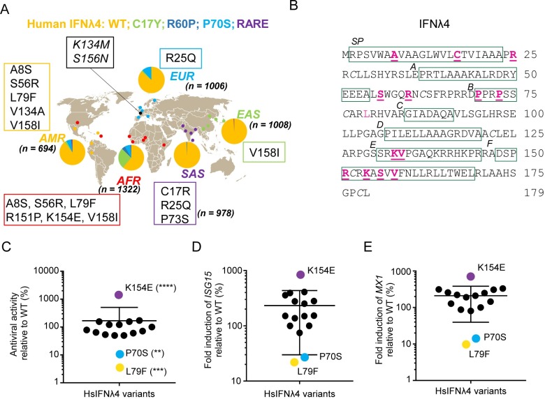
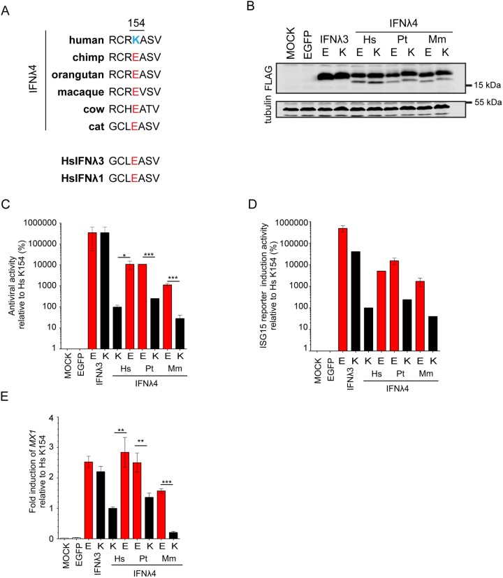
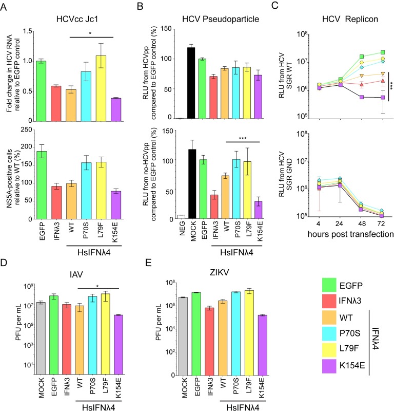
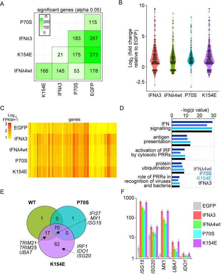
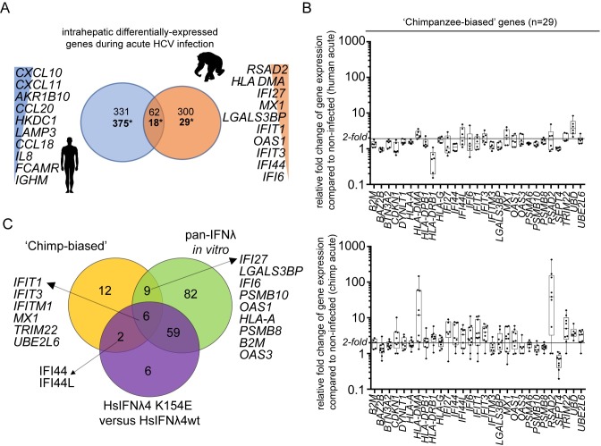
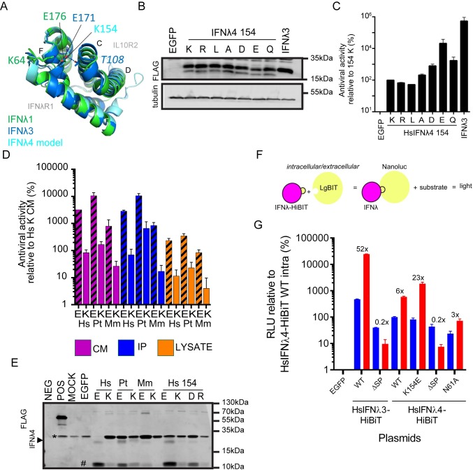
As antimicrobial signalling molecules, type III or lambda interferons (IFNλs) are critical for defence against infection by diverse pathogens, including bacteria, fungi and viruses. Counter-intuitively, expression of one member of the family, IFNλ4, is associated with decreased clearance of hepatitis C virus (HCV) in the human population; by contrast, a natural frameshift mutation that abrogates IFNλ4 production improves HCV clearance. To further understand how genetic variation between and within species affects IFNλ4 function, we screened a panel of all known extant coding variants of human IFNλ4 for their antiviral potential and identify three that substantially affect activity: P70S, L79F and K154E. The most notable variant was K154E, which was found in African Congo rainforest 'Pygmy' hunter-gatherers. K154E greatly enhanced in vitro activity in a range of antiviral (HCV, Zika virus, influenza virus and encephalomyocarditis virus) and gene expression assays. Remarkably, E154 is the ancestral residue in mammalian IFNλ4s and is extremely well conserved, yet K154 has been fixed throughout evolution of the hominid genus Homo, including Neanderthals. Compared to chimpanzee IFNλ4, the human orthologue had reduced activity due to amino acid K154. Comparison of published gene expression data from humans and chimpanzees showed that this difference in activity between K154 and E154 in IFNλ4 correlates with differences in antiviral gene expression in vivo during HCV infection. Mechanistically, our data show that the human-specific K154 negatively affects IFNλ4 activity through a novel means by reducing its secretion and potency. We thus demonstrate that attenuated activity of IFNλ4 is conserved among humans and postulate that differences in IFNλ4 activity between species contribute to distinct host-specific responses to-and outcomes of-infection, such as HCV infection. The driver of reduced IFNλ4 antiviral activity in humans remains unknown but likely arose between 6 million and 360,000 years ago in Africa.


Conflict of interest statement

The authors have declared that no competing interests exist.

Figures

Fig 1
Fig 1. Rare non-synonymous variants of HsIFNλ4 affect antiviral activity.
(A) Ancestry-based localization and frequency of human non-synonymous variants of HsIFNλ4 in African (AFR), South Asian (SAS), East Asian (EAS), European (EUR) and American (AMR) populations within the 1000 Genomes Database. ‘n’ represents the number of alleles tested in each population. Common and rare variants are those which have frequencies of >1% and <1% respectively in the 1000 Genome data. Common variants include: wt (orange), C17Y (light green), R60P (dark blue) and P70S (cyan). Rare variants (purple) include: A8S, C17R, R25Q, S56R, P73S, L79F, K133M, V134A, R151P, K154E, S156N, and V158I. Variants K133M and S156N (black) did not have an associated ethnicity but were found in the dataset from the Netherlands (Genome of the Netherlands cohort) [72]. (B) Location of non-synonymous variants in the HsIFNλ4 polypeptide (underlined pink). Regions of predicted structural significance are boxed (green), including the signal peptide (SP) and helices (A to F) [24]. There is a single N-linked glycosylation site at position 61 (N61). Note that there are 2 non-synonymous changes at C17 (C17R and C17Y). Cysteine residues involved in disulphide bridge formation are italicised. See S1 Data for genetic identifiers for the variants described here. (C) Antiviral activity of all HsIFNλ4 natural variants in an anti-EMCV CPE assay relative to wt protein in HepaRG cells. Cells were stimulated with serial dilutions of HsIFNλ4-containing CM for 24 hrs and then infected with EMCV (MOI = 0.3 PFU/cell) for 24 hrs at which point CPE was assessed by crystal violet staining. After staining, the dilution providing ~50% protection was determined. Mean of combined data from three independent experiments performed on different days (n = 3) are shown. Error bars represent mean and SEM for all variants combined. Data are shown in S2A Fig. **** = <0.0001; *** = <0.001; ** = <0.01 by one-way ANOVA compared to wt with a Dunnett’s test to correct for multiple comparisons. Controls (HsIFNλ4-TT and EGFP) are shown in S2 Fig and gave no protection against EMCV in the assay. Those variants with >2-fold change are highlighted with colours: purple (K154E,), cyan (P70S) and yellow (L79F). (D and E) ISG gene expression determined by RT-qPCR following stimulation of cells with HsIFNλ4 variants. Relative fold change of ISG15 (D) and Mx1 mRNAs (E) in HepaRG cells stimulated with CM (1:4 dilution) from plasmid-transfected cells compared to wt HsIFNλ4. Cells were stimulated for 24 hrs. Data points show mean of biological replicates (n = 3) and the error bar represents mean and SEM for all variants combined. Expanded data are shown in S2B and S2C Fig. Variants are coloured based on antiviral assays described in Fig 1C.
Fig 2
Fig 2. Human IFNλ4 is less active than chimpanzee IFNλ4 due to a substitution at amino acid position 154.
(A) Amino acid alignment from positions 151 to 157 for selected orthologues of HsIFNλ4 from different species as well as 2 human paralogues (HsIFNλ1 and HsIFNλ3). At position 154, HsIFNλ4 encodes a lysine (K; blue) while sequences from all other species predict a glutamic acid at this site (E; red). (B) Western blot analysis of intracellular IFNλ4 from different species encoding E or K at position 154 as well as equivalent E and K variants of HsIFNλ3op. HEK293T cells were transfected with the relevant plasmids for 48 hrs prior to preparation of cell lysates. IFNλ4 variants were detected with anti-FLAG antibody (‘FLAG’) and tubulin was used as a loading control. Mock- and EGFP-transfected cells were used as negative controls. (C) EMCV antiviral assay in HepaRG cells of IFNλ from the different species indicated (human [Hs], chimpanzee [Pt] and macaque [Mm]) encoding an E (red bars) or K (black bars) at position 154 alongside the equivalent amino acid substitutions in HsIFNλ3op. Antiviral activity is shown relative to that for HsIFNλ4 in HepaRG cells. Order denotes wt then variant IFNλ for each species. Data show +/- SD of biological replicates (n = 3) and are representative of two independent experiments performed on different days. *** = <0.001; * = <0.05 by unpaired, two-tailed Student’s T test comparing 154E and 154K for each IFN. (D) IFN signalling reporter assay in HepaRG.EGFP-ISG15 cells of IFNλ from the different species indicated (human [Hs], chimpanzee [Pt] and macaque [Mm]) encoding an E (red bars) or K (black bars) at position 154 alongside the equivalent amino acid substitutions in HsIFNλ3op. Activity is shown relative to that for HsIFNλ4 in HepaRG.EGFP-ISG15 cells. Order denotes wt then variant IFNλ for each species … Serial two-fold dilutions of CM (1:2 to 1:2097152) were incubated with the cells for 24 hrs and EGFP-positive cells (%) were measured by flow cytometry at each dilution and the IC50 value was calculated. Data shown are mean +/- SEM of biological replicates (n = 3) and are representative of two independent experiments carried out on different days. Comparison of all E versus K substituted forms of IFNλ4 within a homologue yielded significant values (p = <0.001 by Two-way ANOVA). (E) MX1 gene expression measured by RT-qPCR for IFNλ4 from different species encoding an E (red bars) or K (black bars) at position 154 alongside the equivalent amino acid substitutions in HsIFNλ3op. Data represent the relative fold change of MX1 mRNA by RT-qPCR in cells stimulated with CM (dilution 1:4) for 24 hrs compared to HsIFNλ4 wt. Data show average +/- SEM of biological replicates (n = 6) combined from two independent experiments performed on different days. *** = <0.001; ** = <0.01 by unpaired, two-tailed Student’s T test comparing 154E and 154K from each species.
Fig 3
Fig 3. HsIFNλ4 E154 has greater antiviral activity compared to wt HsIFNλ4 K154.
(A) Antiviral activity of HsIFNλ4 variants against HCVcc infection in Huh7 cells measured by RT-qPCR of viral RNA (upper panel) and virus antigen-positive cells (HCV NS5A protein; lower panel). HsIFNλ-containing CM (1:3) was incubated with Huh7 cells for 24 hrs before infection with HCVcc Jc1 (MOI = 0.01). HCV RNA was measured by RT-qPCR on RNA isolated at 72 hpi. Results shown are relative to infection in cells treated with EGFP CM (upper panel) or wt HsIFNλ4 (lower panel). Data show +/- SEM (n = 6) combined from two independent experiments performed on different days. * = <0.05 by unpaired, two-tailed Student’s T test comparing wt and K154E. (B) The effect of HsIFNλ4 variants on JFH1 HCV pseudoparticle (pp) infectivity in Huh7 cells. Relative light units (RLU) in the lysate of luciferase-expressing MLV pseudoparticles following inoculation of Huh7 cells stimulated with CM (1:3), relative (%) to CM from EGFP-transfected cells. The upper panel shows data from MLV pseudoparticles containing JFH1 glycoproteins E1 and E2 while the lower panel indicates data from MLV pseudoparticles that lack E1 and E2 (MLV core particles). Luciferase activity was measured at 72 hrs after inoculation. Error bars show +/- SEM of biological replicates (n = 6). (C) The effect of HsIFNλ4 variants on transient HCV RNA replication (upper panel) and translation (lower panel) using a subgenomic replicon assay in Huh7 cells. Huh7 cells were treated with CM (1:3) for 24 hrs before transfection with in vitro transcribed JFH1 HCV-SGR RNA expressing Gaussia luciferase; the upper and lower panels show data from wt (replication competent) and GND (non-replicative) sub-genomic replicons respectively. RLU secreted into the media was measured at 4, 24, 48 and 72 hpt. Error bars show +/- SD of biological replicates (n = 3). Data are representative of two independent experiments performed on different days. *** = <0.001 by two-way ANOVA on wt versus K154E. Comparing wt and K154E RLU at 72h by two-tailed Student’s T-test gave a significant difference ** = <0.01. Antiviral activity of wt and variant HsIFNλ4 on IAV (WSN strain) (D) or ZIKV (strain PE243). (E) infection in A549 cells as determined by plaque assay of virus released from infected cells at 48 hpi for IAV or 72 hpi for ZIKV. HsIFNλ4-, HsIFNλ3- and EGFP-containing CM (1:3) was incubated with A549 cells for 24 hrs before infection with IAV strain (MOI = 0.01 PFU/cell). Supernatant was harvested and titrated on MDCK cells for IAV or Vero cells for ZIKV. Error bars show +/- SEM of biological replicates (n = 3). * = <0.05 by unpaired, two-tailed Student’s T test comparing wt and K154E.
Fig 4
Fig 4. HsIFNλ4 E154 induces more robust antiviral gene expression than the wt K154 variant.
A549 cells were treated with CM (1:3 dilution) for 24 hours from cells transfected with plasmids expressing the different HsIFNλs and EGFP. After isolation of RNA, transcriptome analysis was carried out by RNA-Seq. (A) Total number of significantly differentially-expressed genes in each experimental condition (x-axis) relative to each other condition (y-axis). Colour shaded by differences in numbers of transcripts between sample 1 (x-axis) and sample 2 (y-axis) are shown. (B) Violin plot of all significant, differentially-expressed genes (over two-fold) (log2 fold change for each condition compared to RNA from cells treated with EGFP). CM was obtained from cells transfected with HsIFNλ3op (red); HsIFNλ4 wt (green); HsIFNλ4 P70S (cyan), and HsIFNλ4 K154E (purple). (C) Heat map of all significantly differentially-expressed genes (over two-fold) (log10 Fragments Per Kilobase of transcript per Million mapped reads (FKPM) in each experimental condition including EGFP CM-stimulated cells. Genes shown as columns and values are not normalised to negative control. (D) Pathway analysis using IPA on all significantly differentially-expressed genes (>2 fold) for each variant compared to EGFP. The top five most significantly induced pathways are shown [-log(p value)]. (E) Comparison of differentially-expressed genes (significant and at least 2-fold difference) stimulated by the HsIFNλ4 variants (HsIFNλ4 wt in green, HsIFNλ4 P70S in cyan and HsIFNλ4 K154E in purple) illustrated by a Venn diagram showing shared and unique genes. Three examples in overlapping and unique areas of the Venn diagram are highlighted. (F) Raw gene expression values (FKPM+1) for representative genes from core, shared and K154E-‘specific’ groups for the different treatments. Data are shown as mean +/- SD of biological replicates (n = 3). Exemplary genes selected were: ISG15 and MX1 (core), UBA7 (not significantly induced by P70S), and ISG20 and IDO1 (apparently specific for K154E). All transcriptomic analysis and gene lists are available in S2 Data.
Fig 5
Fig 5. Chimpanzees induce greater levels of antiviral ISG expression during HCV infection in vivo.
(A) Numbers of shared and unique differentially-expressed genes in liver biopsies from HCV-infected humans (blue) and experimentally-infected chimpanzees (orange) during the acute phase of infection represented as a Venn diagram (also see S1 Data). Gene expression during a time period of between 8 and 20 weeks was used where comparable published data for both species exists. The top ten species-‘specific’, differentially-expressed genes are shown ranked by levels of expression. Two sets of values for each comparison are shown; above shows the total differentially-expressed genes from at least one study while * highlights the value relating to the ‘core’ chimpanzee analysis that considered only the genes differentially-expressed in at least two studies of chimpanzee acute HCV infection. (B) Fold change of expression compared to controls (two uninfected individuals) for the 29 ‘chimp-biased’ genes in humans (upper) and chimpanzees (lower) shown as box plot and whiskers. Data are shown as box and whiskers to indicate median and range. Each value is illustrated by a black circle. The chimpanzee values represent an average of all fold changes for each chimpanzee over the time period. (C) Venn diagram analysis comparing the 29 chimpanzee-biased genes to the RNA-Seq data for all IFNs (GFP versus IFN) and for HsIFNλ4 K154E versus wt specifically. Illustrative gene names are shown as examples. All data are available in S2 Data.
Fig 6
Fig 6. Mechanism of action of the IFNλ4 K154E variant.
(A) Modelled structure of HsIFNλ4 showing position 154 at a central location in the molecule with reference to receptor subunit-binding interfaces (IFNλR1 and IL10R2). Overlapping crystal structures for HsIFNλ1 (green) and HsIFNλ3 (dark blue) are overlaid together with a homology model for HsIFNλ4 (light blue). In the overlapping structures, the homologous positions for HsIFNλ4 E154 (E176, IFNλ1; E171, IFNλ3) make intramolecular non-covalent interactions with two distinct regions within IFNλ. (B) Detection of intracellular IFNλ4 154 mutants (K, R, L, A, D, E and Q) by Western blot analysis of lysates from plasmid-transfected producer HEK293T cells. The IFNλ4 variants were detected with an anti-FLAG antibody. Tubulin was used as a loading control. (C) Antiviral activity of HsIFNλ4 IFNλ4 154 mutants (K, R, L, A, D, E and Q) in an anti-EMCV CPE assay relative to CM from wt HsIFNλ4 (K154 variant) in HepaRG cells. Data show mean +/- SEM combined from three independent experiments performed on different days. (D) Antiviral activity of IFNλ4 found in CM, intracellular lysate and immunoprecipitated CM from the different species indicated (human [Hs], chimpanzee [Pt] and macaque [Mm]) encoding an E or K at position 154 in an anti-EMCV CPE assay relative to CM from wt HsIFNλ4 in HepaRG cells. Data show mean +/- SEM from two independent experiments performed on different days. (E) Detection of extracellular IFNλ4 from different species as well as select mutants at position 154 (E, K, D and R) by Western blot analysis of samples of FLAG-tag immunoprecipitated CM (1 ml) from plasmid-transfected producer HEK293T cells. A BAP-FLAG fusion protein was used an immunoprecipitation control (POS). The IFNλ4 variants were detected with an anti-FLAG antibody. A FLAG-positive lower molecular weight product, which is potentially a degradation product is highlighted with a #. An upper band running near to the IFN is shown (*) which is likely antibody fragments from the immunoprecipitation reaction. Blot is representative of three independent experiments. (F) Schematic of the split NanoLuc Luciferase assay to measure the relative abundance of intra- and extracellular abundance of HsIFNλ3 and HsIFNλ4 variants produced in plasmid-transfected cells. (G) Levels of luciferase activity (RLU) relative to intracellular enzyme activity in cells expressing wt IFNλ4 (set at 100%). Intra- and extracellular luciferase activities are shown in blue and red respectively. The constructs used are indicated below the graph. The ratio of intra- to extracellular RLU values are indicated for each construct.

References

    1. Randall RE, Goodbourn S. Interferons and viruses: an interplay between induction, signalling, antiviral responses and virus countermeasures. J Gen Virol. 2008. 89(1):1–47. - PubMed
    1. Schoggins JW, Wilson SJ, Panis M, Murphy MY, Jones CT, Bieniasz P, et al. A diverse range of gene products are effectors of the type I interferon antiviral response. Nature. 2011. 472(7344):481–5. 10.1038/nature09907 - DOI - PMC - PubMed
    1. Shaw AE, Hughes J, Gu Q, Behdenna A, Singer JB, Dennis T, et al. Fundamental properties of the mammalian innate immune system revealed by multispecies comparison of type I interferon responses. PLOS Biol. 2017. 15(12):e2004086 10.1371/journal.pbio.2004086 - DOI - PMC - PubMed
    1. Schoggins JW. Interferon-stimulated genes: roles in viral pathogenesis. Curr Opin Virol. 2014. 6C:40–6. - PMC - PubMed
    1. Kotenko S V., Gallagher G, Baurin V V., Lewis-Antes A, Shen M, Shah NK, et al. IFN-λs mediate antiviral protection through a distinct class II cytokine receptor complex. Nat Immunol. 2003. 4(1):69–77. 10.1038/ni875 - DOI - PubMed

Publication types

MeSH terms