. 2018 Oct 10;10(4):182.
doi: 10.3390/pharmaceutics10040182.

Hydrophobic Amino Acid Tryptophan Shows Promise as a Potential Absorption Enhancer for Oral Delivery of Biopharmaceuticals

Affiliations

Hydrophobic Amino Acid Tryptophan Shows Promise as a Potential Absorption Enhancer for Oral Delivery of Biopharmaceuticals

Noriyasu Kamei et al. Pharmaceutics. .

Abstract

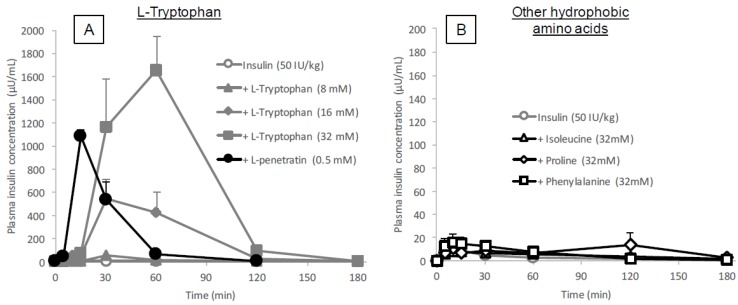
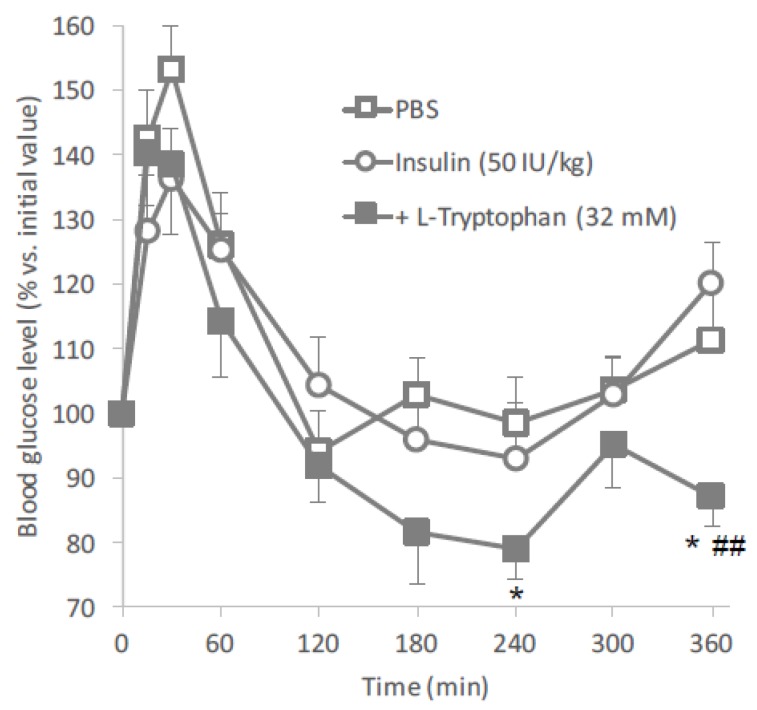
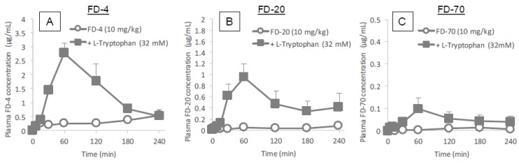
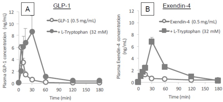
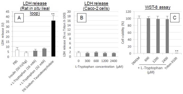
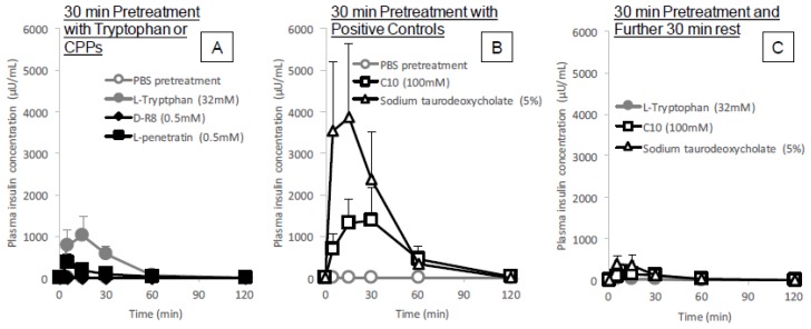
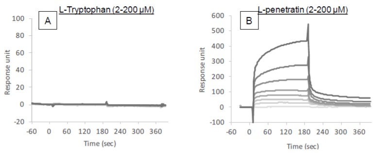
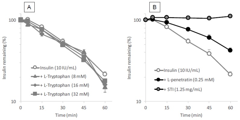
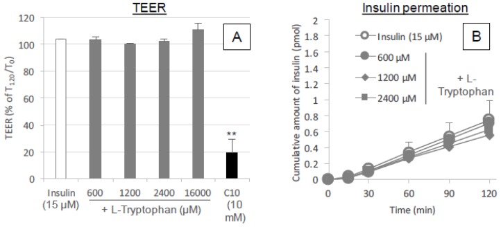
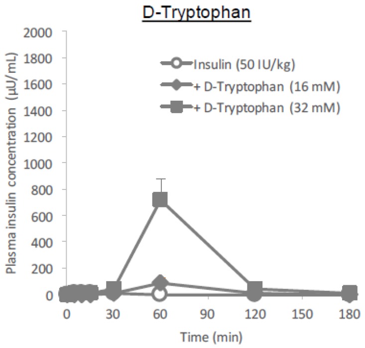
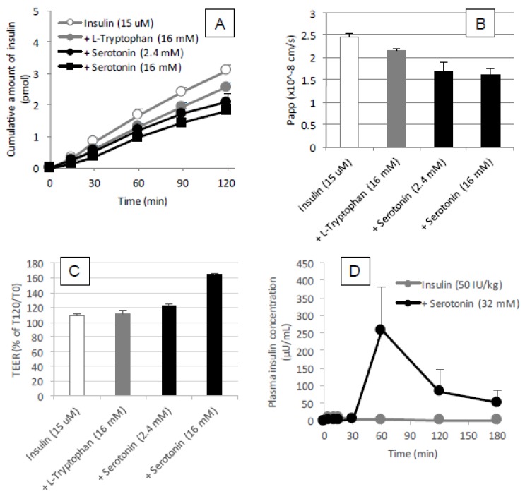
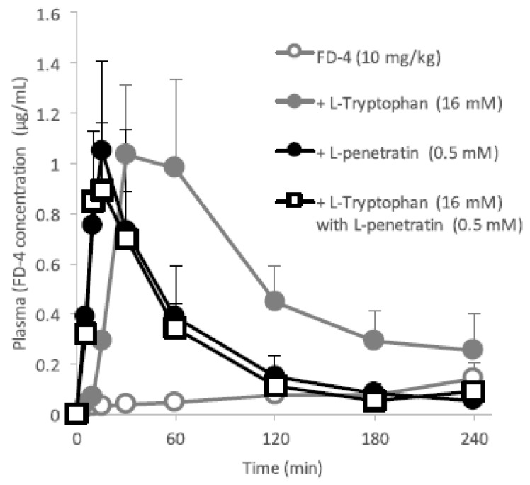
Cell-penetrating peptides (CPPs) have great potential to efficiently deliver drug cargos across cell membranes without cytotoxicity. Cationic arginine and hydrophobic tryptophan have been reported to be key component amino acids for cellular internalization of CPPs. We recently found that l-arginine could increase the oral delivery of insulin in its single amino acid form. Therefore, in the present study, we evaluated the ability of another key amino acid, tryptophan, to enhance the intestinal absorption of biopharmaceuticals. We demonstrated that co-administration with l-tryptophan significantly facilitated the oral and intestinal absorption of the peptide drug insulin administered to rats. Furthermore, l-tryptophan exhibited the ability to greatly enhance the intestinal absorption of other peptide drugs such as glucagon-like peptide-1 (GLP-1), its analog Exendin-4 and macromolecular hydrophilic dextrans with molecular weights ranging from 4000 to 70,000 g/mol. However, no intermolecular interaction between insulin and l-tryptophan was observed and no toxic alterations to epithelial cellular integrity-such as changes to cell membranes, cell viability, or paracellular tight junctions-were found. This suggests that yet to be discovered inherent biological mechanisms are involved in the stimulation of insulin absorption by co-administration with l-tryptophan. These results are the first to demonstrate the significant potential of using the single amino acid l-tryptophan as an effective and versatile bioavailability enhancer for the oral delivery of biopharmaceuticals.

Keywords: GLP-1; amino acid; cell-penetrating peptide; insulin; intestinal absorption; oral delivery; tryptophan.


Conflict of interest statement

Authors declare no conflict of interest. The funders had no role in the design of the study; in the collection, analyses, or interpretation of data; in the writing of the manuscript, and in the decision to publish the results.

Figures

Figure 1
Figure 1
Time profiles of plasma insulin concentration after in situ administration of insulin (50 IU/kg) with or without l-tryptophan, l-penetratin or other hydrophobic amino acid additives into rat ileal loop. Panel (A), l-tryptophan (8–32 mM) or l-penetratin (0.5 mM); panel (B), hydrophobic amino acids (l-isoleucine, l-proline and l-phenylalanine, 32 mM). Each data point represents the mean ± SEM of N = 3–8, except for the group with l-tryptophan (8 mM, N = 2).
Figure 2
Figure 2
Blood glucose levels in mice following oral administration of insulin (50 IU/kg) with or without l-tryptophan (32 mM). Each data point represents the mean ± SEM of N = 6–8. * p < 0.05, ## p < 0.01, significantly different with PBS and insulin (50 IU/kg), respectively.
Figure 3
Figure 3
Time profiles of plasma concentrations of model hydrophilic macromolecules (FD-4, FD-20 and FD-70) after their in situ administration with or without l-tryptophan (32 mM) into rat ileal loop. Panels (AC) show the absorption of FD-4, FD-20 and FD-70, respectively. Each data point represents the mean ± SEM of N = 3–8.
Figure 4
Figure 4
Time profiles of plasma concentrations of peptide drugs (GLP-1 and Exendin-4) after their in situ administration with or without l-tryptophan (32 mM) into rat ileal loop. Panels (A) and (B) show the absorption of GLP-1 and Exendin-4, respectively. Each data point represents the mean ± SEM of N = 4.
Figure 5
Figure 5
Cytotoxicity examinations after the exposure of intestinal epithelium to l-tryptophan. Panel (A), the LDH release in the intestinal fluid collected at 60 min after ileal administration of PBS, insulin (50 IU/kg) with or without l-tryptophan (16–50 mM) and sodium taurodeoxycholate (5%). Panel (B), LDH released from the cytoplasm into the incubation medium (HBSS) after incubation with various concentrations of l-tryptophan (200–2400 μM). The value is expressed as a percentage calculated by dividing the absorbance of the l-tryptophan treated medium sample by that of the sample treated with Triton X-100 (0.8%). Panel C, cell viability after incubation with various concentrations of l-tryptophan (600–2400 μM). Each data point represents the mean ± SEM of N = 3, 3 and 6–11 for panels (AC), respectively. * p < 0.05, ** p < 0.01, significantly different with corresponding control PBS- (panel (A)), or DMEM- (panel (C)) treatment group.
Figure 6
Figure 6
Time profiles of plasma insulin concentration after in situ administration of insulin (50 IU/kg) into rat ileal loop pretreated with l-tryptophan (32 mM), d-R8 (0.5 mM), l-penetratin (0.5 mM), C10 (100 mM), or sodium taurodeoxycholate (5%). In the results shown in Panels (A,B), insulin solution was administered immediately after washing the pretreatment solution from the ileal loop. For the results shown in Panel (C), insulin solution was administered at 30 min after washing out the pretreatment solution. Each data point represents the mean ± SEM of N = 3–4.
Figure 7
Figure 7
Binding sensorgrams obtained by SPR analysis. Various concentrations of l-tryptophan (A) or l-penetratin (B) solutions (pH 7.4) were injected into insulin-immobilized flow cells.
Figure 8
Figure 8
Degradation profiles of insulin in the presence of tryptophan or positive controls (penetratin and STI) in rat intestinal enzymatic fluid. Panel (A), various concentrations of l-tryptophan (8–32 mM); panel (B), l-penetratin (0.25 mM) or STI (positive control, 1.25 mg/mL). Each data point represents the mean ± SEM of N = 3.
Figure 9
Figure 9
Cytotoxicity examinations after the exposure of intestinal epithelium to l-tryptophan. Panel (A), changes in the TEER of Caco-2 cell monolayers after the incubation with insulin (15 μM) and various concentrations of l-tryptophan (600–16,000 μM). The values are expressed as a percentage calculated by dividing the TEER measurement (Ω cm2) at 120 min by the initial value. Panel (B), time courses of permeation of insulin through Caco-2 monolayer in the presence or absence of l-Tryptophan (600–2400 μM). Each data point represents the mean ± SEM of N = 3. * p < 0.05, ** p < 0.01, significantly different with corresponding insulin control group.
Figure 10
Figure 10
Time profiles of plasma insulin concentration after in situ administration of insulin (50 IU/kg) with or without d-tryptophan (16 or 32 mM) into rat ileal loop. Each data point represents the mean ± SEM of N = 3.
Figure 11
Figure 11
Permeation of insulin through the Caco-2 cell monolayer panels (A,B) and TEER values panel (C) during coincubation with l-tryptophan (16 mM) or serotonin (2.4 or 16 mM) and the absorption of insulin after its in situ administration of insulin (50 IU/kg) with or without serotonin (32 mM) into rat ileal loop panel (D). Each data point represents the mean ± SEM of N = 3.
Figure 12
Figure 12
Time profiles of plasma concentrations of FD-4 after its in situ administration with or without l-tryptophan (16 mM) and/or l-penetratin (0.5 mM) into rat ileal loop. Each data point represents the mean ± SEM of N = 3–4.

Similar articles

Cited by

References

    1. Komin A., Russell L.M., Hristova K.A., Searson P.C. Peptide-based strategies for enhanced cell uptake, transcellular transport, and circulation: Mechanisms and challenges. Adv. Drug Deliv. Rev. 2017;110–111:52–64. doi: 10.1016/j.addr.2016.06.002. - DOI - PubMed
    1. Salzano G., Torchilin V.P. Intracellular delivery of nanoparticles with cell penetrating peptides. Methods Mol. Biol. 2015;1324:357–368. - PubMed
    1. Kamei N., Morishita M., Eda Y., Ida N., Nishio R., Takayama K. Usefulness of cell-penetrating peptides to improve intestinal insulin absorption. J. Control. Release. 2008;132:21–25. doi: 10.1016/j.jconrel.2008.08.001. - DOI - PubMed
    1. Kamei N., Morishita M., Takayama K. Importance of intermolecular interaction on the improvement of intestinal therapeutic peptide/protein absorption using cell-penetrating peptides. J. Control. Release. 2009;136:179–186. doi: 10.1016/j.jconrel.2009.02.015. - DOI - PubMed
    1. Khafagy E.-S., Morishita M., Kamei N., Eda Y., Ikeno Y., Takayama K. Efficiency of cell-penetrating peptides on the nasal and intestinal absorption of therapeutic peptides and proteins. Int. J. Pharm. 2009;381:49–55. doi: 10.1016/j.ijpharm.2009.07.022. - DOI - PubMed

LinkOut - more resources