Review
. 2018 Oct 10;19(10):3104.
doi: 10.3390/ijms19103104.

Danger-Associated Molecular Patterns (DAMPs): Molecular Triggers for Sterile Inflammation in the Liver

Affiliations
Review

Danger-Associated Molecular Patterns (DAMPs): Molecular Triggers for Sterile Inflammation in the Liver

Sabine Mihm. Int J Mol Sci. .

Abstract

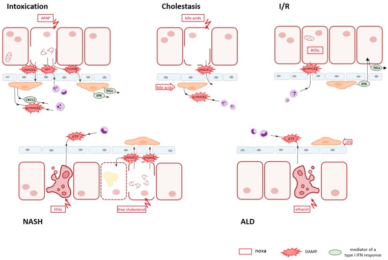
Inflammatory liver diseases in the absence of pathogens such as intoxication by xenobiotics, cholestatic liver injury, hepatic ischemia-reperfusion injury (I/R), non-alcoholic steatohepatitis (NASH), or alcoholic liver disease (ALD) remain threatening conditions demanding specific therapeutic options. Caused by various different noxae, all these conditions have been recognized to be triggered by danger- or death-associated molecular patterns (DAMPs), discompartmentalized self-structures released by dying cells. These endogenous, ectopic molecules comprise proteins, nucleic acids, adenosine triphosphate (ATP), or mitochondrial compounds, among others. This review resumes the respective modes of their release-passively by necrotic hepatocytes or actively by viable or apoptotic parenchymal cells-and their particular roles in sterile liver pathology. It addresses their sensors and the initial inflammatory responses they provoke. It further addresses a resulting second wave of parenchymal death that might be of different mode, boosting the release of additional, second-line DAMPs. Thus, triggering a more complex and pronounced response. Initial and secondary inflammatory responses comprise the activation of Kupffer cells (KCs), the attraction and activation of monocytes and neutrophil granulocytes, and the induction of type I interferons (IFNs) and their effectors. A thorough understanding of pathophysiology is a prerequisite for identifying rational therapeutic targets.

Keywords: acetaminophen (APAP) intoxication; alcoholic liver disease (ALD); cholestasis; danger-associated molecular pattern (DAMP); hepatic ischemia-reperfusion (I/R); high mobility group box-1 (HMGB1); non-alcoholic steatohepatitis (NASH); sterile liver injury; type I interferon (IFN).


Figures

Figure 1
Figure 1
Schematic outline on key events in sterile liver disease conditions. Cell injury of hepatic parenchyma can be provoked by many different noxae such as xenobiotics in intoxication conditions (e.g., APAP), endogenous compounds in cholestatic obstruction (e.g., bile acids) or NASH (e.g., free fatty acids (FFAs), the generation of reactive oxygen species (ROSs) in I/R conditions, or by ethanol in ALD. The release of DAMPs in a first line is common to all conditions. It might be accomplished either actively via secretion from viable or from dying, apoptotic cells or passively via leaking out from necrotic cells. In APAP intoxication, necrotic cell death predominates with the release of HMGB1, mtDNA, and N-formyl peptides (NFP). In cholestasis, bile acids cause a disintegration of hepatocytes, thereby generating a shunt and allowing bile constituents to reach the blood stream and to act also on KCs. In experimental I/R injury, the first insult was demonstrated to be an active secretion of acHMGB1 from intact hepatocytes, while in later stages IFNs become involved in. In the pathogenesis of NASH, concomitant apoptotic and necrotic death of hepatocytes caused by different lipid entities is indicated with the regulated, active release of ATP by apoptotic cells and the passive release of HBMG1 and mtDNA by necrotic cells. In ALD, ethanol-driven apoptosis of parenchymal cells is superposed by microbial products, e.g., LPS, as a result of an impairment of the intestinal mucosa and an increased gut permeability. The first line of DAMPs released by hepatocytes provokes release of a second line of DAMPs and mediators by resident KCs and granulocytes. The composition of second-line DAMPs and mediators governs a secondary innate immune response. This secondary response might comprise cell death of different mode than the initial one, further attraction of neutrophils and monocytes from the circulation, and the induction of an IFN response, thus boosting sterile hepatic inflammation.

Similar articles

  • DAMPs and sterile inflammation in drug hepatotoxicity.
    Yang R, Tonnesseen TI. Yang R, et al. Hepatol Int. 2019 Jan;13(1):42-50. doi: 10.1007/s12072-018-9911-9. Epub 2018 Nov 24. Hepatol Int. 2019. PMID: 30474802 Review.
  • Sterile inflammation in the liver.
    Kubes P, Mehal WZ. Kubes P, et al. Gastroenterology. 2012 Nov;143(5):1158-1172. doi: 10.1053/j.gastro.2012.09.008. Epub 2012 Sep 13. Gastroenterology. 2012. PMID: 22982943 Review.
  • DAMPs and NETs in Sepsis.
    Denning NL, Aziz M, Gurien SD, Wang P. Denning NL, et al. Front Immunol. 2019 Oct 30;10:2536. doi: 10.3389/fimmu.2019.02536. eCollection 2019. Front Immunol. 2019. PMID: 31736963 Free PMC article. Review.
  • Decoding cell death signals in liver inflammation.
    Brenner C, Galluzzi L, Kepp O, Kroemer G. Brenner C, et al. J Hepatol. 2013 Sep;59(3):583-94. doi: 10.1016/j.jhep.2013.03.033. Epub 2013 Apr 6. J Hepatol. 2013. PMID: 23567086 Review.
  • The damage-associated molecular pattern HMGB1 is released early after clinical hepatic ischemia/reperfusion.
    van Golen RF, Reiniers MJ, Marsman G, Alles LK, van Rooyen DM, Petri B, Van der Mark VA, van Beek AA, Meijer B, Maas MA, Zeerleder S, Verheij J, Farrell GC, Luken BM, Teoh NC, van Gulik TM, Murphy MP, Heger M. van Golen RF, et al. Biochim Biophys Acta Mol Basis Dis. 2019 Jun 1;1865(6):1192-1200. doi: 10.1016/j.bbadis.2019.01.014. Epub 2019 Jan 15. Biochim Biophys Acta Mol Basis Dis. 2019. PMID: 30658161

Cited by

References

    1. Eguchi A., Wree A., Feldstein A.E. Biomarkers of liver cell death. J. Hepatol. 2014;60:1063–1074. doi: 10.1016/j.jhep.2013.12.026. - DOI - PubMed
    1. Matzinger P. Tolerance, danger, and the extended family. Annu. Rev. Immunol. 1994;12:991–1045. doi: 10.1146/annurev.iy.12.040194.005015. - DOI - PubMed
    1. Matzinger P. The danger model: A renewed sense of self. Science. 2002;296:301–305. doi: 10.1126/science.1071059. - DOI - PubMed
    1. Dolasia K., Bisht M.K., Pradhan G., Udgata A., Mukhopadhyay S. TLRs/NLRs: Shaping the landscape of host immunity. Int. Rev. Immunol. 2018;37:3–19. doi: 10.1080/08830185.2017.1397656. - DOI - PubMed
    1. Almeda-Valdes P., Aguilar Olivos N.E., Barranco-Fragoso B., Uribe M., Mendez-Sanchez N. The Role of Dendritic Cells in Fibrosis Progression in Nonalcoholic Fatty Liver Disease. BioMed Res. Int. 2015;2015:768071. doi: 10.1155/2015/768071. - DOI - PMC - PubMed