

Correlation of Adventitial Vasa Vasorum with Intracranial Atherosclerosis: A Postmortem Study
- PMID: 30309229
- PMCID: PMC6186920
- DOI: 10.5853/jos.2018.01263
Correlation of Adventitial Vasa Vasorum with Intracranial Atherosclerosis: A Postmortem Study
Abstract
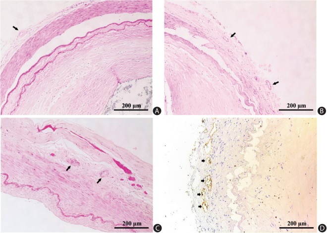
Background and purpose: Vasa vasorum (VV) have been believed to be rare or non-existent in small-caliber intracranial arteries. In a series of human cerebral artery specimens, we identified and examined the distribution of VV in association with co-existing intracranial atherosclerosis.
Methods: We obtained cerebral artery specimens from 32 consecutive autopsies of subjects aged 45 years or above. We scrutinized middle cerebral artery (MCA), vertebral artery (VA), and basilar artery (BA) for the presence of adventitial VV. We described the distribution of VV, and the characteristics of co-existing atherosclerotic lesions.
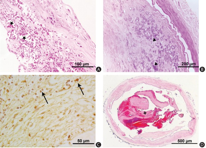
Results: Among 157 intracranial arteries, adventitial VV were present in 74 of the 157 specimens (47%), involving MCA (n=13, 18%), BA (n=14, 19%), and VA (n=47, 64%). Although qualitatively these 74 adventitial VV distributed similarly in arteries with or without atherosclerotic lesions (disease-free arteries n=4/8; arteries of pre-atherosclerosis n=17/42; and arteries of progressive atherosclerosis n=53/107), the presence of adventitial VV in intracranial VA was associated with a heavier plaque load (1.72±1.66 mm2 vs. 0.40±0.32 mm2, P<0.001), severer luminal stenosis (25%±21% vs. 12%±9%, P=0.002), higher rate of concentric lesions (79% vs. 36%, P=0.002), and denser intraplaque calcification (44% vs. 0%, P=0.003). Histologically, intracranial VA with VV had a larger diameter (3.40±0.79 mm vs. 2.34±0.58 mm, P<0.001), thicker arterial wall (0.31±0.13 mm vs. 0.23±0.06 mm, P=0.002), and a larger intima-media (0.19±0.09 mm vs. 0.13± 0.04 mm, P=0.003) than VA without VV.
Conclusion: s Our study demonstrated the distribution of adventitial VV within brain vasculature and association between vertebral VV and progressive atherosclerotic lesions with a heavier plaque load and denser intraplaque calcification.
Keywords: Angiogenesis; Atherosclerosis; Vasa vasorum.
Figures
Similar articles
-
Do human intracranial arteries lack vasa vasorum? A comparative immunohistochemical study of intracranial and systemic arteries.Acta Neuropathol. 1998 Jul;96(1):22-8. doi: 10.1007/s004010050856. Acta Neuropathol. 1998. PMID: 9678510
-
Arterial Adventitial Vasa Vasorum Hyperplasia involved in Atherosclerotic Plaque Formation in a Rabbit Model.Ultrasound Med Biol. 2024 Aug;50(8):1273-1279. doi: 10.1016/j.ultrasmedbio.2024.05.006. Epub 2024 May 25. Ultrasound Med Biol. 2024. PMID: 38796339
-
Arterial Adventitial Vasa Vasorum Density Reflects The Progression Of Unstable Plaques: A Retrospective Clinical Study.Ultrasound Med Biol. 2024 May;50(5):712-721. doi: 10.1016/j.ultrasmedbio.2024.01.011. Epub 2024 Feb 16. Ultrasound Med Biol. 2024. PMID: 38365464
-
Vasa vasorum and atherosclerosis - Quid novi?Thromb Haemost. 2007 Jun;97(6):873-9. Thromb Haemost. 2007. PMID: 17549287 Review.
-
Detection of adventitial vasa vasorum and intraplaque neovascularization in carotid atherosclerotic lesions with contrast-enhanced ultrasound and their role in atherosclerosis.Methodist Debakey Cardiovasc J. 2011 Oct-Dec;7(4):37-40. doi: 10.14797/mdcj-7-4-37. Methodist Debakey Cardiovasc J. 2011. PMID: 22143476 Review.
Cited by
-
Intracranial vessel wall lesions on MRI: anatomical and pathological issues.Neurol Sci. 2025 Jul 16. doi: 10.1007/s10072-025-08366-6. Online ahead of print. Neurol Sci. 2025. PMID: 40668467 Review.
-
Vessel Wall Magnetic Resonance Imaging in Cerebrovascular Diseases.Diagnostics (Basel). 2022 Jan 20;12(2):258. doi: 10.3390/diagnostics12020258. Diagnostics (Basel). 2022. PMID: 35204348 Free PMC article. Review.
-
Outcomes of reconstructive endovascular treatment of vertebrobasilar dissecting aneurysms with intramural hematoma.Front Neurol. 2022 Aug 11;13:914878. doi: 10.3389/fneur.2022.914878. eCollection 2022. Front Neurol. 2022. PMID: 36034310 Free PMC article.
-
Vasa vasorum of proximal cerebral arteries after dural crossing - potential imaging confounder in diagnosing intracranial vasculitis in elderly subjects on black-blood MRI.Eur Radiol. 2022 Feb;32(2):1276-1284. doi: 10.1007/s00330-021-08181-5. Epub 2021 Aug 4. Eur Radiol. 2022. PMID: 34347156 Free PMC article.
-
On vasa vasorum: A history of advances in understanding the vessels of vessels.Sci Adv. 2022 Apr 22;8(16):eabl6364. doi: 10.1126/sciadv.abl6364. Epub 2022 Apr 20. Sci Adv. 2022. PMID: 35442731 Free PMC article. Review.
References
-
- Lu H, Daugherty A. Atherosclerosis: cell biology and lipoproteins. Curr Opin Lipidol. 2015;26:152–153. - PubMed
-
- Wong KS, Huang YN, Gao S, Lam WW, Chan YL, Kay R. Intracranial stenosis in Chinese patients with acute stroke. Neurology. 1998;50:812–813. - PubMed
-
- Wong KS, Li H, Chan YL, Ahuja A, Lam WW, Wong A, et al. Use of transcranial Doppler ultrasound to predict outcome in patients with intracranial large-artery occlusive disease. Stroke. 2000;31:2641–2647. - PubMed
-
- Wang Y, Zhao X, Liu L, Soo YO, Pu Y, Pan Y, et al. Prevalence and outcomes of symptomatic intracranial large artery stenoses and occlusions in China: the Chinese Intracranial Atherosclerosis (CICAS) Study. Stroke. 2014;45:663–669. - PubMed
Grants and funding
LinkOut - more resources
Full Text Sources
