. 2018 Sep;20(3):362-372.
doi: 10.5853/jos.2018.00605. Epub 2018 Sep 30.

Outcome Prediction Using Perfusion Parameters and Collateral Scores of Multi-Phase and Single-Phase CT Angiography in Acute Stroke: Need for One, Two, Three, or Thirty Scans?

Affiliations

Outcome Prediction Using Perfusion Parameters and Collateral Scores of Multi-Phase and Single-Phase CT Angiography in Acute Stroke: Need for One, Two, Three, or Thirty Scans?

Katharina Schregel et al. J Stroke. 2018 Sep.

Abstract

Background and purpose: Collateral status is an important factor determining outcome in acute ischemic stroke (AIS). Hence, different collateral scoring systems have been introduced. We applied different scoring systems on single- and multi-phase computed tomography (CT) angiography (spCTA and mpCTA) and compared them to CT perfusion (CTP) parameters to identify the best method for collateral evaluation in patients with AIS.

Methods: A total of 102 patients with AIS due to large vessel occlusion in the anterior circulation who underwent multimodal CT imaging and who were treated endovascularly were included. Collateral status was assessed on spCTA and mpCTA using four different scoring systems and compared to CTP parameters. Logistic regression was performed for predicting favorable outcome.

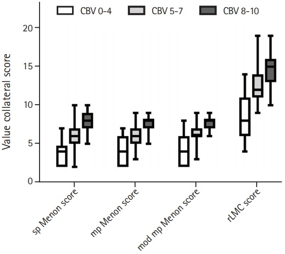
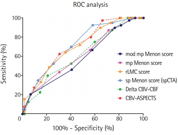
Results: All collateral scores correlated well with each other and with CTP parameters. Comparison of collateral scores stratified by extent of perfusion deficit showed relevant differences between groups (P<0.01 for each). An spCTA collateral score discriminated best between favorable and unfavorable outcome as determined using the modified Rankin Scale 3 months after stroke.

Conclusion: s Collateral status evaluated on spCTA may suffice for outcome prediction and decision making in AIS patients, potentially obviating further imaging modalities like mpCTA or CTP.

Keywords: Acute stroke; Collateral circulation; Computed tomography angiography; Perfusion; Treatment outcome.


Figures

Figure 1.
Figure 1.
Exemplary images of two patients with an occlusion of the left carotid terminus and excellent (top row, A-D) and poor collateral flow (bottom row, E-H). The occlusion of the distal left internal carotid artery can be identified on coronal reformations of the single-phase computed tomography (CT) angiography (spCTA) (A, E: arrowheads). Collateral flow was evaluated on spCTA images (B, F) using the single-phase Menon score (sp Menon score),[14] which compares collaterals within the symptomatic to the contralateral hemisphere. Patient A (top row) had increased prominence and extent of pial vessels within the left symptomatic hemisphere on spCTA images (B, arrows) corresponding to a sp Menon score of 10. In contrast, on the spCTA of patient B (bottom row) just a few pial vessels were visible in the symptomatic hemisphere (F, arrows) resulting in a sp Menon score of 2. Additionally, collateral flow was analyzed on three-phase multi-phase CT angiography (3p-mpCTA) images (C, G) applying the multi-phase Menon score (mp Menon score).[13] The 3p-mpCTA protocol was simulated by choosing the first phase according to the peak arterial phase of the 4D CTA (first image in C, G) and reconstructing the two following phases with a temporal resolution of 7.5 seconds (second and third images in C, G). Patient A exhibited a one-phase delay in filling in of peripheral vessels within the symptomatic hemisphere (arrows in the second and third image in C), but their prominence and extent was similar to the contralateral hemisphere. This resulted in a mp Menon score of 8. Patient B had a one phase delay in filling in of peripheral vessels (arrows in the second and third image in G) and some ischemic regions with no vessels, corresponding to a mp Menon score of 2. Cerebral blood volume (CBV) maps of the two patients are shown in D and H. Patient A had a rather small CBV deficit in the left frontal middle cerebral artery territory and the lentiform nucleus depicted in purple (D). The patient was scored with a CBV-Alberta Stroke Program Early CT Score (ASPECTS) of 7. In contrast, patient B had a CBV deficit in the complete left ACM territory (purple area in H), corresponding to a CBV-ASPECTS of 0.
Figure 2.
Figure 2.
Boxplots of collateral scores stratified by cerebral blood volume (CBV) groups are shown. Patients were trichotomized in three groups ac - cording to their CBV-Alberta Stroke Program Early CT Score (ASPECTS) grades (0 to 4, shown in white; 5 to 7, shown in light grey; and 8 to 10, shown in dark grey) reflecting severity of perfusion deficits and the collat - eral scores were compared between these groups. A P<0.01 for all compar - isons. sp Menon score, single-phase Menon score; mp Menon score, mul - ti-phase Menon score; mod mp Menon score, modified multi-phase Menon score; rLMC score, regional leptomeningeal collateral score.
Figure 3.
Figure 3.
The comparison of receiver operating characteristic (ROC) analyses regarding favorable outcome as defined as a 90-day modified Rankin Scale ≤2 of collateral scores and computed tomography (CT) perfusion parameters is shown. The single-phase Menon score (sp Menon score; blue, solid line), cerebral blood volume (CBV)-Alberta Stroke Program Early CT Score (ASPECTS; red, dashed line), and the regional leptomeningeal collateral score (rLMC score; orange, solid line) discriminate best between favorable and unfavorable outcome. The scores assessed on single-phase CT angiography (spCTA; sp Menon and rLMC score) perform relevantly better than those derived from multi-phase CT angiography (multi-phase Menon score [mp Menon score), pink, solid line; and modified multi-phase Menon score [mod mp Menon score], violet, solid line). CBF, cerebral blood flow.

References

    1. Sheth SA, Liebeskind DS. Collaterals in endovascular therapy for stroke. Curr Opin Neurol. 2015;28:10–15. - PubMed
    1. Elijovich L, Goyal N, Mainali S, Hoit D, Arthur AS, Whitehead M, et al. CTA collateral score predicts infarct volume and clinical outcome after endovascular therapy for acute ischemic stroke: a retrospective chart review. J Neurointerv Surg. 2016;8:559–562. - PubMed
    1. Gersing AS, Schwaiger BJ, Kleine JF, Kaesmacher J, Wunderlich S, Friedrich B, et al. Clinical outcome predicted by collaterals depends on technical success of mechanical thrombectomy in middle cerebral artery occlusion. J Stroke Cerebrovasc Dis. 2017;26:801–808. - PubMed
    1. Higashida R, Furlan A, Roberts H, Tomsick T, Connors B, Barr J, et al. Trial design and reporting standards for intraarterial cerebral thrombolysis for acute ischemic stroke. J Vasc Interv Radiol. 2003;14:S493–S494. - PubMed
    1. Ruland S, Ahmed A, Thomas K, Zhao M, Amin-Hanjani S, Du X, et al. Leptomeningeal collateral volume flow assessed by quantitative magnetic resonance angiography in large-vessel cerebrovascular disease. J Neuroimaging. 2009;19:27–30. - PubMed

LinkOut - more resources