. 2019 Aug;26(8):1045-1053.
doi: 10.1177/1933719118804411. Epub 2018 Oct 11.

Antenatal Microbial Colonization of Mammalian Gut

Affiliations

Antenatal Microbial Colonization of Mammalian Gut

Elisa Borghi et al. Reprod Sci. 2019 Aug.

Abstract

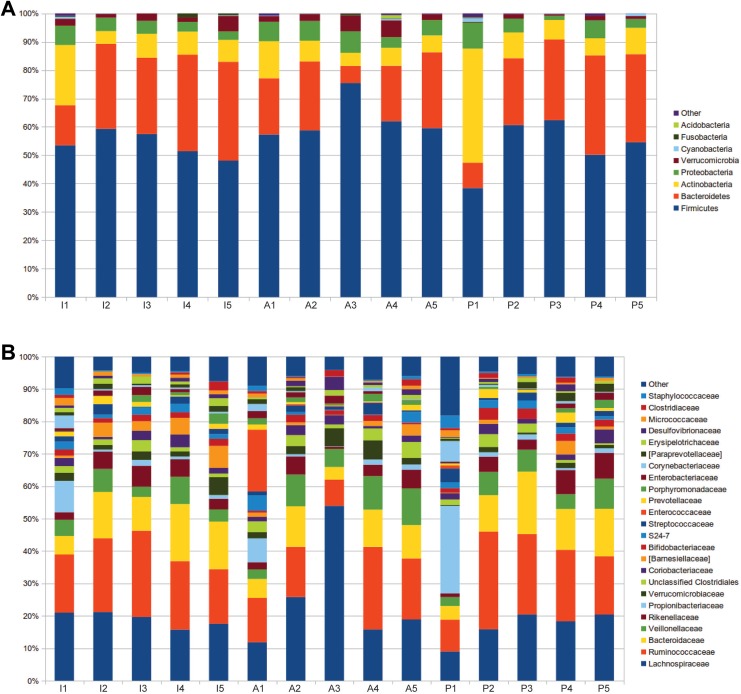
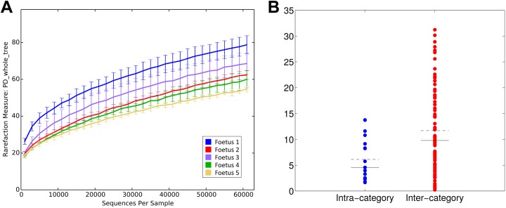
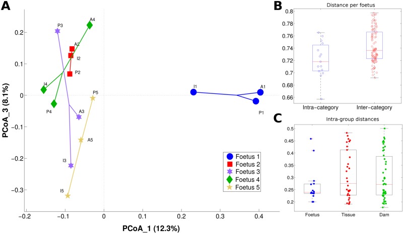
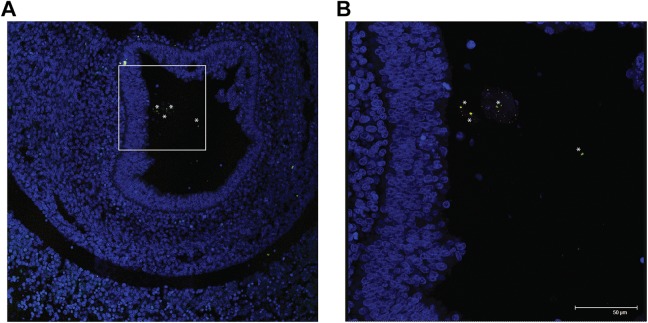
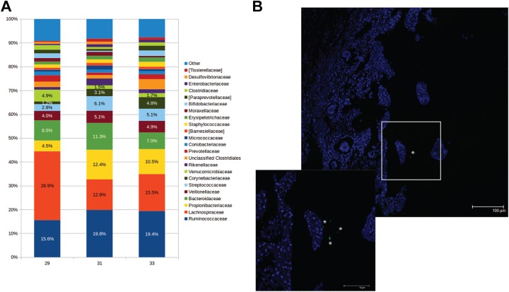
The widely accepted dogma of intrauterine sterility and initial colonization of the newborn during birth has been blurred by recent observations of microbial presence in meconium, placenta, and amniotic fluid. Given the importance of a maternal-derived in utero infant seeding, it is crucial to exclude potential environmental or procedural contaminations and to assess fetal colonization before parturition. To this end, we analyzed sterilely collected intestinal tissues, placenta, and amniotic fluid from rodent fetuses and tissues from autoptic human fetuses. Total bacterial DNA was extracted from collected samples and analyzed by Next Generation Sequencing (NGS) techniques using hypervariable 16S ribosomal RNA (rRNA) regions (V3-V4). Colonizing microbes were visualized in situ, using labeled probes targeting 16S ribosomal DNA by fluorescent in situ hybridization. The NGS analysis showed the presence of pioneer microbes in both rat and human intestines as well as in rodent placentas and amniotic fluids. Microbial communities showed fetus- and dam-dependent clustering, confirming the high interindividual variability of commensal microbiota even in the antenatal period. Fluorescent in situ hybridization analysis confirmed the microbes' presence in the lumen of the developing gut. These findings suggest a possible antenatal colonization of the developing mammalian gut.

Keywords: 16S rRNA gene sequencing; embryonic development; mammalian gut; microbiota.


Conflict of interest statement

Declaration of Conflicting Interests: The author(s) declared no potential conflicts of interest with respect to the research, authorship, and/or publication of this article.

Figures

Figure 1.
Figure 1.
Pioneer microbiota in the developing rodent gut bar charts representing the relative abundance of 5 fetal intestines (I1-I5), amniotic fluids (A1-A5), and placentas (P1-P5). The figure shows relative abundance of bacterial (A) phyla and (B) families.
Figure 2.
Figure 2.
Microbial biodiversity (α-diversity) is fetus-specific (A) α-diversity rarefaction curves according to faith’s phylogenetic distance (“PD whole Tree”). X-axis reports the number of sequences per sample, whereas Y-axis shows the value of the metric. Samples are grouped based on fetus number. (B) Distribution of distances between α-diversity PD whole tree values; Distances are labeled as “intra-” or “inter-category” according to fetus number. Dashed black line represents the mean of the distances, whereas the solid black line represents the median.
Figure 3.
Figure 3.
Microbiota composition (β-diversity) is fetus-specific (A) principal coordinates analysis (PCoA) of the unweighted UniFrac distances; PCoA components 1 and 3 are reported. Samples are connected together on the basis of fetus number. (B) Boxplots of intra- and intercategory unweighted UniFrac distances among samples; categories are based on the fetus number. (C) Boxplots of intracategory weighted UniFrac distances among samples; samples are grouped according to fetus, tissue, or dam.
Figure 4.
Figure 4.
Eubacteria in the developing rodent gut lumen confocal microscopy images showing (A) eubacteria (in green) in the lumen of a 16-day post coitum rat fetus; (B) at a higher magnification (inset is represented as a white box in A) note the typical bacterial morphology and it is possible to identify few bacteroides spp. (yellow). In blue is DAPI (4′,6-diamidino-2-phenylindole) (nuclei) and 50-μm scale bar is reported in B. Asterisks indicate bacterial cells.
Figure 5.
Figure 5.
Eubacteria in the developing human gut (A) bar charts representing the family relative abundance at family level of 3 fetal human intestines (29, 31, and 33 weeks of gestation, respectively). (B) Representative confocal microscopy images of in situ hybridization showing the presence of eubacteria (in green-, inset showing higher magnification) in the lumen human fetuses. In blue is DAPI (nuclei) and 100 μm and 50 μm (inset) scale bars are reported. Asterisks indicate bacterial cells.

References

    1. Brugman S, Perdijk O, van Neerven RJJ, Savelkoul HFJ. Mucosal immune development in early life: setting the stage. Arch Immunol Ther Exp. 2015;63(4):251–268. - PMC - PubMed
    1. Ardissone AN, de la Cruz DM, Davis-Richardson AG, et al. Meconium microbiome analysis identifies bacteria correlated with premature birth. PLoS One. 2014;9(3):e90784. - PMC - PubMed
    1. Aagaard K, Ma J, Antony KM, Ganu R, Petrosino J, Versalovic J. The placenta harbors a unique microbiome. Sci Transl Med. 2014;6(237):237ra65. - PMC - PubMed
    1. Funkhouser LJ, Bordenstein SR. Mom knows best: the universality of maternal microbial transmission. PLoS Biol. 2013;11(8):1–9. - PMC - PubMed
    1. Jiménez E, Fernández L, Marín ML, et al. Isolation of commensal bacteria from umbilical cord blood of healthy neonates born by cesarean section. Curr Microbiol. 2005;51(4):270–274. - PubMed

Publication types

Substances

LinkOut - more resources