. 2018 Oct 11;16(1):79.
doi: 10.1186/s12951-018-0406-6.

Gold nanoparticle distribution in advanced in vitro and ex vivo human placental barrier models

Affiliations

Gold nanoparticle distribution in advanced in vitro and ex vivo human placental barrier models

Leonie Aengenheister et al. J Nanobiotechnology. .

Abstract

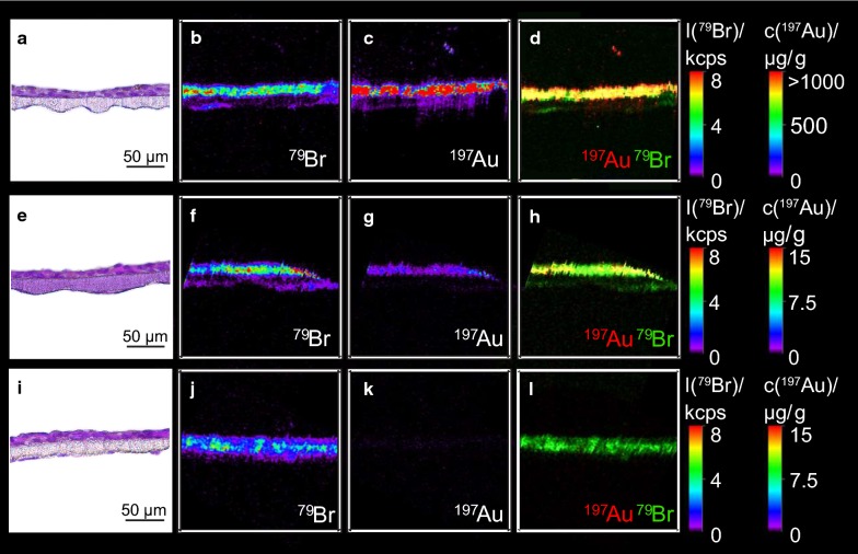
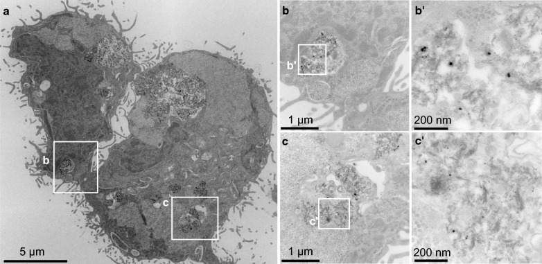
Background: Gold nanoparticles (AuNPs) are promising candidates to design the next generation NP-based drug formulations specifically treating maternal, fetal or placental complications with reduced side effects. Profound knowledge on AuNP distribution and effects at the human placental barrier in dependence on the particle properties and surface modifications, however, is currently lacking. Moreover, the predictive value of human placental transfer models for NP translocation studies is not yet clearly understood, in particular with regards to differences between static and dynamic exposures. To understand if small (3-4 nm) AuNPs with different surface modifications (PEGylated versus carboxylated) are taken up and cross the human placental barrier, we performed translocation studies in a static human in vitro co-culture placenta model and the dynamic human ex vivo placental perfusion model. The samples were analysed using ICP-MS, laser ablation-ICP-MS and TEM analysis for sensitive, label-free detection of AuNPs.

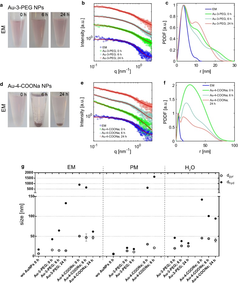
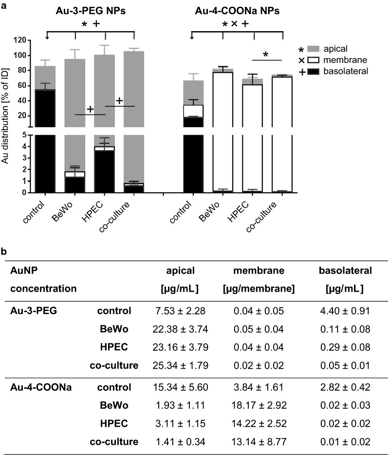
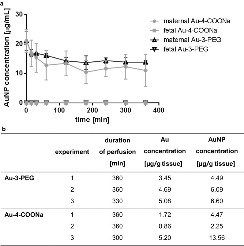
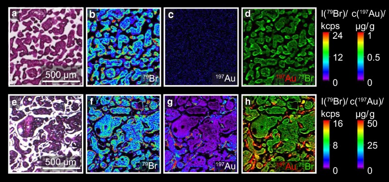
Results: After 24 h of exposure, both AuNP types crossed the human placental barrier in vitro, although in low amounts. Even though cellular uptake was higher for carboxylated AuNPs, translocation was slightly increased for PEGylated AuNPs. After 6 h of perfusion, only PEGylated AuNPs were observed in the fetal circulation and tissue accumulation was similar for both AuNP types. While PEGylated AuNPs were highly stable in the biological media and provided consistent results among the two placenta models, carboxylated AuNPs agglomerated and adhered to the perfusion device, resulting in different cellular doses under static and dynamic exposure conditions.

Conclusions: Gold nanoparticles cross the human placental barrier in limited amounts and accumulate in placental tissue, depending on their size- and/or surface modification. However, it is challenging to identify the contribution of individual characteristics since they often affect colloidal particle stability, resulting in different biological interaction in particular under static versus dynamic conditions. This study highlights that human ex vivo and in vitro placenta models can provide valuable mechanistic insights on NP uptake and translocation if accounting for NP stability and non-specific interactions with the test system.

Keywords: Ex vivo placenta perfusion; Gold nanoparticle; Nanoparticle agglomeration; Placental in vitro co-culture model; Placental uptake and translocation.


Figures

Fig. 1
Fig. 1
Colloidal stability of Au-3-PEG (ac) and Au-4-COONa (df) NPs in EM. a, d Au-3-PEG and Au-4-COONa NP suspensions in EM (19.2 μg/mL Au each) after 0, 6 and 24 h of static incubation (37 °C/5% CO2) in the absence of cells. b, e Scattering intensity as function of scattering vector q and c, f pair distance distribution function (PDDF) of the PEGylated and carboxylated AuNPs in EM. g Diameter of gyration (dgyr; mean ± error) calculated from SAXS data and hydrodynamic diameter from DLS analysis (dhyd; Z-Average)
Fig. 2
Fig. 2
Au distribution in the static in vitro placental barrier model after exposure to Au-3-PEG and Au-4-COONa NPs for 24 h. Control membranes, monocultures or co-cultures were exposed to 19.2 μg/mL Au from Au-3-PEG and Au-4-COONa NPs for 24 h and Au content was determined in apical and basolateral supernatants as well as in the membrane fraction using SF-ICP-MS. Distribution of the Au amount as % of the initial dose (ID; a) and absolute concentrations of the AuNPs (b) in the apical and basolateral chamber and in the membrane fraction, respectively. The latter were calculated from the Au concentrations measured by SF-ICP-MS using the relative Au content of the NPs determined with ICP-OES (see Table 1). Data represent the mean ± SD of 3–4 biologically independent experiments with one technical replicate each. P-value below 0.05 are considered statistically significant (*, x and + are related to apical, membrane and basolateral values)
Fig. 3
Fig. 3
Distribution of Au-4-COONa and Au-3-PEG NPs in the in vitro placental barrier model after 24 h. Co-cultures were exposed to 19.2 μg/mL Au from Au-4-COONa (ad) and Au-3-PEG (eh) NPs for 24 h under static conditions. A co-culture without NP treatment was used as negative control. H&E staining of 5 µm thick sections of the co-culture enables allocation of Au signals (a, e, i; adjacent sections to those for LA-ICP-MS). Elemental distribution of 79Br to visualize cell structure (b, f, j). 197Au was measured to localize Au (c, g, k). Overlay of 79Br and 197Au to co-localize Au signals in cells (d, h, l). Elemental distribution map of 79Br and 197Au is represented in signal intensities and Au concentrations (black: minimum, red: maximum), respectively. Images were obtained from one independent experiment
Fig. 4
Fig. 4
Uptake of Au-4-COONa NPs in BeWo cells after 24 h. a TEM micrographs showing an overview of two BeWo cells. Two different locations with internalized AuNPs (white squares b and c) are presented at higher magnifications (b, b' and c, c')
Fig. 5
Fig. 5
Perfusion kinetics and placental tissue accumulation of Au-3-PEG and Au-4-COONa NPs determined by SF-ICP-MS. a Maternal and fetal AuNP concentration over time (data presented as mean ± SD). AuNP concentrations were calculated from the Au concentrations measured with SF-ICP-MS using the relative Au content of the NPs determined with ICP-OES (see Table 1). b Placental tissue accumulation of Au-3-PEG and Au-4-COONa NPs after 5–6 h of perfusion, shown as Au and AuNP concentration (calculated as indicated before)
Fig. 6
Fig. 6
Distribution of Au-4-PEG NPs in placental tissue after 6 h of perfusion. Placental tissue was perfused with 25 μg/mL Au-3-PEG NPs for 6 h. a, e H&E staining of placental tissue before (a) and after perfusion (e). b, f Elemental distribution of 79Br to visualize tissue structure. c, g 197Au was measured to localize Au. d, h Overlay of 79Br and 197Au to co-localize Au signals in placental tissue. Elemental distribution map of 79Br and 197Au is represented in signal intensities and Au concentrations (black: minimum, red: maximum), respectively. Images were obtained from one independent experiment

References

    1. Dreaden EC, Alkilany AM, Huang X, Murphy CJ, El-Sayed MA. The golden age: gold nanoparticles for biomedicine. Chem Soc Rev. 2012;41(7):2740–2779. doi: 10.1039/C1CS15237H. - DOI - PMC - PubMed
    1. Boisselier E, Astruc D. Gold nanoparticles in nanomedicine: preparations, imaging, diagnostics, therapies and toxicity. Chem Soc Rev. 2009;38(6):1759–1782. doi: 10.1039/b806051g. - DOI - PubMed
    1. Yang Y, Ren L, Wang H. Strategies in the design of gold nanoparticles for intracellular targeting: opportunities and challenges. Ther Deliv. 2017;8(10):879–897. doi: 10.4155/tde-2017-0049. - DOI - PubMed
    1. Keelan JA, Leong JW, Ho D, Iyer KS. Therapeutic and safety considerations of nanoparticle-mediated drug delivery in pregnancy. Nanomedicine. 2015;10(14):2229–2247. doi: 10.2217/nnm.15.48. - DOI - PubMed
    1. Fournier SB, D’Errico JN, Stapleton PA. Engineered nanomaterial applications in perinatal therapeutics. Pharmacol Res. 2018;130:36–43. doi: 10.1016/j.phrs.2018.02.027. - DOI - PMC - PubMed