. 2018 Nov;30(11):2677-2703.
doi: 10.1105/tpc.18.00357. Epub 2018 Oct 11.

A Ycf2-FtsHi Heteromeric AAA-ATPase Complex Is Required for Chloroplast Protein Import

Affiliations

A Ycf2-FtsHi Heteromeric AAA-ATPase Complex Is Required for Chloroplast Protein Import

Shingo Kikuchi et al. Plant Cell. 2018 Nov.

Abstract

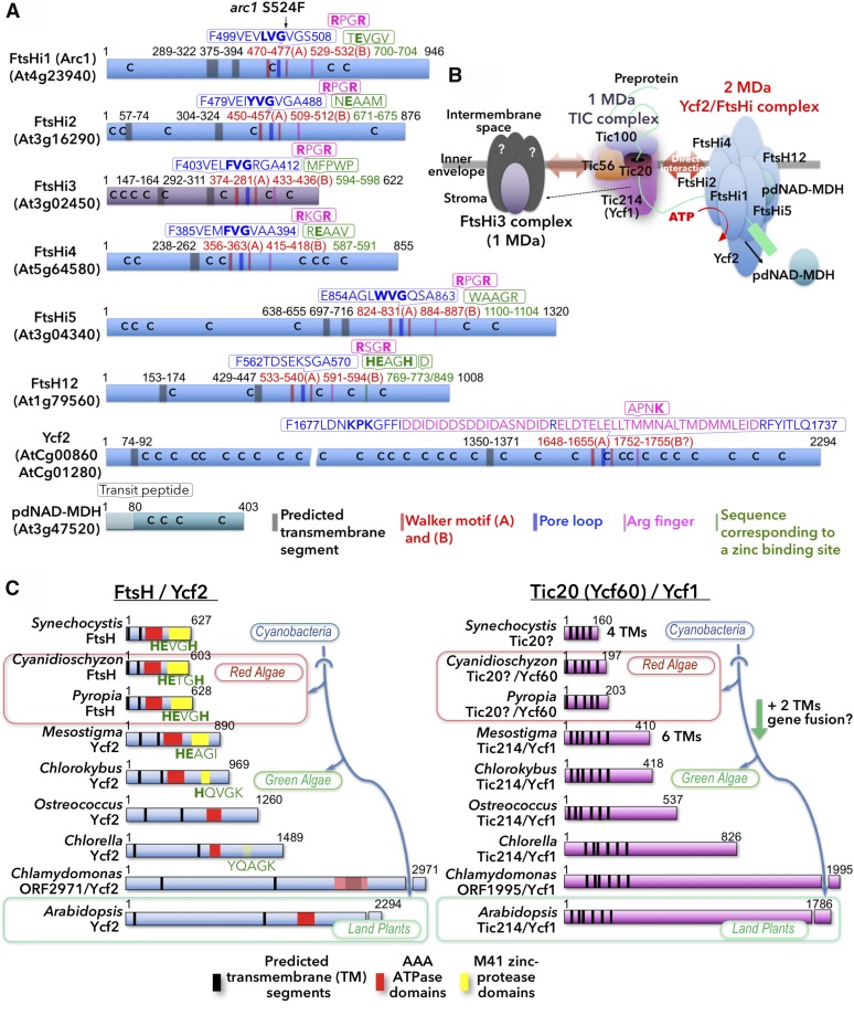
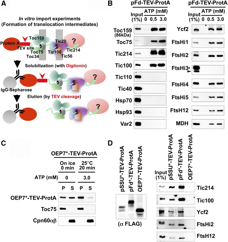
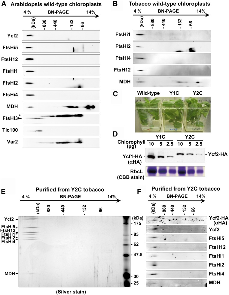
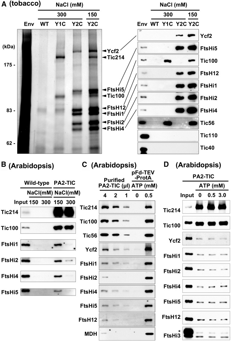
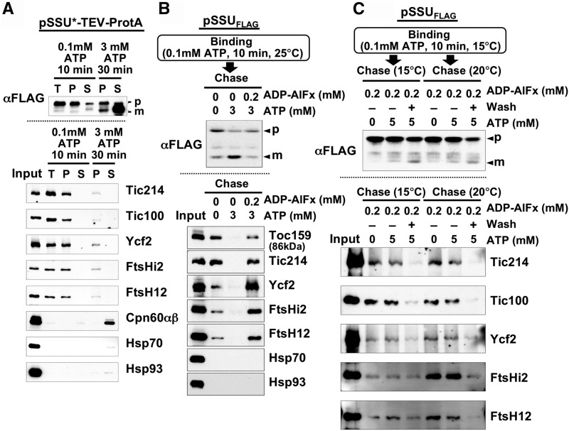
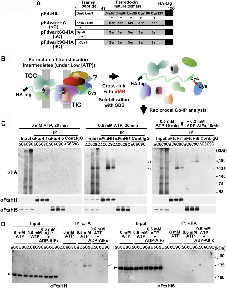
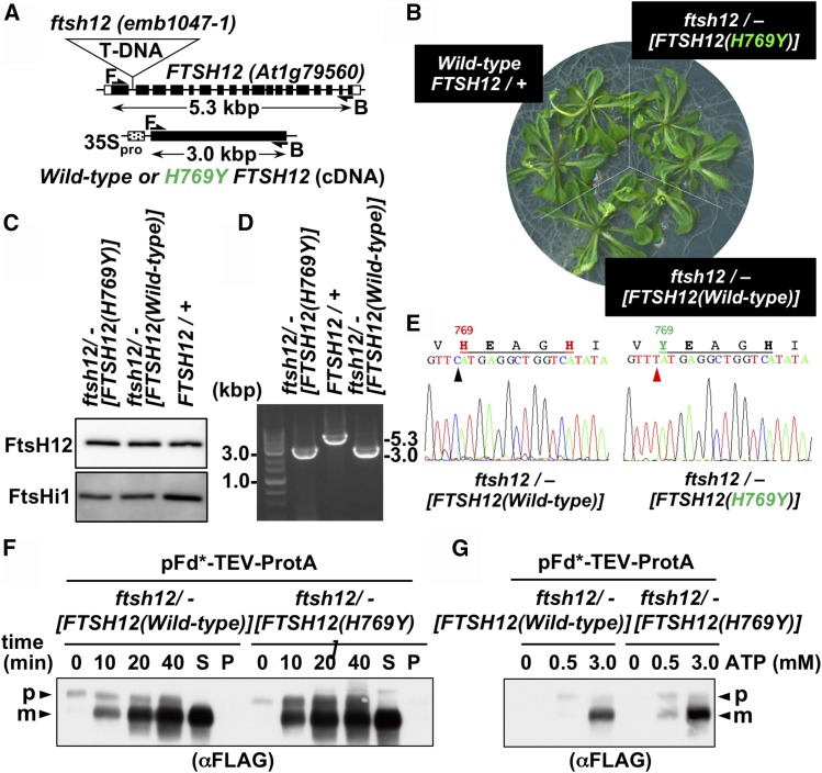
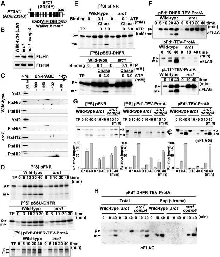
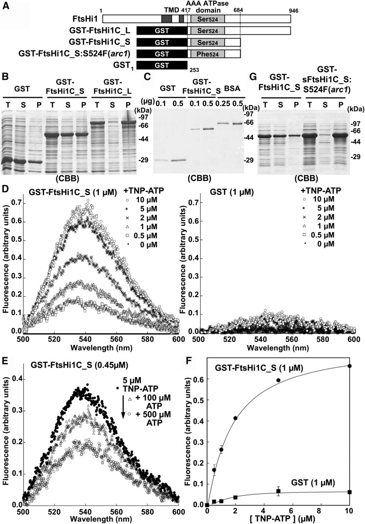
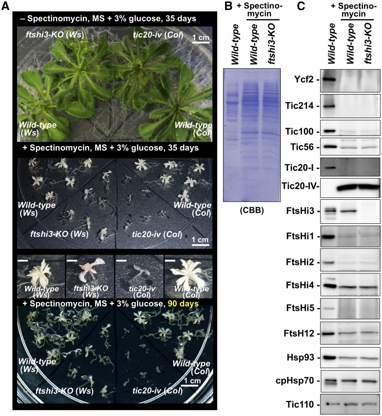
Chloroplasts import thousands of nucleus-encoded preproteins synthesized in the cytosol through the TOC and TIC translocons on the outer and inner envelope membranes, respectively. Preprotein translocation across the inner membrane requires ATP; however, the import motor has remained unclear. Here, we report that a 2-MD heteromeric AAA-ATPase complex associates with the TIC complex and functions as the import motor, directly interacting with various translocating preproteins. This 2-MD complex consists of a protein encoded by the previously enigmatic chloroplast gene ycf2 and five related nuclear-encoded FtsH-like proteins, namely, FtsHi1, FtsHi2, FtsHi4, FtsHi5, and FtsH12. These components are each essential for plant viability and retain the AAA-type ATPase domain, but only FtsH12 contains the zinc binding active site generally conserved among FtsH-type metalloproteases. Furthermore, even the FtsH12 zinc binding site is dispensable for its essential function. Phylogenetic analyses suggest that all AAA-type members of the Ycf2/FtsHi complex including Ycf2 evolved from the chloroplast-encoded membrane-bound AAA-protease FtsH of the ancestral endosymbiont. The Ycf2/FtsHi complex also contains an NAD-malate dehydrogenase, a proposed key enzyme for ATP production in chloroplasts in darkness or in nonphotosynthetic plastids. These findings advance our understanding of this ATP-driven protein translocation system that is unique to the green lineage of photosynthetic eukaryotes.


Figures

Figure 1.
Figure 1.
The Plastome-Encoded AAA Protein Ycf2 and Evolutionarily Related FtsH-Like Proteins. (A) Schematic presentation of the Ycf2/FtsHi complex components and FtsHi3 from Arabidopsis. Positions of predicted transmembrane domains (gray), the Walker motifs (red), the pore loop (blue), the Arg finger (magenta), the zinc binding region (green), and Cys residues (C) are depicted. Amino acid sequences corresponding to the pore loop region and the zinc binding region are shown. (B) Inferred model (deduced by this study) of the 2-MD Ycf2/FtsHi complex function in preprotein translocation across the inner envelope membrane of chloroplasts as a TIC-associated import motor. (C) Coevolution of chloroplast-encoded FtsH/Ycf2 and Tic20 (Ycf60)/Tic214 (Ycf1) of cyanobacterial origin. Schematic representation of size expansion of chloroplast genome-encoded Ycf2 (upper) and Tic214 (Ycf1; lower) proteins along with the evolution of red and green lineages from their most probable ancestral proteins encoded by the cyanobacterial genomes. It should be noted that the zinc binding motif (HEXXH) critical for protease activity of FtsH-type metalloprotease appears to have been gradually lost from Ycf2 proteins during evolution. Sequences from NCBI (http://www.ncbi.nlm.nih.gov/protein): Synechocystis FtsH (BAA10230), Cyanidioschyzon FtsH (BAC76202), Pyropia FtsH (AGH27655), Mesostigma Ycf2 (AAF43852), Chlorokybus Ycf2 (ABD62184), Ostreococcus Ycf2 (CAL36342), Chlorella Ycf2 (ADZ04994), Chlamydomonas Ycf2 (ACJ50152), Arabidopsis Ycf2 (BAA84428), Synechocystis Tic20 homolog (BAA17427), Cyanidioschyzon Tic20/Ycf60 (BAC76161), Pyropia Tic20/Ycf60 (AGH27688), Mesostigma Ycf1 (AAF43878), Chlorokybus Ycf1 (ABD62257), Ostreococcus Ycf1 (CAL36331), Chlorella Ycf2 (ADZ05009), Chlamydomonas Ycf1 (CAA63385)(Boudreau et al., 1997), and Arabidopsis Ycf1 (BAA84445).
Figure 2.
Figure 2.
Association of the Ycf2 and FtsH-Like Proteins with a Model Translocating Preprotein together with TOC and TIC Components. Inferred model of Ycf2/FtsHi complex function. (A) Schematic diagram showing how translocation intermediate-associating proteins/complexes are purified (Kikuchi et al., 2013). First, we performed in vitro import experiments with isolated Arabidopsis chloroplasts using a purified model preprotein consisting of an entire authentic preprotein containing a transit peptide and a C-terminal Protein A-tag (ProtA; 289 amino acids). A cleavage site of TEV protease (12 amino acids) was introduced in between the preprotein and the Protein A-tag. After import, translocation intermediates were purified after solubilization by digitonin using IgG-Sepharose beads. Bound translocating preprotein-associated proteins were eluted under nondenaturing conditions by TEV cleavage and specifically eluted by TEV cleavage under nondenaturing conditions. (B) Affinity purification of translocation intermediate complexes via a model preprotein pFd-TEV-ProtA (ferredoxin preprotein, 146 amino acids; TEV-ProtA, 301 amino acids) after in vitro import reactions under indicated ATP concentration (0, 0.5, and 3 mM) with isolated Arabidopsis chloroplasts for 10 min at 25°C. Purification was performed under high-salt conditions (250 mM NaCl) using digitonin as detergent. Association of various chloroplast proteins with the pFd-TEV-ProtA was assessed by immunoblotting using respective specific antibodies. Input lanes contained 1% equivalent of digitonin-solubilized chloroplast extracts used for each purification. Asterisks, cross-reactions to abundant thylakoidal Var2, a typical FtsH homolog. MDH, pdNAD-MDH. Tic110 and Tic40, well-known classical Tic proteins that are not included in the TIC20/56/100/214 complex did not associate at all with translocating pFd-TEV-ProtA. (C) Insertion of OEP7FLAG-TEV-ProteinA (a 7-kD outer envelope membrane protein, 64 amino acids) (OEP7*-TEV-ProtA) into the outer envelope membrane in vitro. The purified OEP7*-TEV-ProtA was incubated with isolated Arabidopsis chloroplasts in the absence of ATP on ice or in the presence of ATP for 20 min at 25°C. After import, chloroplasts were treated by alkaline extraction with 0.1 M Na2CO3, pH 11.5, and separated into integral membrane proteins (P) and soluble or peripherally-bound membrane proteins (S) by centrifugation. Integration of OEP*-TEV-ProtA was assessed by immunoblotting using anti-FLAG-tag antibody. Toc75 was also analyzed as an integral membrane protein control whereas Cpn60αβ (chaperonin α and β proteins) as soluble proteins. (D) Specific binding of the Ycf2/FtsHi complex components to the translocating preproteins, namely, pS*-TEV-ProtA and pFd*-TEV-ProtA, but not to OEP7*-TEV-ProtA. pS*-TEV-ProtA, pFd*-TEV-ProtA, and OEP7*-TEV-ProtA were separately incubated with isolated Arabidopsis chloroplasts in the presence of 0.1 mM ATP (pS*-TEV-ProtA and pFd*-TEV-ProtA) or 3 mM ATP (OEP7*-TEV-ProtA) for 20 min at 25°C. Chloroplasts were reisolated and washed several times and were analyzed by immunoblotting using anti-FLAG tag antibody to confirm the presence of bound preproteins (left panel). After solubilization of chloroplasts by digitonin, preprotein-associating proteins were purified and analyzed as in (B, right panel).
Figure 3.
Figure 3.
Purification of the 2-MD Ycf2/FtsHi Complex from Transplastomic Tobacco Plants Expressing the C-Terminal HA-Tagged Ycf2 Protein. (A) Ycf2 and FtsH-like proteins migrate 2-MD area upon BN-PAGE analysis of both Arabidopsis and tobacco chloroplasts. 2D-BN/SDS-PAGE analysis of Arabidopsis chloroplasts followed by immunoblotting. Chloroplasts were solubilized with 1% Triton X-100 in the presence of 300 mM NaCl, and solubilized proteins/complexes were separated by 4 to 14% BN-PAGE as the first dimension and SDS-PAGE as the second dimension followed by immunoblotting using antisera against indicated proteins. MDH, pdNAD-MDH. Asterisk indicates cross-reactions to abundant thylakoidal Var2 (FtsH2). (B) Isolated tobacco chloroplasts were analyzed as in (A). (C) Generation of transplastomic tobacco lines expressing C-terminal HA-tagged Ycf2 (Y2C) or Ycf1 (Y1C) proteins. See Supplemental Figure 4 for details. The transplastomic lines (Y2C and Y1C) exhibited normal growth. The Y2C-11 and Y1C-3 lines and their parental tobacco line (Wild-type) were cultivated on MS media containing 3% sucrose for 1 month under the normal growth condition. (D) Comparison of protein levels of Ycf2-HA and Ycf1-HA proteins is shown. Chloroplasts (equivalents to indicated amounts of chlorophylls) isolated from the Y2C-11 (Y2C) and the Y1C-3 (Y1C) lines were analyzed by SDS-PAGE followed by immunoblotting using anti-HA-tag antibody (upper) or by Coomassie Brilliant Blue (CBB) staining to compare the plastome-encoded Rubisco large subunit protein levels. (E) The 2-MD Ycf2/FtsHi complex contains five FtsH-like proteins and pdNAD-MDH. The purified 2-MD Ycf2-HA complex analyzed by silver staining. Chloroplasts isolated from the Y2C-11 line were solubilized by 1% Triton X-100 in the presence of 300 mM NaCl and purified using anti-HA-tag agarose beads. Specifically bound proteins were eluted by HA-peptides and analyzed by 2D-BN/SDS-PAGE analysis followed by silver staining. MDH, pdNAD-MDH. (F) As in (E) but after 2D-BN/SDS-PAGE separation, indicated proteins were analyzed by immunoblotting using corresponding antisera raised against Arabidopsis counterparts.
Figure 4.
Figure 4.
Physical Interaction between the TIC Complex and the Ycf2/FtsHi Complex Observed in Tobacco as well as in Arabidopsis Chloroplasts. (A) Copurification of the TIC complex with the Ycf2/FtsHi complex from tobacco chloroplasts. Chloroplasts isolated from the Y1C and Y2C transplastomic tobacco lines expressing the HA-tagged forms of Tic214(Ycf1) and Ycf2, respectively, together with those from the parental control wild-type line were solubilized with 1% dodecylmaltoside containing 300 mM NaCl and were separately subjected to purification using anti-HA-agarose beads under the presence of 300 mM NaCl. The similar purification was also performed from Y2C chloroplasts but with the presence of physiological salt concentrations (150 mM NaCl) throughout the solubilization and the purification instead of 300 mM NaCl. The purified fractions eluted from the anti-HA agarose beads by the addition of HA peptides were analyzed by silver staining (left) and by immunoblotting (right). Envelope membranes (Env) isolated from the chloroplasts of the parental wild-type line were also analyzed for reference. (B) Purification of the Arabidopsis TIC complex from the Protein A-tagged Tic20 (PA2-TIC)-TIC-expressing chloroplasts under the presence of NaCl and association of Ycf2/FtsHi complex components. Chloroplasts isolated from transgenic Arabidopsis plants expressing the Protein A-tagged Tic20 (PA2-TIC) were used for purification of TIC after solubilization of 1% dodecylmaltoside under the presence of 150 or 300 mM NaCl. The wild-type chloroplasts were used as control. The purified proteins were analyzed by SDS-PAGE followed by immunoblotting. Input, 1% of solubilized extracts. (C) Comparison of amounts of Ycf2/FtsHi complex components associated with the TIC complex with those associated with the translocating preproteins. After in vitro import of pFd-TEV-ProtA in the absence (0 mM) or presence (0.5 mM) of ATP (at 25°C for 10 min) using the wild-type Arabidopsis chloroplasts, the translocation intermediates were purified under high-salt conditions (250 mM NaCl) and analyzed as shown in Figure 3 (right two lanes). The TIC complex was purified from Arabidopsis plants expressing Protein A-tagged Tic20 (PA2-TIC) under the same conditions (i.e., in the presence of 250 mM NaCl), and purified fractions (4, 2, and 1 µL) were analyzed for comparison. MDH, pdNAD-MDH. (D) Absence of increased level of association of Ycf2/FtsHi complex components with TIC complex after simple incubation of chloroplasts with ATP without any addition of preproteins. Chloroplasts isolated from the transgenic Arabidopsis plants expressing PA2-TIC were incubated with the indicated amount of ATP (0–3.0 mM) at 25°C for 10 min and subjected for the purification of the PA2-TIC complex as in (C) in the presence of 250 mM NaCl. The purified proteins were analyzed by SDS-PAGE and immunoblotting. Input, 1% equivalent of the solubilized extracts of PA2-TIC chloroplasts. Asterisk indicates cross-reactions to abundant thylakoidal Var2, a typical FtsH homolog.
Figure 5.
Figure 5.
The Ycf2/FtsHi Complex Is Directly Involved in the ATP-Dependent Preprotein Translocation at the Inner Envelope of Chloroplasts. (A) Association of stromal chaperones occurs at a rather later stage mostly after preprotein translocation into the stroma. In vitro import experiments were performed using Arabidopsis chloroplasts and the purified pSSU*-TEV-ProtA in the presence of 0.1 mM ATP (for 10 min) or 3 mM ATP (for 30 min). After import, chloroplasts (T, total) were ruptured in hypotonic solution (10 mM HEPES-KOH, pH 7.8, and 4 mM MgCl2) and fractionated into the membrane pellets (P) and the soluble supernatants containing stroma (S). Equivalent aliquots of each fraction were analyzed directly by SDS-PAGE followed by immunoblotting with anti-FLAG-tag antibodies (upper panel). p, precursor form; m, mature form. The obtained membrane pellets and supernatant together with total chloroplasts before fractionation (T) were subjected to purification of preprotein-associating proteins as shown in Figure 2. Equivalent amounts of each purified fraction were analyzed by immunoblotting (lower panel). Input, 1% equivalent of digitonin-solubilized total chloroplast extracts used for purification. (B) The ADP-AlFx causes accumulation of the Ycf2/FtsHi complex-bound form of preproteins. The pSSUFLAG (203 amino acids) synthesized in S30 extracts was incubated with isolated Arabidopsis chloroplasts in the presence of 0.1 mM ATP to accumulate early translocation intermediates (Binding, at 25°C for 10 min). Subsequent chase incubation (at 25°C for 20 min) was performed without any addition or in the presence of 3 mM ATP alone or 3 mM ATP plus 0.2 mM ADP-AlFx. After incubation, aliquots of each reaction were directly analyzed for import of pSSUFLAG by immunoblotting using anti-FLAG-tag antibody (upper panel). p, precursor form; m, mature form. The remainders were subjected to purification of preprotein-associating proteins using anti-FLAG-tag M2 affinity gel instead of IgG-Sepharose beads and analyzed by immunoblotting as in (A) (lower panel). Input, 1% equivalent of digitonin-solubilized chloroplast extracts used for purification. (C) Preproteins arrested as translocation intermediates in the presence of ADP-AlFx can be imported into the chloroplast to form a mature protein by washing out the ADP-AlFx and subsequent addition of excess ATP. After binding in the presence of 0.1 mM ATP (at 15°C for 10 min), translocation intermediates were accumulated in the presence of 0.2 mM ADP-AlFx as in (B) but by incubating for 10 min at low temperature (15°C or 20°C) to slow down the translocation process. Complete translocation (formation of mature form) was observed by further chase incubation for additional 20 min in the presence of excess 5 mM ATP only after washing out the ADP-AlFx (Wash). After incubation, aliquots of each reaction were directly analyzed for import of pSSUFLAG as in (B) (upper panel). The remainder was subjected to purification of preprotein-associating proteins using anti-FLAG-tag M2 affinity gel and analyzed as in (B) (lower panel).
Figure 6.
Figure 6.
Direct Interaction between the Transit Peptide Moiety of Translocating Preproteins and the Ycf2/FtsHi Complex Components Revealed by Site-Specific Chemical Cross-Linking. (A) Schematic presentations of the model preproteins used for site-specific cross-linking. All Cys residues in pFd-HA, a HA-tagged ferredoxin preprotein (156 amino acids), were substituted by Ser residues to construct pFdvari-HA(ΔC). Then, Ser-6 and Leu-9 residues were individually mutated to Cys and resulting constructs were named pFdvari-HA(6C) and pFdvari-HA(9C), respectively. (B) Procedures of chemical cross-linking. Translocation intermediates were accumulated under low ATP conditions and the reisolated chloroplasts were subjected to chemical cross-linking with the membrane-permeable cross-linker BMH, which covalently cross-links sulfuhydryl groups of two adjacent Cys residues. After complete solubilization and denaturation by addition of 1% SDS, immunoprecipitation was performed using the anti-HA agarose or using the purified IgGs specific to the Ycf2/FtsHi complex components that had been conjugated to Protein A Sepharose. Immunoprecipitated proteins were analyzed for coimmunoprecipitation of directly interacting proteins (Reciprocal Co-IP) by SDS-PAGE followed by immunoblotting. (C) Arabidopsis chloroplasts were incubated with pFd-HA derivatives, ΔC, 6C, and 9C, separately in the absence (0 mM) (left panels) or presence (0.5 mM) (central panels) of ATP at 25°C for 20min. For experiments shown in the right panels, import reactions were performed in the presence of 0.5 mM ATP at 25°C for 10 min followed by further 10 min incubation at 25°C after addition of 0.2 mM ADP-AlFx. After import reactions, cross-linking with BMH and subsequent SDS-solubilization/denaturation were performed as described above. Immunoprecipitation (IP) was performed using either the purified anti-FtsHi1(αFtsHi1), anti-FtsHi5(αFtsHi5), or control rabbit IgGs (Cont.IgG). Immunoprecipitates were analyzed by SDS-PAGE followed by immunoblotting using anti-HA-tag antibody(αHA) to detect cross-linked and coimmunoprecipitated pFdvari-HA derivatives (white arrowheads, top panels). Specificities of immunoprecipitations were confirmed by immunoblotting using anti-FtsHi1 (middle panels) or anti-FtsHi5 antibodies (bottom panels). Input, 1% equivalent of digitonin-solubilized total chloroplast extracts used for purification. (D) As in (C), but anti-HA-tag(αHA) agarose was used for immunoprecipitation and the cross-linked and coimmunoprecipitated FtsHi1 (left panel) and FtsHi5 (right panel, arrowheads) were detected by immunoblotting. Note that apparent sizes of ATP-dependent cross-linked products observed in the reciprocal co-IP analysis shown in (C) and (D) were perfectly matched with each other and well correspond to the estimated size of 1:1 cross-linked product between a pFdvari-HA and FtsHi1 or FtsHi5 proteins.
Figure 7.
Figure 7.
A Remaining Zinc Binding Motif of FtsH12 Is Dispensable for the Essential Function of FtsH12 in Arabidopsis. (A) The position of T-DNA insertion in the FTSH12 locus on the Arabidopsis genome. Transgene cDNA constructs placed under the 35S promoter (35Spro) together with the positions of PCR primers used for genotyping were also depicted. (B) Functional complementation of the embryo-lethal homozygous ftsH12 null mutant (emb1047-1) by expressing the FtsH12(H769Y) mutant. Individuals carrying the indicated genotypes were grown on the MS media containing 2% sucrose for 25 d. (C) Protein levels in the transformants and in the wild type were analyzed by SDS-PAGE and immunoblotting. Ten micrograms of total protein extracts of each seedling was analyzed. (D) PCR genotyping confirmed the homozygous T-DNA insertions in the ftsh12 locus of the obtained transformants. The forward (F) and backward (B) primers shown in (A) were used. (E) The genomic PCR fragments obtained from the transformants were directly sequenced. Only the mutated region was shown. (F) The FtsH12(H769Y) mutant showed normal chloroplast protein import. Chloroplasts were isolated from the homozygous T-DNA inserted line expressing the wild-type FtsH12 or the mutant FtsH12(H769Y) and used for the in vitro import experiments with pFd*-TEV-ProtA as a model preprotein. Import was performed in the presence of 3 mM ATP for 0, 10, 20, and 40 min and analyzed by SDS-PAGE followed by immunoblotting using anti-FLAG antibody. After 40 min incubation, another aliquot of chloroplasts was ruptured to separate the soluble supernatant containing stroma (S) from the membrane pellet (P) and analyzed similarly. m, mature form; p, precursor form. (G) ATP-dependent import of pFd*-TEV-ProtA into the chloroplasts expressing the wild-type FtsH12 or FtsH12(H769Y) mutant was analyzed as in (F).
Figure 8.
Figure 8.
The arc1 Mutation (S524F) Near the Walker B Motif of FtsHi1 Causes Severe Protein Import Defects. (A) The position of arc1 mutation in the FTSHi1 locus on the Arabidopsis genome. The arc1 encodes a mutant form of FtsHi1 whose Ser-524 was substituted with Phe. (B) The protein expression levels in the arc1 mutant were compared with those in the wild type by SDS-PAGE followed by immunoblotting. Forty micrograms of proteins of total cell extracts was loaded. The wild-type genomic copy of FTSHi1-complemented chloroplasts (arc1 comp4) was also analyzed. (C) The arc1 (ftsHi1-1) mutation does not affect the formation of the Ycf2/FtsHi complex. Chloroplasts (20 µg chlorophyll) isolated either from the wild-type or the arc1 seedlings were subjected to 2D-BN/SDS-PAGE analysis followed by the immunoblotting after solubilization with 1% Triton X-100 in the presence of 300 mM NaCl as shown in Figure 3A. (D) The arc1 chloroplasts impair the ATP-dependent in vitro import of various preproteins. In vitro import experiments were performed in the presence of 3 mM ATP using chloroplasts (50 µg chlorophyll) isolated either from the wild-type or the arc1 seedlings. Import time-course analysis with [35S]-labeled preproteins synthesized in the cell-free lysates is shown. Chloroplasts isolated from the import reactions after the indicated time of incubation at 25°C were analyzed by SDS-PAGE followed by autoradiography to detect [35S]-labeled proteins. p, precursor form; m, mature form. TP, 10% of translated preproteins used for the import reaction. (E) The import defects of the arc1 mutant occur at the high ATP-requiring preprotein translocation step across the inner membrane rather than the initial binding step. In vitro import experiments were performed as in (D) but initially without (0 mM) or with 0.1 mM ATP for 10 min at 25°C to form early translocation intermediates (Binding). After reisolation of chloroplasts from each reaction, chase incubations were performed in the absence (0 mM) or the presence (3 mM) of ATP for 40 min at 25°C and analyzed as in (D). (F) Import time-course analysis with the E. coli-expressed purified preproteins, pFd*-DHFR-TEV-ProtA, pFd*-TEV-ProtA, and pL11*-TEV-ProtA, were performed as in (D). Urea-denatured purified preproteins (1–2 µg) were incubated with chloroplasts (50 µg chlorophyll) isolated either from the wild-type or from the arc1 mutant seedlings in the presence of 3 mM ATP at 25°C. At indicated time points, chloroplasts were reisolated, washed, and analyzed by SDS-PAGE followed by immunoblotting using anti-FLAG tag antibody. (G) Comparison of import of the purified preproteins with that of [35S]-labeled preproteins synthesized in the cell-free lysates. Import experiments were performed with the [35S]-labeled pFNR (left) or pFd*-TEV-protA (center) synthesized in the cell-free lysates or with the purified pFd*-TEV-ProtA (right) and analyzed by autoradiography or immunoblotting as in (D) and (F), respectively. The wild-type FTSHi1-complemented chloroplasts (arc1 comp4) (Kadirjan-Kalbach et al., 2012) were also analyzed as controls. Processed mature proteins (M) were quantified as described in Methods and are indicated by bar graphs at the bottom (n = 3). The amount of mature proteins observed after 40 min import with the wild-type chloroplasts was set to 100%. (H) Precursor form accumulated after import reactions in the arc1 chloroplasts remained membrane-bound. Import time-course analysis with the purified preproteins, pFd*-DHFR-TEV-ProtA, was performed as in (F). After import, chloroplasts (Total) were ruptured in hypotonic solution (10 mM HEPES-KOH, pH 7.8, and 4 mM MgCl2) and the soluble supernatants containing stroma (Sup) were obtained by centrifugation to remove membrane pellets and membrane-bound preproteins. Equivalent amounts of the total and Sup fractions were analyzed by SDS-PAGE followed by immunoblotting using anti-FLAG-tag antibody.
Figure 9.
Figure 9.
Analysis of ATP Binding to FtsHi1. (A) Schematic representation of domain structure of Arabidopsis FtsHi1 and the GST-fusion proteins used for the analysis of ATP binding. The corresponding fusion genes were cloned into a pCold-GST vector. GST, glutathione S-transferase (from Schistosoma japonicum); TMD, transmembrane domain. FtsHi1C_L and FtsHi1C_S are long and short form of the C-terminal domain of FtsHi1. (B) GST, GST-FtsHiC_L, and GST-FtsHiC_S were expressed in E. coli cells at 15°C. Cells were suspended in 50 mM Tris-HCl, pH 7.5, and 150 mM NaCl and disrupted by freeze-thaw cycles with bath sonication and separated into soluble (S) and insoluble pellet (P) fractions. Equivalent amounts of each fraction and total cell extracts (T) were analyzed by SDS-PAGE followed by Coomassie blue staining. (C) Purified GST and GST-FtsHi1C proteins were analyzed by as in (B) with BSA for comparison. (D) TNP-ATP binding assay of the purified GST-FtsHiC_S (left panel) and the GST (right panel). Fluorescence emission spectra (excitation 410 nm) of TNP-ATP bound to the purified proteins (1 µM). Reference spectra obtained with the corresponding concentration of TNP-ATP without any addition of protein and those with 1 µM proteins but no added TNP-ATP were subtracted as background. (E) Displacement of bound TNP-ATP from GST-FtsHi1C_S with ATP. GST-FtsHi1C_S (0.45 µM) was preincubated with TNP-ATP (5 µM) in a cuvette to form TNP-ATP:GST-FtsHi1C_S complex (closed circles). Subsequently, indicated concentration of ATP was added into a cuvette and, after 1 min, fluorescence emission spectra were recorded (triangles, open circles). (F) Determination of TNP-ATP:GST-FtsHi1C_S dissociation constant. Fluorescence intensities of bound TNP-ATP to GST-FtsHi1C_S (1 µM) (closed circles) and to GST (1 µM) (closed squares) as a function of TNP-ATP concentration were measured at 540 nm as in (D) (n = 3). Data are shown as an average of three independent measurements ± sd.
Figure 10.
Figure 10.
Inhibition of Chloroplast Translation by Spectinomycin Causes Deficiency in Chloroplast-Encoded Tic214(Ycf1) and Ycf2 Accumulation and Abolishes Growth of Arabidopsis Seedlings. (A) Homozygous knockout mutants for FtsHi3 (ftsHi3-KO) or Tic20-IV (tic20-iv), a minor isoform of Tic20, were grown on MS media for 35 d with (middle) or without (top) 50 µg/mL spectinomycin together with corresponding wild types (Parker et al., 2016). No further growth on spectinomycin-containing media from 35 d after sowing onward (90 d, at the bottom). (B) and (C) Proteins extracted from the 35-d-old seedlings of wild-type and ftshi3-KO mutant were analyzed by Coomassie Brilliant Blue (CBB) staining (B) and immunoblotting (C). Five micrograms of total proteins of the wild-type seedlings grown in the absence of spectinomycin and 20 µg of total proteins of the wild-type or the ftshi3-KO seedlings grown in the presence of spectinomycin were loaded. Remarkably high expression of Tic20-IV was observed in the seedlings grown in the presence of spectinomycin.

Comment in

Similar articles

Cited by

References

    1. Asakura Y., Hirohashi T., Kikuchi S., Belcher S., Osborne E., Yano S., Terashima I., Barkan A., Nakai M. (2004). Maize mutants lacking chloroplast FtsY exhibit pleiotropic defects in the biogenesis of thylakoid membranes. Plant Cell 16: 201–214. - PMC - PubMed
    1. Beeler S., Liu H.C., Stadler M., Schreier T., Eicke S., Lue W.L., Truernit E., Zeeman S.C., Chen J., Kötting O. (2014). Plastidial NAD-dependent malate dehydrogenase is critical for embryo development and heterotrophic metabolism in Arabidopsis. Plant Physiol. 164: 1175–1190. - PMC - PubMed
    1. Bodnar N.O., Kim K.H., Ji Z., Wales T.E., Svetlov V., Nudler E., Engen J.R., Walz T., Rapoport T.A. (2018). Structure of the Cdc48 ATPase with its ubiquitin-binding cofactor Ufd1-Npl4. Nat. Struct. Mol. Biol. 25: 616–622. - PMC - PubMed
    1. Boudreau E., Turmel M., Goldschmidt-Clermont M., Rochaix J.-D., Sivan S., Michaels A., Leu S. (1997). A large open reading frame (orf1995) in the chloroplast DNA of Chlamydomonas reinhardtii encodes an essential protein. Mol. Gen. Genet. 253: 649–653. - PubMed
    1. Bruce B.D., Perry S., Froehlich J., Keegstra K. (1994). In vitro Import of Proteins into Chloroplasts. In Plant Molecular Biology Manual, Gelvin S.B., Schilperoot R.A., eds (Dordrecht: Springer; ), pp. 1–15.

Publication types

MeSH terms

LinkOut - more resources