. 2018 Sep 27:11:2095-2106.
doi: 10.2147/JPR.S144852. eCollection 2018.

PKCβII-induced upregulation of PGP9.5 and VEGF in postoperative persistent pain in rats

Affiliations

PKCβII-induced upregulation of PGP9.5 and VEGF in postoperative persistent pain in rats

Xiang Zhu et al. J Pain Res. .

Abstract

Purpose: Postoperative pain is a common clinical problem. In this study, we aimed to investigate the role of protein kinase C βII (PKCβII) in the progression of postoperative pain following skin/muscle incision and retraction (SMIR) surgery.

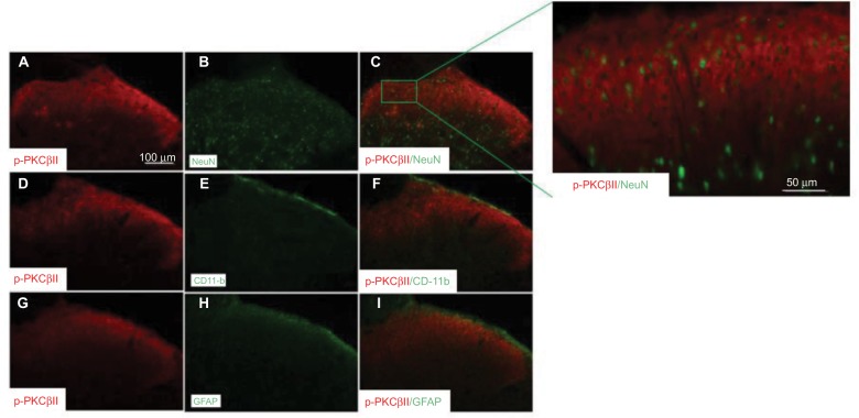
Materials and methods: SMIR postoperative pain model was established in rats, akin to a clinical procedure. The expression level and location of p-PKCβII were observed in dorsal root ganglion (DRG) or spinal cord from SMIR-operated rats by Western blotting and immunofluorescence. In addition, the effects of PKCβII on the expression of protein gene product 9.5 (PGP9.5) or vascular endothelial growth factor (VEGF) were assessed by using pharmacological activator and inhibitor of PKCβII. Moreover, mechanical withdrawal threshold (MWT) was assessed before or after SMIR-operated rats were treated with inhibitor or activator of PKCβII.

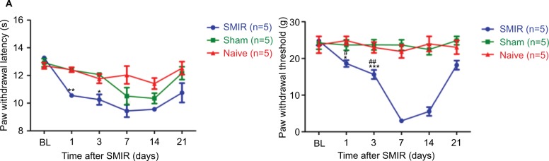
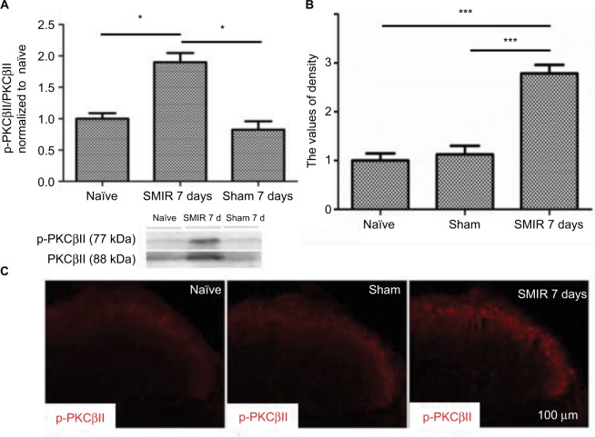
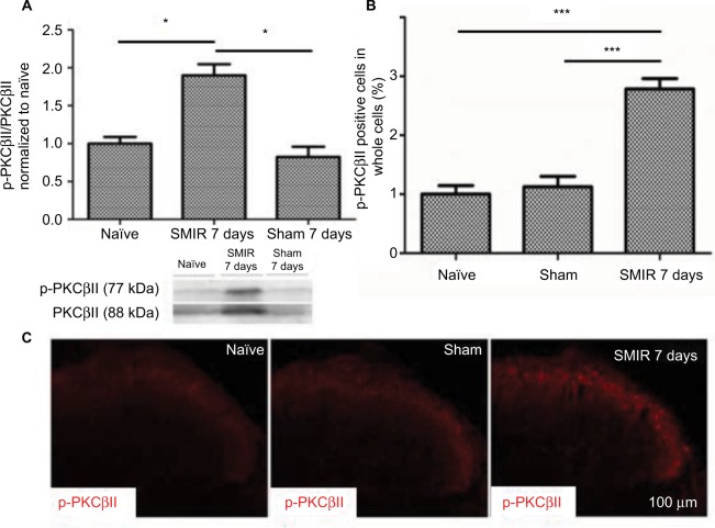
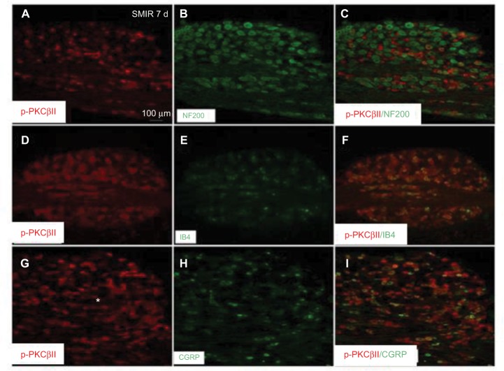
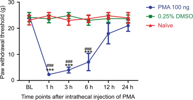
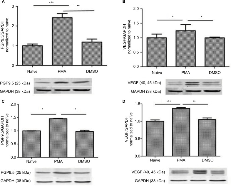
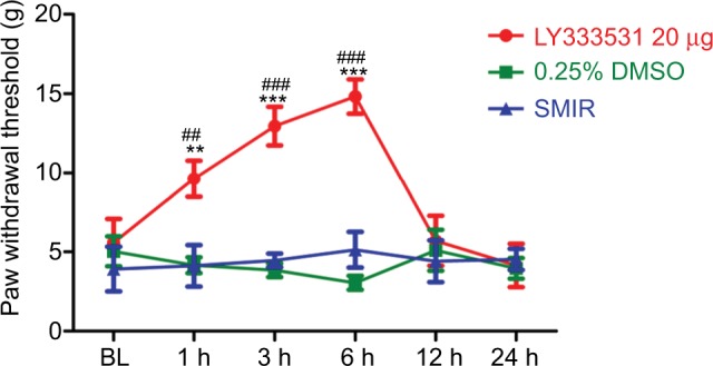
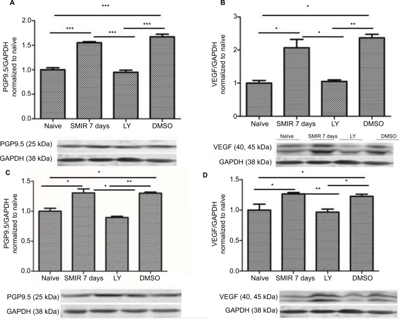
Results: The expression of PKCβII in DRG and spinal cord was significantly increased after SMIR surgery (P < 0.001, P < 0.01) and expression of PKCβII was located in the neurons of the spinal cord, and magnocellular neurons, non-peptide neurons, and peptide neurons in DRG. Besides, compared with skin/muscle incision group, retraction caused a marked increase in the expression of PKCβII and a significant decrease of MWT (P < 0.001, P < 0.05). The activator of PKCβII greatly increased the expression of PGP9.5 and VEGF (P < 0.05, P < 0.01) and enhanced MWT (P < 0.001), while inhibitor of PKCβII decreased the expression of PGP9.5 and VEGF and attenuated MWT (P < 0.05, P < 0.01, P < 0.001).

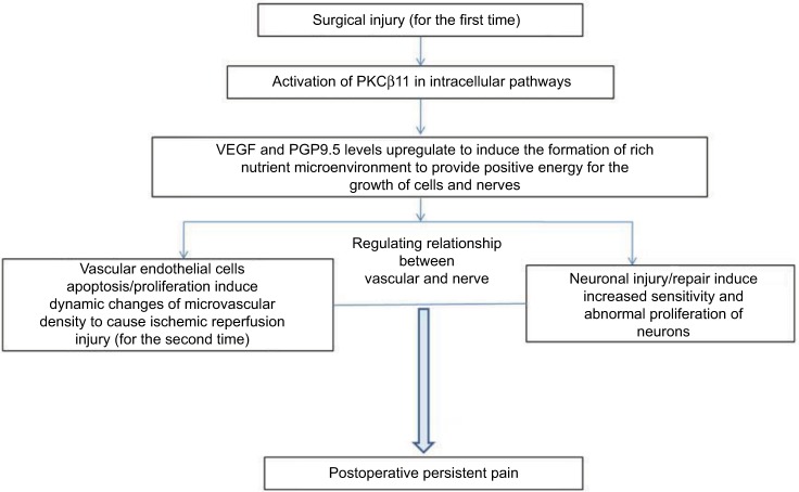
Conclusion: Activation of PKCβII signaling pathways might be an important mechanism in the progression of postoperative pain.

Keywords: PGP9.5; PKCβII; VEGF; neurons; postoperative persistent pain.


Conflict of interest statement

Disclosure The authors report no conflicts of interest in this work.

Figures

Figure 1
Figure 1
(A) Skin/muscle incision and retraction (SMIR)-induced thermal response to pain. Compared with the control group, #P < 0.05; compared with the Sham group, *P < 0.05, **P < 0.01. (B) SMIR-induced allodynia (mechanical pain sensitization) in response to hind paw mechanical stimulation. The mechanical withdrawal thresholds (MWTs) in the control (naïve), sham, and SMIR groups are plotted at baseline and at different time points after SMIR. The decreased MWT after SMIR is indicative of allodynia. Compared with the control (naïve) group, #P < 0.05, ##P < 0.01, ###P < 0.001; compared with the Sham group, *P < 0.05, ***P < 0.001.
Figure 2
Figure 2
Upregulation of p-PKCβII in the lumbar spinal cord after SMIR. (A) Expression of p-PKCβII 7 days after SMIR was determined by densitometric analysis on Western blots using PKCβII as control. Compared with the untreated control and Sham groups, expression level of p-PKCβII was significantly higher at 7 days after SMIR (*P < 0.05); n=5 for each group. (B) Average values of density of p-PKCβII-positive cells in the lumbar spinal cord in SMIR, naïve, and Sham groups. SMIR and Sham values were compared for the same time and segment by one-way analysis of variance (***P < 0. 001); n=5 for each group. (C) Immunostaining pictures of the p-PKCβII expression in the lumbar spinal cord in naïve, sham, and SMIR 7 days animals. Abbreviations: SMIR, skin/muscle incision and retraction; PKCβII, protein kinase C βII.
Figure 3
Figure 3
Upregulation of p-PKCβII in the DRG after SMIR. (A) Expression of p-PKCβII 7 days after SMIR was determined by densitometric analysis on Western blots using PKCβII as control. Compared with the untreated control and Sham groups, expression levels of p-PKCβII were significantly higher 7 days after SMIR (*P < 0.05); n=5 for each group. (B) Average values (SD) of densitometric analysis of p-PKCβII-positive cells in the DRG in SMIR, naïve, and sham groups. SMIR and Sham values were compared for the same time and segment by one-way analysis of variance (***P < 0.001); n=5 for each group. (C) Immunostaining pictures of the p-PKCβII expression in the DRG in naïve, sham, and SMIR animals. Abbreviations: SMIR, skin/muscle incision and retraction; DRG, dorsal root ganglion; PKCβII, protein kinase C βII.
Figure 4
Figure 4
Neuronal expression of p-PKCβII in the lumbar spinal cord. Spinal cord sections (L4–L5) excised 7 days after SMIR were double-labeled with p-PKCβII and the neuron marker (NeuN) (AC) antibodies, or p-PKCβII and microglial marker (CD11b) (DF) antibodies, or p-PKCβII and glial fibrillary acidic protein (GFAP) (GI) to detect the expression of p-PKCβII. Double staining shows that p-PKCβII is co-localized with NeuN, but not with CD11b or GFAP. Abbreviations: SMIR, skin/muscle incision and retraction; PKCβII, protein kinase C βII.
Figure 5
Figure 5
Expression of p-PKCβII in the L5 DRG in SMIR, naïve, and Sham groups. L5 DRG sections excised 7 days after SMIR were double-labeled with p-PKCβII and DRG magnocellular neurons marker (NF200) antibodies (AC), or p-PKCβII and DRG non-peptide neuron marker (IB4) antibodies (DF), or p-PKCβII and DRG peptide neuron marker (CGRP) antibodies (GI) to detect the expression of p-PKCβII. Double staining shows that p-PKCβII is colocalized with NF200 (C), IB4 (F), and CGRP (I). Abbreviations: SMIR, skin/muscle incision and retraction; DRG, dorsal root ganglion; CGRP, calcitonin gene-related peptide; PKCβII, protein kinase C βII.
Figure 6
Figure 6
At day 7 after SMIR, intrathecal injection of p-PKCβII agonist PMA at a dose of 100 ng increases mechanical allodynia. Compared with the 0.25% DMSO group, ***P < 0.001; compared with the control group, ###P <0.001; n=5 for each group. Abbreviations: SMIR, skin/muscle incision and retraction; PKCβII, protein kinase C βII; PMA, TPA, Phorbol 12-myristate 13-acetate; BL, baseline.
Figure 7
Figure 7
Upregulation of PMA-induced PGP9.5 and VEGF expression in the spinal cord and the DRG. (A, B) Expression of PGP9.5 and VEGF in the spinal cord after PMA treatment was determined by densitometric analysis on Western blots using GAPDH as the internal control. Compared with the control and DMSO group, *P < 0.05, **P < 0.01, ***P < 0.001; n=5 for each group. (C, D) Expression of PGP9.5 and VEGF in the DRG after PMA Treatment was determined by densitometric analysis on Western blots using GAPDH as the internal control. Compared with the control and DMSO group, *P < 0.05, **P < 0.01, ***P < 0.001; n=5 for each group. Abbreviations: DRG, dorsal root ganglion; PGP, protein gene product; PGP9.5, protein gene product 9.5; VEGF, vascular endothelial growth factor; PMA, TPA, Phorbol 12-myristate 13-acetate.
Figure 8
Figure 8
Intrathecal injection of p-PKCβII inhibitor LY333531, at 7 days after SMIR, reverses SMIR-induced mechanical allodynia. Compared with the 0.25% DMSO group, **P < 0.01, ***P < 0.001; Compared with the SMIR group, ##P < 0.01, ###P < 0.001; n=5 for each group. Abbreviations: SMIR, skin/muscle incision and retraction; BL, baseline.
Figure 9
Figure 9
Downregulation of LY333531-induced PGP9.5 and VEGF expression in the spinal cord and the DRG. (A, B) Expression of PGP9.5 and VEGF in the spinal cord after LY333531 treatment was determined by densitometric analysis on Western blots using GAPDH as the internal control. Compared with the control and DMSO group, *P < 0.05, **P < 0.01, ***P < 0.001; n=5 for each group. (C, D) Expression of PGP9.5 and VEGF in the DRG after LY333531 treatment was determined by densitometric analysis on Western blots using GAPDH as the internal control. Compared with the control group, LY group, and DMSO group, *P < 0.05, **P < 0.01; n=5 for each group. Abbreviations: SMIR, skin/muscle incision and retraction; DRG, dorsal root ganglion; VEGF, vascular endothelial growth factor; GAPDH, glyceraldehyde phosphate dehydrogenase; LY, LY333531; PGP, protein gene product.
Figure 10
Figure 10
Schematic diagram shows potential relationships among p-PKCβII, VEGF, and PGP9.5 to cause postoperative persistent pain. Abbreviations: VEGF, vascular endothelial growth factor; PGP, protein gene product; PKCβII, protein kinase C βII.

Similar articles

Cited by

References

    1. Wang H, Heijnen CJ, van Velthoven CT, et al. Balancing GRK2 and EPAC1 levels prevents and relieves chronic pain. J Clin Invest. 2013;123(12):5023–5034. - PMC - PubMed
    1. Steinberg SF. Structural basis of protein kinase C isoform function. Physiol Rev. 2008;88(4):1341–1378. - PMC - PubMed
    1. Melemedjian OK, Tillu DV, Asiedu MN, et al. BDNF regulates atypical PKC at spinal synapses to initiate and maintain a centralized chronic pain state. Mol Pain. 2013;9:12. - PMC - PubMed
    1. Mo G, Grant R, O’Donnell D, Ragsdale DS, Cao CQ, Séguéla P. Neuropathic Nav1.3-mediated sensitization to P2X activation is regulated by protein kinase C. Mol Pain. 2011;7:14. - PMC - PubMed
    1. Roh DH, Cho SR, Yoon SY, et al. Spinal neuronal NOS activation mediates sigma-1 receptor-induced mechanical and thermal hypersensitivity in mice: involvement of PKC-dependent GluN1 phosphorylation. Br J Pharmacol. 2011;163(8):1707–1720. - PMC - PubMed

LinkOut - more resources