Review
. 2018 Dec;60(12):1253-1266.
doi: 10.1007/s00234-018-2103-4. Epub 2018 Oct 11.

CT and MR imaging of orbital inflammation

Affiliations
Review

CT and MR imaging of orbital inflammation

Teresa A Ferreira et al. Neuroradiology. 2018 Dec.

Abstract

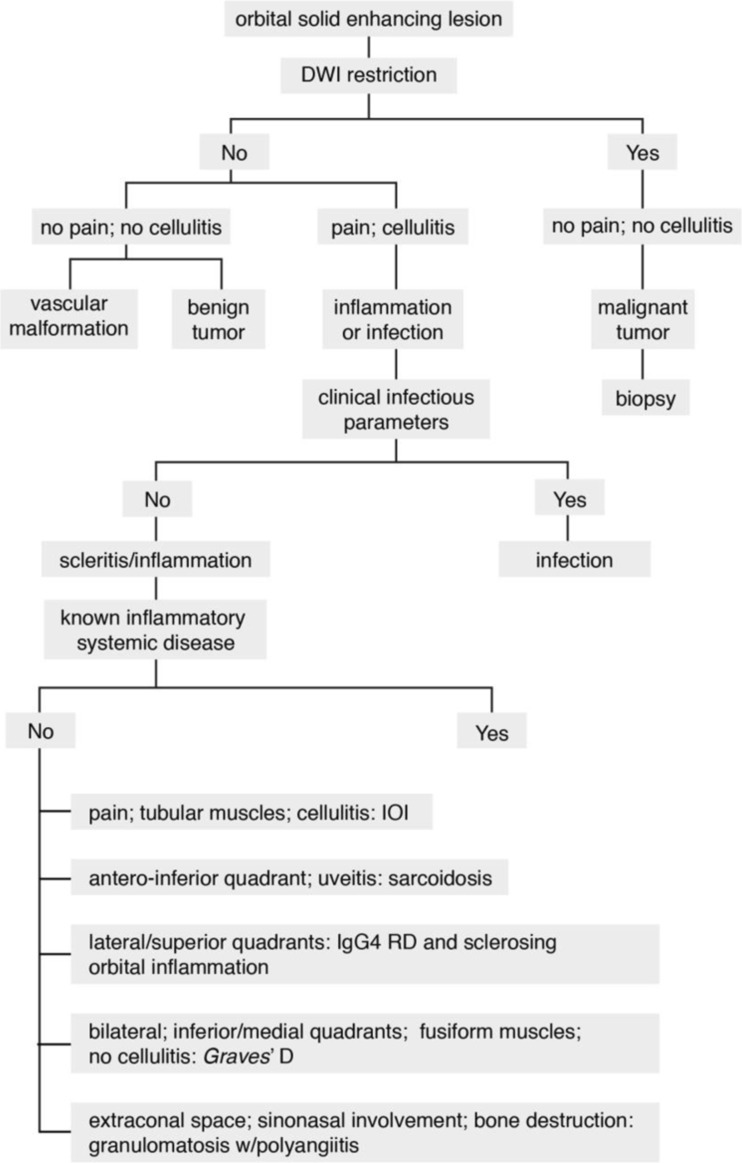
Purpose: Orbital inflammation can be idiopathic or in the context of a specific disease and it can involve different anatomical orbital structures. On imaging, inflammatory disease is frequently mistaken for infection and malignant tumors, and its underlying cause is often not determined. Through this article we aim to improve orbital inflammation diagnosis and underlying inflammatory diseases recognition.

Methods: The imaging protocols and characteristics of orbital inflammation were reviewed.

Results: A decision tree for the evaluation of these patients is provided. First, a combination of clinical and radiological clues is used to recognize inflammation, in particular to differentiate it both from orbital infection and tumor. Subsequently, different radiological patterns are recognized, often allowing the differentiation of the several orbital inflammatory diseases.

Conclusion: The use of adequate imaging protocols and subsequent evaluation allow the recognition of an orbital lesion as inflammatory and the diagnosis of the underlying inflammatory disease. All in all, a proper treatment can be established, and at times, a biopsy can be avoided.

Keywords: CT; Diffusion-weighted imaging; MRI; Orbital inflammation; Orbital inflammatory diseases.


Conflict of interest statement

Conflict of interest

The authors declare that they have no conflict of interest.

Ethical approval

All procedures were performed in accordance with the ethical standards of the institutional and/or national research committee and with the 1964 Helsinki declaration and its later amendments or comparable ethical standards.

Informed consent

For this type of study formal consent is not required.

Figures

Fig. 1
Fig. 1
a 69-year-old female. Inflammatory scleritis on the left. Axial contrast enhanced CT (CECT): focal eccentric outward thickening and enhancement of the globe wall (arrow) and minimal blurring of the adjacent fat (dashed arrow), both consistent with scleritis. b 38-year-old male. Idiopathic inflammatory scleritis on the right. Enhanced sagittal T1-WI with fat signal suppression: scleral thickening and enhancement (dashed arrow) and slight suprachoroidal effusion (arrowhead), consistent with scleritis. Contrary to CT, MRI clearly depicts which ocular layer is enhancing
Fig. 2
Fig. 2
8-year-old male with graft versus host disease after stem cell transplant. Extensive orbital cellulitis, myositis involving the lateral rectus, and sclerouveitis on the right. Axial T2-WI (a), axial T1-WI (c), enhanced axial T1-WI with fat signal suppression (b), and axial FLAIR (d): increased thickness and enhancement of the whole uvea consistent with uveitis (long arrow), subretinal effusions (black arrow), and slightly increased signal intensity of the vitreous humor signal on T1 and FLAIR consistent with vitritis (asterisk). Extensive periscleral cellulitis due to scleritis (empty arrowhead), preseptal cellulitis (arrowhead), myositis of the lateral rectus (short arrow) and perioptic neuritis (double headed arrows)
Fig. 3
Fig. 3
a 68-year-old male with Stills’ disease. Inflammatory dacryoadenitis on the left. Axial CECT: enlarged lacrimal gland (dashed arrow) with slight blurred margin and preseptal cellulitis (arrow) consistent with dacryoadenitis. The coexistence of preseptal cellulitis makes the diagnosis of tumor not probable. b 54-year-old female with Sjögren’s disease. Bilateral inflammatory dacryoadenitis. Coronal T2-WI with fat signal suppression: bilateral enlargement of the lacrimal gland, involving both the orbital (dashed arrow) and palpebral (arrowhead) lobes, a feature typical of dacryoadenitis but that can also occur in lymphomas. The levator palpebrae tendon separating the orbital and palpebral lobes (arrows)
Fig. 4
Fig. 4
40-year-old male with Tolosa-Hunt disease. a Optic perineuritis on the left at the orbital apex. Enhanced axial T1-WI with fat signal suppression: tram-track sign (arrow) and slight streaky enhancement of the surrounding fat consistent with perioptic neuritis. Slight orbital apical enhancement (dashed arrow) keeping with Tolosa Hunt disease. b Enhanced axial T1-WI with fat signal suppression at the level of the carotid siphons: smaller internal carotid artery on the left (arrowhead), due to pericarotid inflammatory tissue, a known finding in Tolosa-Hunt disease
Fig. 5
Fig. 5
68-year-old male. Inflammatory myositis of the right superior oblique muscle. Enhanced axial T1-WI with fat signal suppression (a, d), axial ADC (b), and enhanced coronal T1-WI with fat signal suppression (c): enlarged and with marked contrast enhancement right superior oblique muscle (arrow), with no DWI restriction (arrowhead), and slight surrounding cellulitis (dashed arrow), favoring inflammation. Clinically not suspicious for infection. Improvement after corticosteroids (d) avoiding biopsy (double-headed arrow)
Fig. 6
Fig. 6
a 46-year-old female with idiopathic orbital inflammation on the right. Axial CECT: pre (arrow) and postseptal (arrowhead) cellulitis, scleritis (dashed arrow), and dacryoadenitis (double-headed arrow). Scleritis and no sinusitis favored inflammation over infection. b 5-year-old male. Axial CECT: pre- and postseptal cellulitis on the left. Ethmoiditis (dashed arrow) and subperiosteal abscess (arrow), both favoring an infectious etiology
Fig. 7
Fig. 7
a 41-year-old female. Idiopathic inflammatory nodular scleritis on the left. Sagittal CECT: posterior wall globe focal mass. Deviation of the choroid-retinal layer internally (dashed arrow) suggesting a scleral or periocular origin. Painful periscleral cellulitis (arrow) favoring the diagnosis of scleritis. b 57-year-old female. Periocular breast cancer metastasis. Sagittal CECT: posterior wall globe focal mass, similar to (a), but note the normal aspect of the fat (arrow). Clinically there was no pain. Both favoring the diagnosis of a tumor
Fig. 8
Fig. 8
a, b 89-year-old male with idiopathic orbital inflammation. Enhanced coronals T1-WI with fat signal suppression (a, b): unilateral multifocal disease on the right involving the complex levator palpebrae-superior rectus and lacrimal gland (arrow) and the cavernous sinus (arrowhead). The bright signal around the inferior recti is due to inhomogeneous fat saturation. c, d 46-year-old man with idiopathic orbital inflammation. Axial (c) and sagittal (d) CECT: myositis of the right superior muscle complex with surrounding cellulitis (dashed arrows). Muscle involvement encompasses tendon and muscle belly giving the muscle a tubular configuration (d). e 43-year-old male with sarcoidosis. Sagittal CECT: typical involvement of the antero-inferior quadrant of the left orbit with mass in the inferior eyelid (arrow)
Fig. 9
Fig. 9
a, b 40-year-old female with thyroid-associated orbitopathy. Axial (a) and coronal T1-WI (b): bilateral and symmetric enlargement of the inferior, medial and complex levator palpebrae-superior rectus muscles. Notice fusiform configuration of the muscles because sparing of the tendon (arrow) and no surrounding infiltration of the fat. ce 35-year-old male with IgG4-related disease. Coronals enhanced T1-WI with fat signal suppression (c) and DWI (d) and axial enhanced T1-WI with fat signal suppression (e): bilateral involvement of the superolateral quadrants of the orbits, including bilateral enlargement of the lacrimal glands with involvement of some of the adjacent muscles. Involvement of both the orbital (arrowhead) and palpebral (dashed arrow) lobes of the lacrimal gland. Fusiform configuration (double-headed arrow) of the muscle. No diffusion restriction favoring an inflammatory process
Fig. 10
Fig. 10
67-year-old male with granulomatosis with polyangiitis involving the orbit, nose, and sinuses. Coronals (a, b) and axial (c) CT: chronic pansinusitis with sclerosis of the bony walls and intrasinusal calcifications coexisting with nasal septum (arrowhead) and lamina papiracea (arrows) erosions. Bilateral orbital involvement in the extraconal compartment (dashed arrows)
Fig. 11
Fig. 11
48-year-old female with idiopathic sclerosing orbital inflammation. Axial T2-WI (a), axial DWI (b) and enhanced axial T1-WI with fat signal suppression (c): large intraconal mass on the left (arrow), with no restriction diffusion, hypointense on T2, with marked enhancement after contrast. Notice enophthalmus, very unusual for a retrobulbar mass, together with absence of restriction diffusion, making it suspicious for sclerosing orbital inflammation
Fig. 12
Fig. 12
47-year-old female with Erdheim-Chester disease. Axial T2-WI (a, b), axial DWI (c) and enhanced axial T1-WI with fat signal suppression (d): bilateral intraconal masses surrounding the optic nerves (dashed arrows), slightly heterogeneous on T2, no diffusion restriction, enhancing after contrast. Bilateral involvement of cerebellum, middle cerebellar peduncles, and pons (arrows)
Fig. 13
Fig. 13
Decision tree in an orbital solid-enhancing lesion. IOI, idiopathic orbital inflammation; IgG4 RD immunoglobulin G4-related disease

References

    1. Gordon LK. Orbital inflammatory disease: a diagnostic and therapeutic challenge. Eye. 2006;20:1196–1206. doi: 10.1038/sj.eye.6702383. - DOI - PubMed
    1. Pakdaman Michael N. Orbital inflammatory disease: Pictorial review and differential diagnosis. World Journal of Radiology. 2014;6(4):106. doi: 10.4329/wjr.v6.i4.106. - DOI - PMC - PubMed
    1. Wallace ZS, Khosroshahi A, Jakobiec FA, Deshpande V, Hatton MP, Ritter J, Ferry JA, Stone JH. IgG4-related systemic disease as a cause of “idiopathic” orbital inflammation, including orbital myositis, and trigeminal nerve involvement. Surv Ophthalmol. 2012;57:26–33. doi: 10.1016/j.survophthal.2011.07.004. - DOI - PubMed
    1. Kahaly GJ. Imaging in thyroid-associated orbitopathy. Eur J Endocrinol. 2001;145:107–118. doi: 10.1530/eje.0.1450107. - DOI - PubMed
    1. Edrich CL. Posterior scleritis mimicking orbital cellulitis. Eye. 2005;19:1229–1232. doi: 10.1038/sj.eye.6701738. - DOI - PubMed

MeSH terms