Review
. 2018 Dec;9(6):1097-1106.
doi: 10.1007/s13244-018-0660-z. Epub 2018 Oct 11.

Enteric duplication cysts in children: varied presentations, varied imaging findings

Affiliations
Review

Enteric duplication cysts in children: varied presentations, varied imaging findings

Cinta Sangüesa Nebot et al. Insights Imaging. 2018 Dec.

Abstract

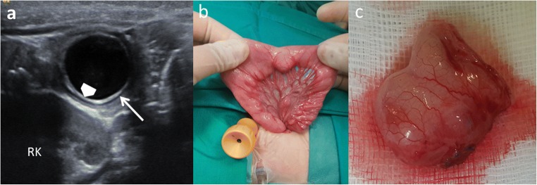
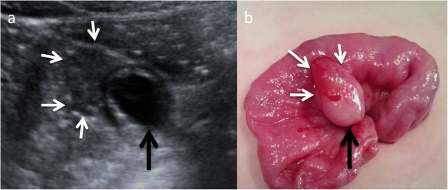
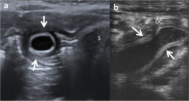
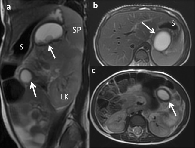
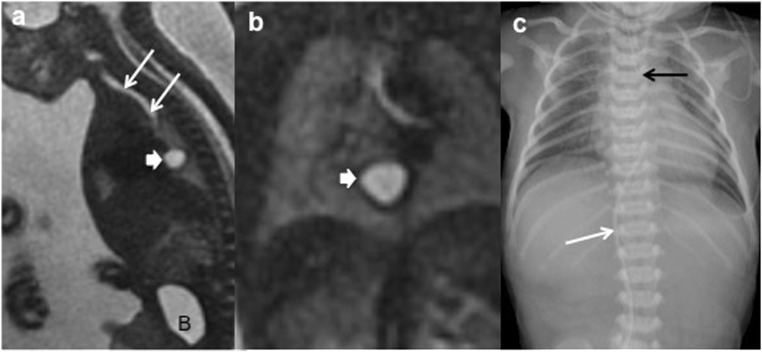
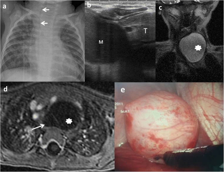
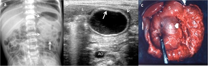
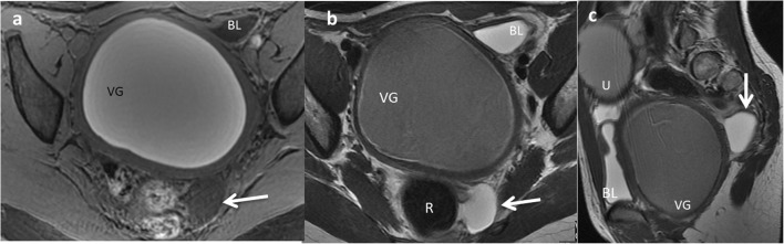
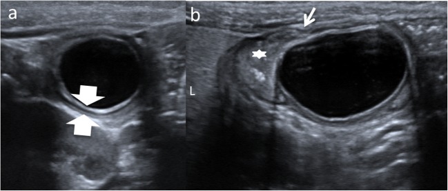
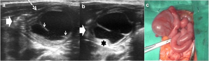
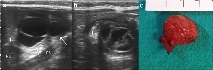
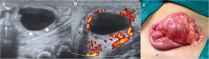
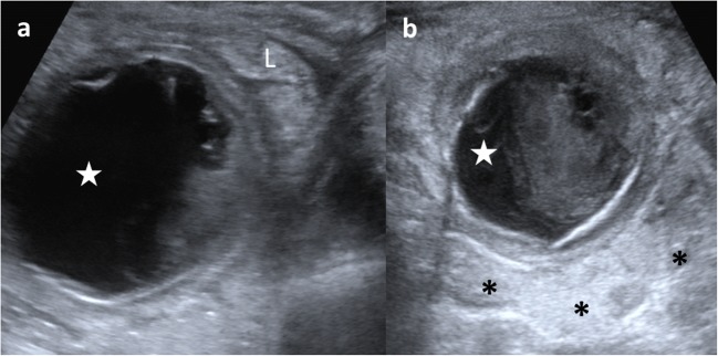
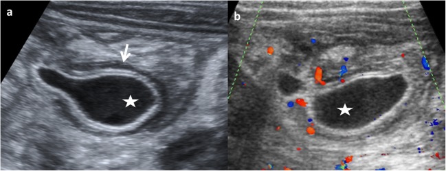
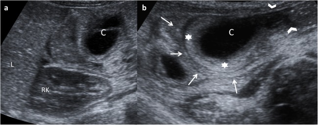
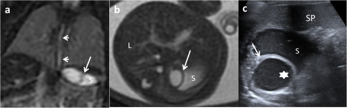
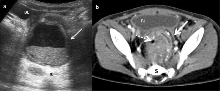
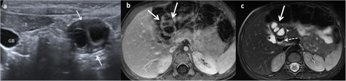
Enteric duplication cysts (EDCs) are rare congenital malformations formed during the embryonic development of the digestive tract. They are usually detected prenatally or in the first years of life. The size, location, type, mucosal pattern and presence of complications produce a varied clinical presentation and different imaging findings. Ultrasonography (US) is the most used imaging method for diagnosis. Magnetic resonance (MR) and computed tomography (CT) are less frequently used, but can be helpful in cases of difficult surgical approach. Conservative surgery is the treatment of choice. Pathology confirms the intestinal origin of the cyst, showing a layer of smooth muscle in the wall and an epithelial lining inside, resembling some part of the gastrointestinal tract (GT). We review the different forms of presentation of the EDCs, showing both the typical and atypical imaging findings with the different imaging techniques. We correlate the imaging findings with the surgical results and the final pathological features. TEACHING POINTS: • EDCs are rare congenital anomalies from the digestive tract with uncertain pathogenesis. • More frequently, diagnosis is antenatal, with most EDCs occurring in the distal ileum. • Ultrasonography is the method of choice for diagnosis of EDCs. • Complicated EDCs can show atypical imaging findings. • Surgery is necessary to avoid complications.

Keywords: Children; Cyst; Gastrointestinal tract; Magnetic resonance; Ultrasound.


Figures

Fig. 1
Fig. 1
An 11-month-old boy with abdominal pain is studied. a US view showing the typical US features of an EDC: an inner hyperechoic epithelial lining containing the mucosa of the alimentary tract (wide arrow) and the outer hypoechoic layer of smooth muscle (white long arrow), closely attached to the gastrointestinal tract by sharing a common wall. RK right kidney. b Surgical findings: typical ileal EDC. c Detailed picture of the EDC after resection from the ileal wall
Fig. 2
Fig. 2
A 4-year-old boy in a routinary US control of a horseshoe kidney. a Abdominal ultrasound view of an EDC (black arrow) with a peripheral eccentric hypoechoic cap (white arrows). b Surgical findings: the gastric mucosa was visible as a polypoid mass (white arrows) arising from the external surface of the EDC (black arrow)
Fig. 3
Fig. 3
Types of duplication cysts seen in the abdominal US. a Spherical EDC in gastric antrum (arrows). S stomach. b Tubular EDC (arrows) next to the descending colon (DC)
Fig. 4
Fig. 4
A 2-year-old girl with a splenic lesion (not shown) is studied with abdominal MR. a FSE T2 sagittal MRI: two similar cystic lesions are found (arrows). S stomach, LK left kidney, SP spleen. b, c FSE T2 axial MRI: a gastric and a jejunal duplication cyst are shown (arrows) in both images. Surgical findings: multiple EDCs
Fig. 5
Fig. 5
A 21-week-old fetus with polyhydramnios and absent normal gastric bubble in the US is studied. a Sagittal FIESTA fetal MRI showing a mediastinal cyst (thick arrow). Oesophageal atresia without fistula is suspected. B bladder. Trachea (arrows). b Coronal HASTE fetal MRI: detailed view of the mediastinal cyst (thick arrow). c Postnatal thoraco-abdominal radiograph: the gastric line tip is seen (black arrow) confirming the oesophageal atresia. The absence of air in the abdomen indicates a type-I or -II oesophageal atresia (without fistula). Venous umbilical catheter (white arrow)
Fig. 6
Fig. 6
A 10-month-old boy with a congenital cardiopathy presents respiratory distress. a Chest X-ray: a left cervicothoracic mass is suspected displacing the trachea to the right (arrows). b Chest US: cystic mass (M) with slightly echogenic content inside is seen next to the thymus (T). Its origin is unclear. c Coronal view SSFSE T2 MRI: a well-delineated and hyperintense lesion (star) is seen. d Transversal view of the lesion in a gadolinium-enhanced VIBE MRI confirms the cystic nature of the mass (star) next to the anterior oesophageal wall (arrow). e Thoracoscopic findings: a 6 × 4-cm lesion with close contact with the trachea originated from the muscular wall of the oesophagus
Fig. 7
Fig. 7
An 18-month-old girl with an abdominal mass in the physical exam is studied. a Abdominal X-ray: round, dense mass is discovered (arrows) in the left upper quadrant. b Transversal US view of the lesion: a cyst with the “double-wall” sign: the mucosa is hyperechoic (arrow) and the muscular layer is hypoechoic (dashed-line arrow). A gastric duplication cyst was suspected. L liver, P pancreas, AO aorta. c Laparotomy: antral duplication cyst was found (arrow). S stomach
Fig. 8
Fig. 8
A 12-year-old girl with a gynaecological malformation and haematometrocolpos. a A hypointense lesion is seen (white arrow) next to the rectum in axial T1 weighted pelvic MR. b On T2-weighted pelvic MR, the cyst next to the left wall of the rectum is seen. c Sagittal plane of the T2 MRI showing the posterior location of the cyst (arrow). VG vagina, BL bladder, U uterus. Imaging findings of a rectal duplication cyst. Note the haemorrhagic content of the uterus and vagina because of haematometrocolpos
Fig. 9
Fig. 9
A 3-month-old boy with vomiting is admitted to the emergency room. a US shows a cystic round-shape lesion with the “five-layers sign” (between arrows). b The “Y sign” is seen (long arrow). Star ileum, L liver. Laparoscopic findings: a non-complicated ileal duplication cyst
Fig. 10
Fig. 10
An 8-month-old boy with abdominal pain. a Longitudinal grey-scale US image showing a cystic lesion with an incomplete septum inside (small white arrows), next to the terminal ileum (big arrow). The “Y” sign is shown (dashed-line arrow). b US image obtained a few seconds later: peristalsis of the cyst causes small angulation of the contour and changes shape (black star). c Surgical findings: ileal duplication cyst
Fig. 11
Fig. 11
An 8-month-old boy with abdominal pain and abdominal mass on physical exam. a Longitudinal US view of a multiseptated cystic mass in the right flank with the “Y” sign (white arrows). I ileum, L liver, Ps psoas, RK right kidney. b Transversal US of the same mass demonstrates the relation with the ileal walls. c Surgical findings: ileal duplication cyst
Fig. 12
Fig. 12
A 3-week-old term newborn with abdominal distention and gastric intolerance. a Longitudinal US view of the right low quadrant: thickened wall (stars) cystic lesion next to a bowel loop (dashed-line arrow). The “Y” sign between the bowel and the lesion (arrow). b Power Doppler US demonstrates the significant vascularisation in the cyst wall. c Surgical findings: a cystic tumour next to the ileocecal valve. Pathological findings: ileal duplication cyst with heterotopic pancreatic tissue
Fig. 13
Fig. 13
A 3-year-old boy with fever and abdominal pain is studied. a US shows a cystic mass (star) with internal debris and next to an ileal loop (L). b The lesion (star) is surrounded by echogenic mesenteric fat (*) as an inflammatory sign. Surgical findings: a 5-cm ileal complicated duplication cyst was found with gastric mucosa with haemorrhagic and ulcerated walls
Fig. 14
Fig. 14
An 8-month-old boy baby with continuous crying is taken to the emergency department. Because of high suspicion of intussusception, a US exam is required. a Abdominal pear-shaped cystic lesion (star) in the left flank was found in a coronal US view. It shows a typical outer hypoechoic wall and inner hyperechoic layer, with hypoechoic content inside. b Doppler US showing light hyperaemia of the lesion wall that was also thickened. Surgical findings: complicated duplication cyst in the colonic splenic flexure
Fig. 15
Fig. 15
A 14-month-old girl with acute gastroenteritis and continuous crying. a Abdominal US shows an intestinal intussusception with a cyst (C) as the leading cause. L liver, RK right kidney. b A detailed US view: the intussuscipiens (arrows) and intussuscepted bowel (arrowheads) with the cyst inside (C) and the hyperechoic and thickened walls. Surgical findings: ileal duplication cyst as the cause of the intussusception
Fig. 16
Fig. 16
A 22-week-old fetus with an abdominal cyst seen on ultrasound is studied with MR. a Coronal fetal FIESTA MR: a cystic lesion is seen next to the stomach (S). Oesophageal lumen is seen (arrows). b FIESTA transversal MR: the hypointense wall of the suspected gastric duplication cyst (arrow). L liver, S stomach. c Postnatal abdominal US view: the lesion (star) imprinting the gastric wall (arrow). The content of the stomach is seen (S). SP spleen. Surgical findings: duplication cyst of the oesophageal-gastric transition
Fig. 17
Fig. 17
A 22-month-old boy with fever and abdominal pain. a Transversal US view of the pelvis shows a cystic mass between the bladder (BL) and the sacrum (S). The lesion presents anterior and left wall thickening (arrow) and contains a fluid-fluid level suggesting the presence of a complicated rectal mass. b Contrast-enhanced CT confirmed the presence of a complicated rectal cyst with remarkable inflammatory findings (white arrow) and gas (dashed-line arrow). Surgical findings: rectal duplication cyst with mucous and purulent content. Rectum muscular wall was oedematous and it was shared with the duplication cyst
Fig. 18
Fig. 18
A 2-year-old girl with fever and abdominal pain. a Abdominal US view of a complex cystic lesion (white arrows) in the duodenal area, next to the liver (L). GB gallbladder. b Axial gadolinium-enhanced GRE T1 demonstrating the presence of a multilocular cystic mass (white arrows) with wall thickening. c Axial T2 MRI: the mass (white arrow) shows three cystic cavities inside, close to the anterior wall of the duodenum (dashed-line arrow) and lateral to the gallbladder (star). Surgical findings: multiple duodenal duplication cyst, without connection with the lumen and with ectopic gastric mucosa

References

    1. Hur J, Yoon CS, Kim MJ, Kim OH. Imaging features of gastrointestinal tract duplications in infants and children: from esophagus to rectum. Pediatr Radiol. 2007;37:691–699. doi: 10.1007/s00247-007-0476-3. - DOI - PubMed
    1. Berrocal T, Hidalgo P, Gutiérrez J, De Pablo L, Rodríguez-Lemos R. Imagen radiológica de las duplicaciones del tubo digestivo. Radiología. 2004;46:282–292. doi: 10.1016/S0033-8338(04)77978-3. - DOI
    1. Sharma S, Yadav AK, Mandal AK, Zaheer S, Yadav D, Samie A. Enteric duplication cysts in children: a clinicopathological dilemma. J Clin Diagn Res. 2015;9:8–11. - PMC - PubMed
    1. Macpherson RI. Gastrointestinal tract duplications: clinical, pathologic, etiologic, and radiologic considerations. Radiographics. 1993;13:1063–1080. doi: 10.1148/radiographics.13.5.8210590. - DOI - PubMed
    1. Tiwari C, Shah H, Waghmare M, Makhija D, Khedkar K. Cysts of gastrointestinal origin in children: varied presentation. Pediatr Gastroenterol Hepatol Nutr. 2017;20:94–99. doi: 10.5223/pghn.2017.20.2.94. - DOI - PMC - PubMed

LinkOut - more resources