. 2019 Apr 1;527(5):942-956.
doi: 10.1002/cne.24557. Epub 2018 Dec 11.

Midline thalamic inputs to the amygdala: Ultrastructure and synaptic targets

Affiliations

Midline thalamic inputs to the amygdala: Ultrastructure and synaptic targets

Alon Amir et al. J Comp Neurol. .

Abstract

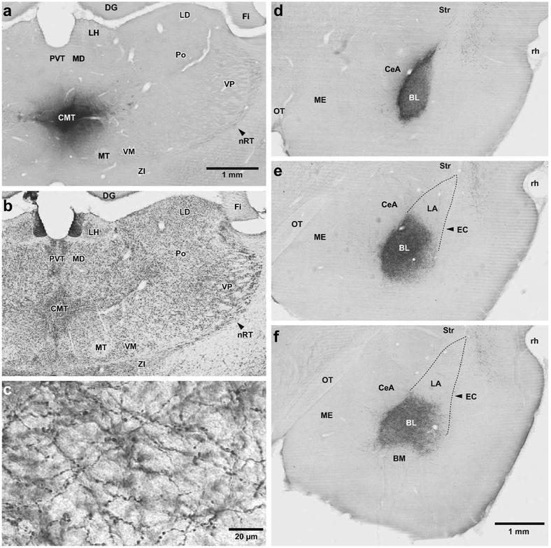
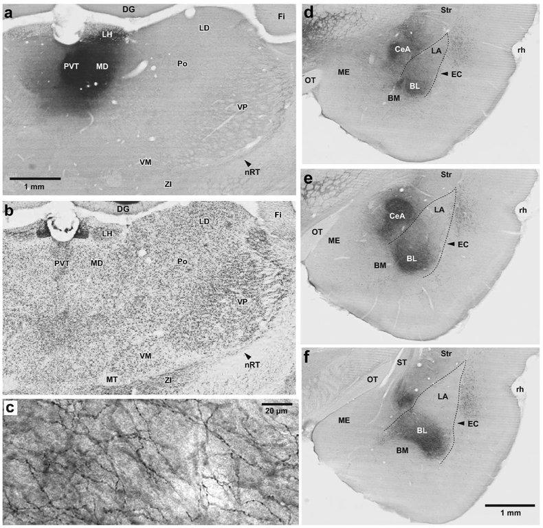
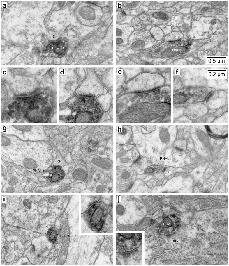
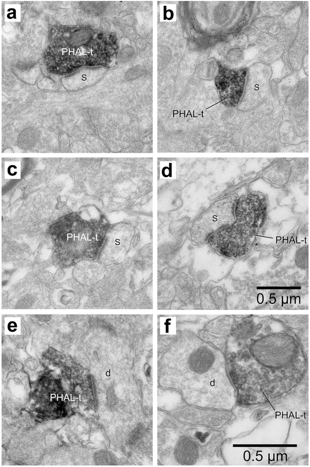
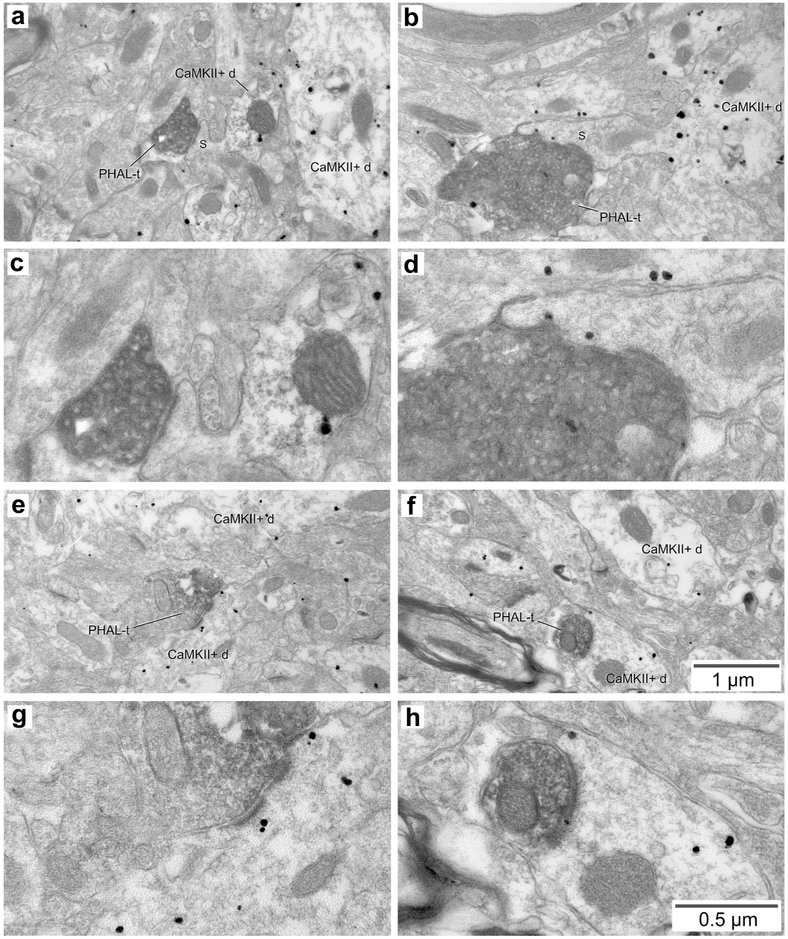
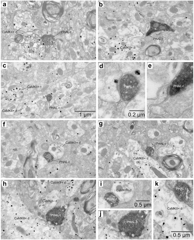
One of the main subcortical inputs to the basolateral nucleus of the amygdala (BL) originates from a group of dorsal thalamic nuclei located at or near the midline, mainly from the central medial (CMT), and paraventricular (PVT) nuclei. Although similarities among the responsiveness of BL, CMT, and PVT neurons to emotionally arousing stimuli suggest that these thalamic inputs exert a significant influence over BL activity, little is known about the synaptic relationships that mediate these effects. Thus, the present study used Phaseolus vulgaris-leucoagglutinin (PHAL) anterograde tracing and electron microscopy to shed light on the ultrastructural properties and synaptic targets of CMT and PVT axon terminals in the rat BL. Virtually all PHAL-positive CMT and PVT axon terminals formed asymmetric synapses. Although CMT and PVT axon terminals generally contacted dendritic spines, a substantial number ended on dendritic shafts. To determine whether these dendritic shafts belonged to principal or local-circuit cells, calcium/calmodulin-dependent protein kinase II (CAMKIIα) immunoreactivity was used as a selective marker of principal BL neurons. In most cases, dendritic shafts postsynaptic to PHAL-labeled CMT and PVT terminals were immunopositive for CaMKIIα. Overall, these results suggest that CMT and PVT inputs mostly target principal BL neurons such that when CMT or PVT neurons fire, little feed-forward inhibition counters their excitatory influence over principal cells. These results are consistent with the possibility that CMT and PVT inputs constitute major determinants of BL activity.

Keywords: CAMKIIα; RRID AB_2313606; RRID AB_2313686; RRID AB_2336656; RRID AB_2637031; RRID AB_447192; RRID RGD_734476; amygdala; electron microscopy; thalamus; tract-tracing.


Conflict of interest statement

CONFLICT OF INTEREST STATEMENT: The authors state that they have no conflict of interest.

Figures

Figure 1.
Figure 1.
CMT projections to the amygdala are largely confined to the basolateral nucleus (BL). (a,b) Two adjacent coronal sections processed to revealed the PHAL injection site in CMT (a) or the localization of thalamic nuclei in a Nissl-stained section (c) adjacent to that shown in a. (c) High power photomicrograph showing varicose PHAL-containing CMT axons ramifying in BL. (d-f) Three coronal sections showing the distribution of anterogradely labeled PHAL-immunoreactive CMT axons at rostral (d), intermediate (e), and caudal (f) levels of BL. Abbreviations: BL, basolateral nucleus of the amygdala; BM, basomedial nucleus of the amygdala; CeA, central nucleus of the amygdala CMT, central medial thalamic nucleus; DG, dentate gyrus; EC, external capsule; Fi, fimbria; LA, lateral nucleus of the amygdala; LD, laterodorsal thalamic nucleus; LH, Lateral habenula; MD, mediodorsal thalamic nucleus; ME, medial nucleus of the amygdala; MT, mammillothalamic tract; OT, Optic tract; nRT, reticular thalamic nucleus; Po, posterior thalamic nuclear group; PVT, paraventricular thalamic nucleus; rh, rhinal sulcus; Str, striatum; VM, ventromedial thalamic nucleus; VP, ventroposterior thalamic nucleus; ZI, zona incerta. Scale bar in a also applies to b. Scale bar in f also applies to d and e.
Figure 2.
Figure 2.
PVT projections to the amygdala target both, the central (CeA) and basolateral (BL) nuclei. (a,b) Two adjacent coronal sections processed to revealed the PHAL injection site in PVT (a) or the localization of thalamic nuclei in a Nissl-stained section (b) adjacent to that shown in a. (c) High power photomicrograph showing varicose PVT axons ramifying in BL. (d-f) Three coronal sections showing the distribution of anterogradely labeled PVT axons at rostral (d), intermediate (e), and caudal (f) levels of the CeA and BL. Abbreviations: BL, basolateral nucleus of the amygdala; BM, basomedial nucleus of the amygdala; CeA, central nucleus of the amygdala CMT, central medial thalamic nucleus; DG, dentate gyrus; EC, external capsule; Fi, fimbria; LA, lateral nucleus of the amygdala; LD, laterodorsal thalamic nucleus; LH, Lateral habenula; MD, mediodorsal thalamic nucleus; ME, medial nucleus of the amygdala; MT, mammillothalamic tract; OT, Optic tract; nRT, reticular thalamic nucleus; Po, posterior thalamic nuclear group; PVT, paraventricular thalamic nucleus; rh, rhinal sulcus; Str, striatum; VM, ventromedial thalamic nucleus; VP, ventroposterior thalamic nucleus; ZI, zona incerta. Scale bar in a also applies to b. Scale bar in f also applies to d and e.
Figure 3.
Figure 3.
Examples of synapses formed by PHAL-labeled CMT axon terminals in BL. (a,b,g,h) Four examples of PHAL-labeled axon terminals (PHAL-t) forming asymmetric synapses with dendritic spines (s). They are also shown at high magnification (c,d,e,f). In the cases shown in a and g, the spine targeted by the CMT axon terminal emerges from a large dendritic profile. In b, a long spine neck is evident. (i) Rare example of a CMT axon terminal forming an asymmetric synapse with a dendritic profile (d). Inset in i shows the same synapse at a higher magnification. (j) Sole example of a CMT axon terminal forming an asymmetric synapse with a somatic profile (soma). Inset in j shows the same synapse at a higher magnification. Scale bars in b and f apply to all low and high power electron micrographs, respectively.
Figure 4.
Figure 4.
Examples of synapses formed by PHAL-labeled PVT axons terminals in BL. (a-d) Four examples of PHAL-labeled axon terminals (PHAL-t) forming asymmetric synapses with dendritic spines (s). (e) Example of PVT axon terminal forming an asymmetric synapse with a dendritic profile (d). (f) The sole example of PVT axon terminal forming a symmetric synapse with a dendritic profile (d). Scale bar in d applies to panels a-d. Scale bar in f applies to panels e and f.
Figure 5.
Figure 5.
Relationship between PHAL-labeled CMT axon terminals and CaMKIIa-immunopositive elements in BL. (a, b) Two examples of PHAL-positive CMT axon terminals (PHAL-t) forming asymmetric synapses with dendritic spines that emerge from CaMKIIa-immunopositive dendritic profiles (CaMKII+d). These synapses are also shown at high magnification (c,d). In a, the spine is devoid of gold particles, but it emerges from a labeled dendritic profile. In b, gold particles are present in both the spine and dendrite. (e, f) Two examples of PHAL-positive CMT axon terminals (PHAL-t) forming asymmetric synapses with CaMKIIa-immunopositive dendritic profiles. These synapses are also shown at high magnification (g,h). Scale bars in f and h apply to all low and high power electron micrographs, respectively.
Figure 6.
Figure 6.
Relationship between PHAL-labeled PVT axon terminals and CaMKIIa-immunopositive elements in BL. (a-c) Three examples of PHAL-positive PVT axon terminals (PHAL-t) forming asymmetric synapses with dendritic spines that emerge from CaMKIIa-immunopositive dendritic profiles (CaMKII+d). Synapses in b and c are also shown at high magnification (d,e). (f, g) Two examples of PHAL-positive PVT axon terminals (PHAL-t) forming asymmetric synapses with CaMKIIa-immunopositive dendritic profiles. These synapses are shown at a higher magnification (i,k). (h) Example of a PHAL-positive PVT axon terminal (PHAL-t) forming an asymmetric synapse with CaMKIIa-immunonegative dendritic profile (CaMKII– d). This synapse is shown at a higher magnification (j). Scale bar in c also applies to a, b f, g, h. Scale bar in d also applies to e. Scale bar in i also applies to j.
Figure 7.
Figure 7.
Examples of synapses formed by PHAL-labeled PVT axons terminals in CeA. (a-b) Two examples of PHAL-labeled axon terminals (PHAL-t) forming asymmetric synapses with dendritic spines (s). (c) Example of PHAL-t forming an asymmetric synapse with a dendritic shaft (d). (d) Rare example of PHAL-t forming a symmetric synapse with a dendritic shaft (d). A spine (s) can be seen to emerge from this dendrite. Inset in d, magnified view of the same synapse.

References

    1. Ahmed B, Anderson JC, Douglas RJ, Martin KA, & Whitteridge D (1998). Estimates of the net excitatory currents evoked by visual stimulation of identified neurons in cat visual cortex. Cereb Cortex, 8(5), 462–76. 10.1093/cercor/8.5.462 - DOI - PubMed
    1. Alkire MT, McReynolds JR, Hahn EL, & Trivedi AN (2007). Thalamic microinjection of nicotine reverses sevoflurane-induced loss of righting reflex in the rat. Anesthesiology, 107(2), 264–272. 10.1097/01.anes.0000270741.33766.24 - DOI - PubMed
    1. Amaral DG, Price JL, Pitkanen A, & Carmichael ST (1992). In Aggleton JP (Ed.), The amygdala: neurobiological aspects of emotion, memory, and mental dysfunction. (pp.1–66). New York, NY: Wiley-Liss.
    1. Amir A, Lee SC, Headley DB, Herzallah MM, & Paré D (2015). Amygdala signaling during foraging in a hazardous environment. J Neurosci, 35(38), 12994–3005. 10.1523/jneurosci.0407-15.2015 - DOI - PMC - PubMed
    1. Amir S, & Stewart J (2009). Motivational modulation of rhythms of the expression of the clock protein PER2 in the limbic forebrain. Biol Psychiatry, 65(10), 829–34. 10.1016/j.biopsych.2008.12.019 - DOI - PubMed

Publication types

Substances