. 2018 Oct 12;14(10):e1007698.
doi: 10.1371/journal.pgen.1007698. eCollection 2018 Oct.

Genome amplification and cellular senescence are hallmarks of human placenta development

Affiliations

Genome amplification and cellular senescence are hallmarks of human placenta development

Philipp Velicky et al. PLoS Genet. .

Abstract

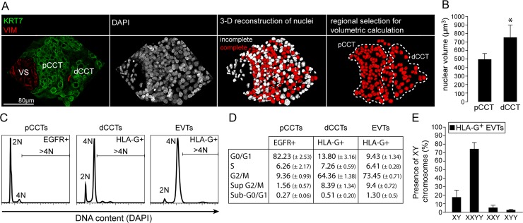
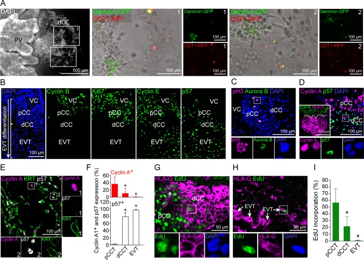
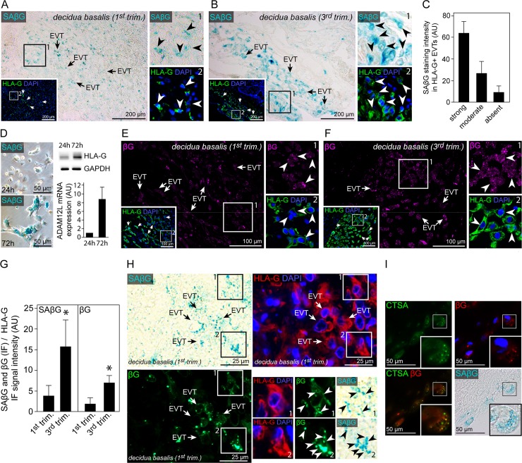
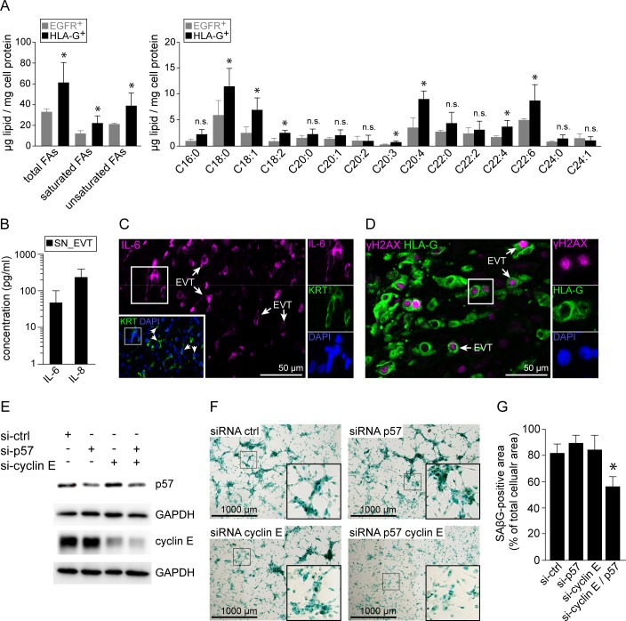
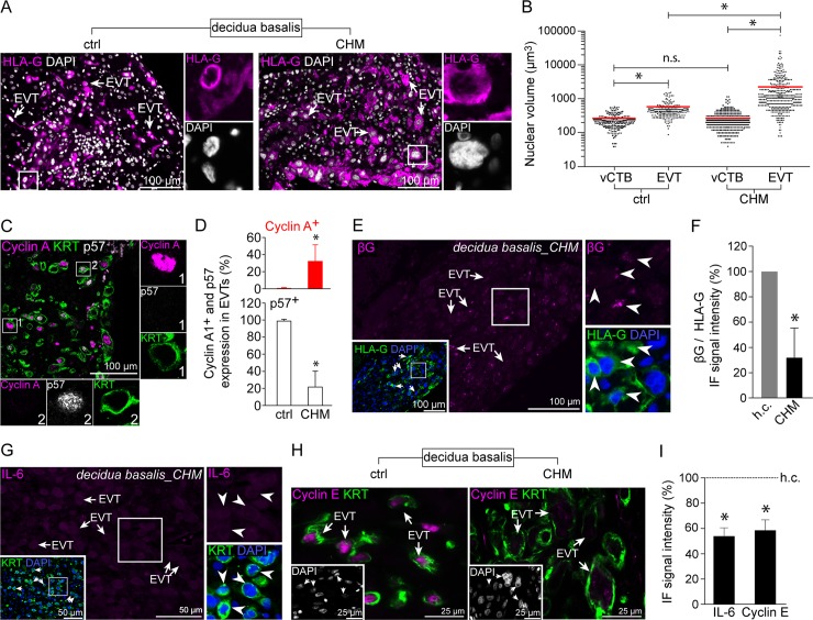
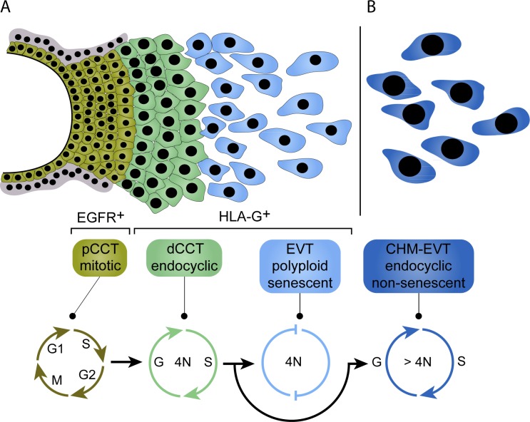
Genome amplification and cellular senescence are commonly associated with pathological processes. While physiological roles for polyploidization and senescence have been described in mouse development, controversy exists over their significance in humans. Here, we describe tetraploidization and senescence as phenomena of normal human placenta development. During pregnancy, placental extravillous trophoblasts (EVTs) invade the pregnant endometrium, termed decidua, to establish an adapted microenvironment required for the developing embryo. This process is critically dependent on continuous cell proliferation and differentiation, which is thought to follow the classical model of cell cycle arrest prior to terminal differentiation. Strikingly, flow cytometry and DNAseq revealed that EVT formation is accompanied with a genome-wide polyploidization, independent of mitotic cycles. DNA replication in these cells was analysed by a fluorescent cell-cycle indicator reporter system, cell cycle marker expression and EdU incorporation. Upon invasion into the decidua, EVTs widely lose their replicative potential and enter a senescent state characterized by high senescence-associated (SA) β-galactosidase activity, induction of a SA secretory phenotype as well as typical metabolic alterations. Furthermore, we show that the shift from endocycle-dependent genome amplification to growth arrest is disturbed in androgenic complete hydatidiform moles (CHM), a hyperplastic pregnancy disorder associated with increased risk of developing choriocarinoma. Senescence is decreased in CHM-EVTs, accompanied by exacerbated endoreduplication and hyperploidy. We propose induction of cellular senescence as a ploidy-limiting mechanism during normal human placentation and unravel a link between excessive polyploidization and reduced senescence in CHM.


Conflict of interest statement

The authors have declared that no competing interests exist.

Figures

Fig 1
Fig 1. Polyploidization of human trophoblasts correlates with HLA-G expression.
(A) Representative IF co-staining showing keratin7 (green) and vimentin (VIM, red), DAPI (grey) and a 3D reconstruction of DAPI signals of a placental villous tissue section (30μm) of the same image. Complete and incompletely reconstructed nuclei are shown in red and white, respectively. (B) Quantification of nuclear volumes obtained as demonstrated in A of proximal and distal cell column trophoblast nuclei (n = 5 cell columns of 3 placentas). (C) Flow cytometry analyses of DAPI signals representing DNA content of MACS-sorted EGFR+ and HLA-G+ CCTs or HLA-G-positive EVTs (n = 3). (D) Quantification and segmentation in cell cycle phases of data obtained from FC analyses (n = 3). (E) FISH analysis using probes against sex chromosomes X and Y of MACS-sorted HLA-G+ EVTs from decidua basalis tissue (n = 4 male placentas).
Fig 2
Fig 2. EVTs show cell cycle patterns of endoreduplication.
(A) FUCCI cell cycle sensor analyses of outgrowing placental explants. Two different outgrowth-areas (boxes 1 and 2) of a representative placental explant (left picture) are shown in detail (n = 9 explants of 3 first trimester placentas). (B) IF stainings of serial tissue sections of a first trimester placental anchoring villous showing (from left to right) DAPI, Cyclin B, Ki67, Cyclin E and p57. (n = 3) (C) IF co-staining of first trimester placental tissue (n = 4) showing pH3 (magenta) and Aurora B (green) expressing mitotic figures (blue) in pCCTs. (D) IF co-staining of first trimester placental tissue (n = 4) showing Cyclin A (magenta) and p57 (green) in CCTs. EGFR (green) and HLA-G (magenta) double staining of a serial section is shown in the boxed insert (left bottom). (E) IF triple-staining of a first trimester decidua basalis tissue section (n = 5) showing Cyclin A (magenta), p57 (grey) and KRT7 (green) in EVTs. (F) Percentages of Cyclin A+ and p57+ in KRT+ pCCTs, dCCTs and EVTs (G) IF co-staining of first trimester tissue (n = 3) showing HLA-G (magenta) and EdU (green) incorporation into CCTs. (H) IF co-staining of first trimester tissue showing HLA-G (magenta) expression and EdU (green) incorporation into EVTs in decidua basalis tissue (n = 3). (I) Quantification of EdU incorporation into pCCT, dCCT and EVTs of cultivated first trimester tissue explants (n = 8). Digitally zoomed insets display a split-channel-depiction of the boxed area. DAPI was used to visualize nuclei.
Fig 3
Fig 3. Senescence-associated β-galactosidase activity in HLA-G positive invasive, decidual EVTs co-localizes with beta-galactosidase protein and lysosomal cathepsin A.
(A) Cryo-section of first trimester decidua basalis tissue (n = 7) showing co-stained SAβG activity (blue, large image) and HLA-G (green, insert). (B) Cryo-section of third trimester decidua basalis tissue (n = 6) showing co-stained SAβG activity (blue, large image) and HLA-G (green, insert). (C) Quantification of SAßG staining intensity in first trimester HLA-G+, deciudal EVTs. (D) SAβG activity in isolated primary human trophoblasts after 24 hrs (top) and 72 hrs (bottom) of cultivation (left panel). Western blot analysis (n = 3) of the same cultures showing increasing HLA-G+ and ADAM12L expression in differentiating EVTs after 72 hrs of cultivation. (E) Section of the same first trimester decidua basalis tissue (n = 3) depicted in A showing βG (magenta, large image) and HLA-G (green, boxed insert) co-staining. (F) Section of the same third trimester decidua basalis tissue (n = 3) depicted in B showing βG (magenta, large image) and HLA-G (green, boxed insert) co-staining. (G) Ratio of SaβG and βG relative to HLA-G signal intensities in first (n = 6) and third (n = 7) trimester decidua basalis tissue sections. (H) Section of first trimester paraffin-embedded decidua basalis tissue (n = 3) showing SAβG activity (blue), HLA-G (red) and βG (green) co-staining. (I) IF staining of SAβG (blue) expressing, decidual EVTs with antibodies against βG (red) and CTSA (green) (n = 3). The picture in the lower left corner represents a merged image of the green (CTSA) and red (βG) channel. Zoomed insets on the right show image details of the boxed area, arrowheads indicate SAβG (A and B) or βG (E and F) and HLA-G double positive cells. In (H) zoomed insets represent HLA-G, βG and SAβG triple positive regions marked by arrow heads. DAPI was used to visualize nuclei.
Fig 4
Fig 4. Decidual EVTs upregulate senescence-associated markers.
(A) Gas chromatography-assisted analysis of fatty acid contents in isolated EGFR+ and HLA-G+ trophoblasts (n = 3). (B) Levels of IL-6 and IL-8 secreted by cultivated EVTs. (C) Section of first trimester decidua basalis tissue (n = 3) showing IL-6 (magenta, large image) and KRT co-staining (green, insert). (D) IF co-staining of paraffin embedded decidua basalis tissue sections (n = 3) with antibodies against γH2AX (magenta) and HLA-G (green). (E) Western blot analysis of p57 and cyclin-E single- and double-knockdowns (si-) performed in primary EVTs. Glyceraldehyde 3-phosphate dehydrogenase (GAPDH) served as loading control. One out of three independent experiments is shown. (F) SAßG expression in cultivated EVTs treated with siRNA targeting p57 or cyclin E. (G) Quantification of SAßG staining intensity in cultivated EVTs (n = 4). Zoomed insets on the right show image details of the boxed area, arrowheads indicate IL-6 (C) or γH2AX (D) and KRT or HLA-G double positive cells, respectively. DAPI was used to visualize nuclei.
Fig 5
Fig 5. Complete hydatidiform mole EVTs exhibit increased ploidy and decreased cellular senescence.
(A) IF co-staining of healthy (left image) and CHM (right image) first trimester decidua basalis tissues showing HLA-G+ (magenta) invasive EVTs. (B) Volumes calculated on the basis of the cross-section radius of vCTB and EVT nuclei of healthy (n = 20) and CHM (n = 23) diagnosed first trimester placentas. (C) IF triple-staining of first trimester decidua basalis tissue showing Cyclin A (magenta), p57 (grey) and Keratin7 (KRT7, green) in CHM-EVTs. (D) Rates of Cyclin A+ and p57+ EVTs in CHM (n = 18) and age-matched healthy control tissues (n = 15). (E) Section of CHM diagnosed first trimester decidua basalis tissue (n = 6) showing beta-galactosidase (βG) (magenta, large image) and HLA-G co-staining (green, insert). (F) Ratio of βG to HLA-G signal intensities in CHM diagnosed first trimester tissues (n = 23) age-matched healthy control sections (n = 15). (G) IF co-staining of CHM first trimester dedicuda basalis tissue showing IL-6 (magenta) and KRT (green, insert). (H) IF stainings of healthy (left image) and CHM (right image) decidua basalis tissues demonstrating cyclin E (magenta) and KRT (green) expression in invasive EVTs. (I) Ratio of IL-6 (n = 12) and cyclin E (n = 10) to KRT signal intensities in CHM diagnosed first trimester tissues age-matched healthy control sections (n = 22). Digitally zoomed insets display a split-channel-depiction of the boxed area. DAPI was used to visualize nuclei.
Fig 6
Fig 6. Proposed model of tetraploidization in HLA-G+ trophoblasts and induction of senescence during EVT differentiation.
(A) During early pregnancy, EGFR+ pCCTs form a highly proliferative so-called cell column, which give rise to HLA-G+ CCTs at the distal end of the cell column. During this transition, a endoreduplicative cell cycle provokes tetraploidization in dCCTs. Endoreduplication in human HLA-G+ trophoblasts is likely characterized by an cyclin A+/p57- expression pattern, expressed by a subset of dCCTs. Upon invasion into the deciuda, EVTs lose their cyclic activity marked by a dominant cylclin A-/p57+ phenoptye and undergo cellular senescence. Induction of cellular senescence is at least partly regulated by p57 and cyclin E. (B) The balance between endocycle and senescence is disturbed in CHM. CHM-EVTs show exacerbated endocyclic activity characterized by an increase in cyclin A+/p57- EVTs, enhanced polyploidization and reduced signs for cellular senescence.

Similar articles

Cited by

References

    1. Hamilton WJ, Boyd JD. Trophoblast in human utero-placental arteries. Nature. 1966;212(5065):906–8. . - PubMed
    1. Pijnenborg R, Dixon G, Robertson WB, Brosens I. Trophoblastic invasion of human decidua from 8 to 18 weeks of pregnancy. Placenta. 1980;1(1):3–19. . - PubMed
    1. Madeja Z, Yadi H, Apps R, Boulenouar S, Roper SJ, Gardner L, et al. Paternal MHC expression on mouse trophoblast affects uterine vascularization and fetal growth. Proceedings of the National Academy of Sciences of the United States of America. 2011;108(10):4012–7. 10.1073/pnas.1005342108 ; PubMed Central PMCID: PMC3053985. - DOI - PMC - PubMed
    1. Tilburgs T, Evans JH, Crespo AC, Strominger JL. The HLA-G cycle provides for both NK tolerance and immunity at the maternal-fetal interface. Proceedings of the National Academy of Sciences of the United States of America. 2015;112(43):13312–7. 10.1073/pnas.1517724112 ; PubMed Central PMCID: PMC4629323. - DOI - PMC - PubMed
    1. Zeldovich VB, Robbins JR, Kapidzic M, Lauer P, Bakardjiev AI. Invasive extravillous trophoblasts restrict intracellular growth and spread of Listeria monocytogenes. PLoS pathogens. 2011;7(3):e1002005 10.1371/journal.ppat.1002005 ; PubMed Central PMCID: PMC3048367. - DOI - PMC - PubMed

Publication types