Review
. 2018 Oct 11;19(10):3110.
doi: 10.3390/ijms19103110.

CEACAM1 in Liver Injury, Metabolic and Immune Regulation

Affiliations
Review

CEACAM1 in Liver Injury, Metabolic and Immune Regulation

Andrea Kristina Horst et al. Int J Mol Sci. .

Abstract

Carcinoembryonic antigen-related cell adhesion molecule 1 (CEACAM1) is a transmembrane glycoprotein that is expressed on epithelial, endothelial and immune cells. CEACAM1 is a differentiation antigen involved in the maintenance of epithelial polarity that is induced during hepatocyte differentiation and liver regeneration. CEACAM1 regulates insulin sensitivity by promoting hepatic insulin clearance, and controls liver tolerance and mucosal immunity. Obese insulin-resistant humans with non-alcoholic fatty liver disease manifest loss of hepatic CEACAM1. In mice, deletion or functional inactivation of CEACAM1 impairs insulin clearance and compromises metabolic homeostasis which initiates the development of obesity and hepatic steatosis and fibrosis with other features of non-alcoholic steatohepatitis, and adipogenesis in white adipose depot. This is followed by inflammation and endothelial and cardiovascular dysfunctions. In obstructive and inflammatory liver diseases, soluble CEACAM1 is shed into human bile where it can serve as an indicator of liver disease. On immune cells, CEACAM1 acts as an immune checkpoint regulator, and deletion of Ceacam1 gene in mice causes exacerbation of inflammation and hyperactivation of myeloid cells and lymphocytes. Hence, hepatic CEACAM1 resides at the central hub of immune and metabolic homeostasis in both humans and mice. This review focuses on the regulatory role of CEACAM1 in liver and biliary tract architecture in health and disease, and on its metabolic role and function as an immune checkpoint regulator of hepatic inflammation.

Keywords: CEACAM1; immune checkpoint receptor; insulin clearance; liver disease.


Conflict of interest statement

The authors declare no conflict of interest.

Figures
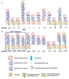
Figure 1
Figure 1
Structural representation of human CEACAM proteins and CEACAM1 splice variants. (A) In humans, there are 12 different CEACAM genes that encode functional proteins: CEACAM1, CEACAM3, CEACAM4, CEACAM5 (CEA), CEACAM6, CEACAM7, CEACAM8, CEACAM16, and CEACAM18-CEACAM21. Amongst these, CEACAM16 is expressed as a soluble protein. CEACAM1L, 3L and 4L as well as CEACAM18-21 possess a transmembrane anchor with a cytoplasmic tail (represented by L), whereas CEACAM5-8 are GPI-linked. (B) Schematic representation of the 12 CEACAM1 protein isoforms as products of alternative splicing of the human CEACAM1 gene: The most prominent and best-studied CEACAM1 isoforms are highlighted in a blue box. They either comprise a long (L) or a short cytoplasmic tail (S) and four or three extracellular immunoglobulin-like domains (1-4 or 1-3, respectively). Amongst the 12 isoforms, further 4-domain variants are found with a modified short cytoplasmic tail (CEACAM1-4S2) or a soluble isoform (CEACAM1-4C1). Additional soluble isoforms include CEACAM1-3 and CEACAM1-C2. The membrane-bound CEACAM1-1L and CEACAM1-1S as well as CEACAM1-AL and CEACAM1-AS only comprise one extracellular domain. Their functions remain elusive. Further details and hyperlinks to protein databases are found on www.carcinoembryonic-antigen.de; a full list of genes encoding CEACAM proteins in humans and rodents can be found in [29]. Adapted in modified form from www.carcinoembryonic-antigen.de, with permission.
Figure 2
Figure 2
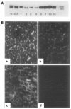
Dynamic expression of CEACAM1 in regenerating rat livers after partial hepatectomy. (A) A Western Blot showing CEACAM1 expression before (N) and after partial hepatectomy at the indicated timepoints (days) in rats. The decline in CEACAM1 expression starts 1 day after the procedure and returns back to near normal levels approximately after 7–10 days. (B) Analysis of CEACAM1-expression by indirect fluorescence (white signals) in a healthy rat liver (a), 48 h (b), and 15 days (c) after partial hepatectomy. CEACAM1 is expressed on all membranes of the hepatocytes with especially strong staining present at the apical surface of the hepatocytes. Weaker staining is present on the hepatocellular surface facing the sinusoidal space (a,c: white arrows). (d) negative control. (A,B) reprinted with modifications from [53] with permission from Elsevier.
Figure 3
Figure 3
Regulation of insulin clearance by CEACAM1-L. (1.) Insulin binding to the insulin receptor (IR) induces its autophosphorylation and the phosphorylation of downstream substrates such as the insulin receptor substrate-1 (IRS-1) and CEACAM1-L. Phosphorylation of CEACAM1-L on Tyr488, which requires an intact Ser503 residue, mediates its indirect association with IR to increase the rate of endocytosis of the insulin-insulin receptor complex into clathrin pits/vesicles of the hepatocytes, facilitating subsequent insulin degradation and clearance. (2.) Phosphorylated CEACAM1 can bind to SHP-2 to sequester the phosphatase and subsequently, mediate more sustained phosphorylation of the IRS-1/PI3K/AKT pathway that transduces the metabolic effects of insulin, such as inhibiting gluconeogenesis (via phosphorylating and nuclear exclusion of forkhead box protein O1 (Foxo1), among others [86,93,94]). The physiologic correlation of this CEACAM1 expression is underlined by the negative effect of Foxo1 activation on Ceacam1 expression [95]. Furthermore, CEACAM1-L–mediated IR internalization via clathrin-coated pits (3.) enhances the exposure of the insulin receptor tyrosine kinase to its endogenous substrates (IRS-1), supporting sustained IRS-1/PI3K/AKT signaling until insulin is degraded and removed. Single-headed black arrows indicate a chain of phosphorylation/dephosphorylation events by the indicated insulin receptor tyrosine kinase (IR), Ser/Thr kinases (PI3K/AKT), and SHP-2 phosphatase. Double-headed black arrows denote the physical (indirect) interaction between phosphorylated-CEACAM1-L (pCEACAM1) and the IR to stabilize the insulin-receptor endocytosis complex and increase its uptake into the endocytotic vesicles.
Figure 4
Figure 4
Pathophysiological consequences of loss of CEACAM1 expression and function. Hepatic inactivation or global loss of functional CEACAM1 expression impairs insulin clearance to cause insulin resistance and fatty liver disease in addition to disturbed energy homeostasis. Initially, these Ceacam1 mutations impair insulin clearance to promote hyperinsulinemia and insulin resistance (1). Hyperinsulinemia, in turn, promotes lipogenesis in the liver, accompanied by elevated fatty acid synthase (FASN) expression and activity, followed by hepatic fat accumulation (2a). Subsequently, NAFLD and NASH emerge, accompanied by elevation of inflammatory cytokine production (TNFα, IL-6, IL1β). The emergence of macrosteatosis as well as fibrosis can be accelerated by high-fat-diet feeding of Ceacam1 mutant mice. Chronic liver inflammation can ultimately lead to more progressive liver fibrosis and cirrhosis. Paralleling insulin resistance, hyperinsulinemia, fatty liver disease and disturbed energy homeostasis, visceral adiposity and systemic inflammation emerge (3). Additionally, leptin resistance, elevation of hypothalamic FASN expression, hyperphagia and reduction of energy expenditure (2b/4) fuel the vicious cycle that ultimately contributes to the cardio-metabolic abnormalities in Ceacam1-deficient mice. Upward or downward red arrows indicate increase or decrease, respectively, in cardio-metabolic or inflammatory events/parameters. Single-headed black arrows indicate hierarchical chains of events in the emergence of metabolic dysfunction. Double-headed black arrows depict feedback loops pointing towards the connection between visceral obesity and adipose tissue inflammation on the one hand and hepatosteatitis (3.) and central control circuits in obesity (4.) on the other.
Figure 5
Figure 5
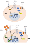
Specific functions of CEACAM1-S and CEACAM1-L isoforms in the regulation of T cell activation. (A) Different features of the CEACAM1-isoforms, CEACAM1-S and CEACAM1-L, in T cell signaling: CEACAM1-S and the TCR complex trigger T cell activation, and NFAT-mediated signaling [176,231]. Subsequently, NFAT nuclear translocation and transcription of Il-2 are initiated; CEACAM1-S can act as an independent signaling unit in T cells. CEACAM1-L with two ITIM motifs is phosphorylated by Src family kinases, and subsequently binds SHP-1 that dephosphorylates ZAP-70 and consequently blocks TCR-mediated activation of MAPK signaling in T cells [5,177,178]. The molecular adaptors that relay CEACAM1-S signaling via NFAT1 have not been defined yet. The initiation of CEACAM1-S-mediated IL-2 production is a key event in Treg induction and stability (B). Upon CEACAM1-S and TCR-induced IL-2 production, signaling of the IL-2R complex induces STAT5 phosphorylation; phosphorylated STAT5 translocates into the nucleus and acts as a transcription factor in a heterodimeric or heterotetrameric complex. pSTAT5 controls the transcription of key functional Treg genes, such as CD25, Foxp3, and Bcl-2, indicated by a curved black arrow ([204], and references therein). Single-headed black arrows indicate a chain of downstream phosphorylation and dephosphorylation events: Phosphorylation is elicited by the IL-2R, as well as as TCR in conjuction with CEACAM1-S and CEACAM1-L; in turn, SHP-2 recruitment by phosphylated CEACAM1-L localizes ZAP-70 phosphatase to TCR, which consequently blocks MAPK activation (black T bar). The question mark downstream of CEACAM1-S indicates that direct activation and phosphorylation of NFAT by CEACAM1-S via potential physical interactions remains to be determined. Reprinted with adaptations from [195] with permission from Wiley and Sons.

References

    1. Paxton R.J., Mooser G., Pande H., Lee T.D., Shively J.E. Sequence analysis of carcinoembryonic antigen: Identification of glycosylation sites and homology with the immunoglobulin supergene family. Proc. Natl. Acad. Sci. USA. 1987;84:920–924. doi: 10.1073/pnas.84.4.920. - DOI - PMC - PubMed
    1. Hinoda Y., Neumaier M., Hefta S.A., Drzeniek Z., Wagener C., Shively L., Hefta L.J., Shively J.E., Paxton R.J. Molecular cloning of a cDNA coding biliary glycoprotein I: Primary structure of a glycoprotein immunologically crossreactive with carcinoembryonic antigen. Proc. Natl. Acad. Sci. USA. 1988;85:6959–6963. doi: 10.1073/pnas.85.18.6959. - DOI - PMC - PubMed
    1. Aurivillius M., Hansen O.C., Lazrek M.B.S., Bock E., Öbrink B. The cell adhesion molecule Cell-CAM 105 is an ecto-ATPase and a member of the immunoglobulin superfamily. FEBS Lett. 1990;264:267–269. doi: 10.1016/0014-5793(90)80264-J. - DOI - PubMed
    1. Obrink B. CEA adhesion molecules: Multifunctional proteins with signal-regulatory properties. Curr. Opin. Cell Biol. 1997;9:616–626. doi: 10.1016/S0955-0674(97)80114-7. - DOI - PMC - PubMed
    1. Gray-Owen S.D., Blumberg R.S. CEACAM1: Contact-dependent control of immunity. Nat. Rev. Immunol. 2006;6:433–446. doi: 10.1038/nri1864. - DOI - PubMed

MeSH terms