. 2018 Oct 12;8(1):15215.
doi: 10.1038/s41598-018-33192-6.

Bioactive Hydrogel Marbles

Affiliations

Bioactive Hydrogel Marbles

Álvaro J Leite et al. Sci Rep. .

Abstract

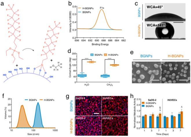
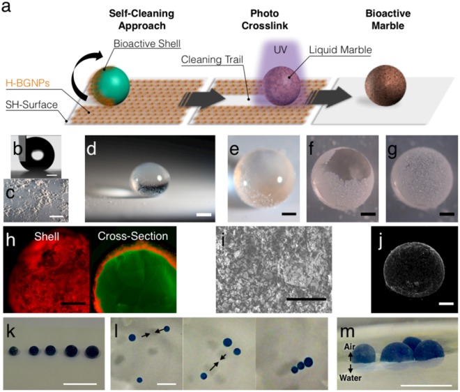
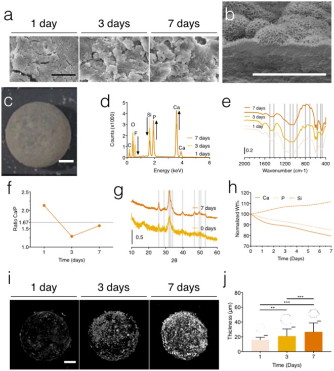
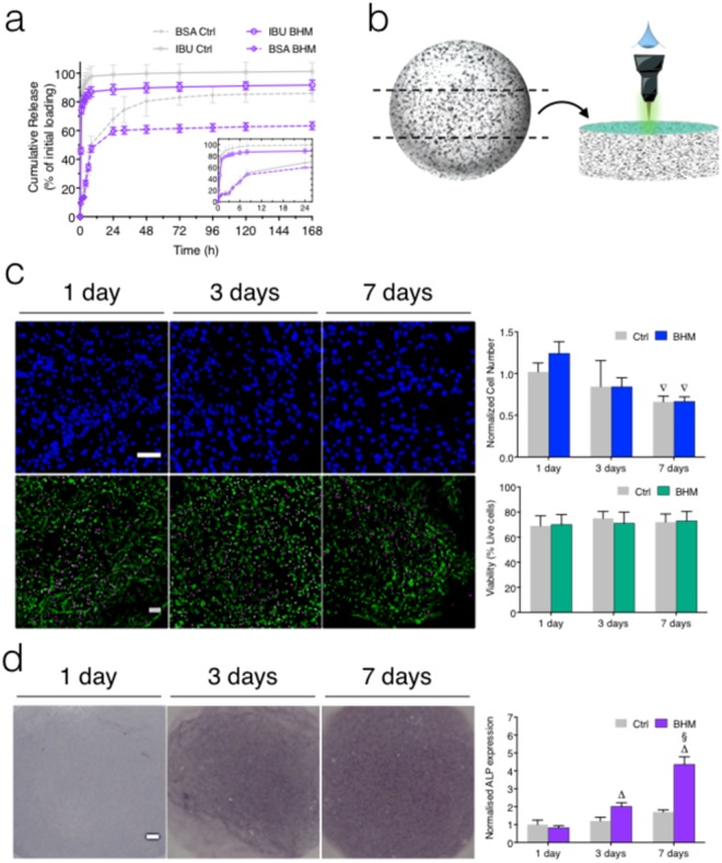
Liquid marbles represented a significant advance in the manipulation of fluids as they used particle films to confine liquid drops, creating a robust and durable soft solid. We exploit this technology to engineering a bioactive hydrogel marble (BHM). Specifically, pristine bioactive glass nanoparticles were chemically tuned to produce biocompatible hydrophobic bioactive glass nanoparticles (H-BGNPs) that shielded a gelatin-based bead. The designed BHM shell promoted the growth of a bone-like apatite layer upon immersion in a physiological environment. The fabrication process allowed the efficient incorporation of drugs and cells into the engineered structure. The BHM provided a simultaneously controlled release of distinct encapsulated therapeutic model molecules. Moreover, the BHM sustained cell encapsulation in a 3D environment as demonstrated by an excellent in vitro stability and cytocompatibility. The engineered structures also showed potential to regulate a pre-osteoblastic cell line into osteogenic commitment. Overall, these hierarchical nanostructured and functional marbles revealed a high potential for future applications in bone tissue engineering.


Conflict of interest statement

The authors declare no competing interests.

Figures

Figure 1
Figure 1
Production and characterization of the hydrophobic bioactive glass nanoparticles (H-BGNPs). (a) Illustration of the synthesis of the novel H-BGNPs through nature-inspired chemistry: the theoretical grafting of PFDTS molecules on the BGNPs surface. (b) X-ray photoelectron spectroscopy (XPS) analysis of the nanoparticles before and after grafting by PFDTS (H-BGNPs), which confirmed the chemical functionalization. An EDS spectra are also present in Fig. S1. (c) Water contact angle (WCA) of the BGNPs and H-BGNPs. Scale bar = 1 mm. The BGNPs could be wetted by water due to the hydroxyl groups on its surface. The fluorosilanization of the BGNPs renders the initial hydrophilic nanoparticles into hydrophobic bioactive glass nanoparticles. (c) The contact angle of BGNPs and H-BGNPs. (e) SEM micrographs of H-BGNPs and BGNPs. Scale bar = 500 nm. (f) Evaluation of the size distribution by dynamic light scattering (DLS). (g) Representative photographs of SaOS-2 and HUVECs at 7days of cell culture. Scale bar = 200 µm. The full comparison with non-modified BGNPs is presented in Fig. S3. (h) AlamarBlue assay results, presenting the metabolic activity of both cell types for 1, 3, and 7 days of cell culture.
Figure 2
Figure 2
Fabrication of the hydrogel marble. (a) Schematic drawing of the synthesis procedure of the bioactive hydrogel marble by a biomimetic approach based on the self-cleaning of the lotus leaf. (b) Profile of a water drop in contact with the superhydrophobic surface. Water contact angle of 162 ± 1.3°. Scale bar = 500 µm. (c) Superhydrophobic surface covered with H-BGNPs. The observed clusters could be attributed to powder electrostatic effects. Scale bar = 1 cm. (d) Polymeric drop on the superhydrophobic surface. Scale bar = 500 µm. (e) Detail of the polymeric drop rolling on the superhydrophobic surface. Scale bar = 500 µm. (f) Formation of the bioactive shell. Scale bar = 500 µm. (g) Bioactive marble: polymeric sphere covered with H-BGNPs. Scale bar = 500 µm. (h) Fluorescent micrographs detail of the shell and cross-section of the bioactive marble. Before the fabrication of the bioactive marble, the gelatin and the H-BGNPs were first stained with fluorescein (green) and rhodamine (red), respectively. Scale bar = 500 µm. (i) Representative SEM micrographs of the H-BGNPs at the surface of the BHM. Scale Bar = 5 µm. (i) μCT 3D reconstruction images of the BHM. (k) Photographs showing BHM (dyed in blue) on top of a superhydrophobic surface. The dispensed volumes were 1.5, 2, 4, 8, and 16 μL. The approximate diameters of the spheres after crosslinking were 1, 1.2, 2.5, 4; 5; 6 mm, respectively. Scale bar = 1 cm. (l) BHM floating at the surface of the water. Scale bar = 0.5 cm. (m) Representative image of the floating performance of the BHM cluster that could be manipulated and self-assemble on the surface of the water. Scale bar = 1 cm.
Figure 3
Figure 3
The bioactive behavior of the produced marbles. (a) Representative SEM micrographs of the bioactive marbles soaked in SBF for 1, 3, and 7 days. Scale bar = 5 µm. (b) Cross-section SEM micrograph of the hydrogel marbles soaked in SBF after 7 days, exhibiting the aggregates of nanometric needle-like crystals that characterized cauliflower morphology of hydroxyapatite. Scale bar = 5 µm. (c) Detail of the hydrogel marbles soaked in SBF after 7 days. Scale bar = 500 µm. (d) Identification of chemical elements performed by EDS following testing of BHM in SBF during 1, 3, and 7 days. (e) FTIR spectra of the apatite development on the surface of the bioactive marble. The chemical groups and the bandwidths used for identification are specified in Table S3. (f) Ca/P ratio during 1, 3, and 7 days of immersion in SBF. (g) XRD spectra of the BHM obtained before and after the immersion in SBF (0 and 7 days). The main characteristic hydroxyapatite peaks are shown at 2θ = 25.9°, 29°, 31.8°, 32.2°, 32.9°, 34°, 39.8°, 46.7°, 49.5°, 50.5° and 53.1° . (h) ICP study about the variation of Ca, Si and P in the SBF along of 7 days. (i) μCT 3D reconstruction images of the bioactive marbles after 1, 3, and 7 days of immersion in SBF. Scale bar = 500 µm. (j) The thickness of the bioactive marbles based on the μCT during 1, 3, and 7 days of immersion in SBF and the representative cross-sections images. Scale bar = 500 µm.
Figure 4
Figure 4
Biological functional performance of the produced bioactive hydrogel marbles and the comparison with non-coated hydrogel spheres (Ctrl). (a) BSA and Ibuprofen accumulative release profiles from the constructs. The inset graphic amplifies the release profiles in the first 24 hours. (b) Slice of the BHM for core imaging purposes to eliminate the shell artifacts. (c) DAPI (on top) and live-dead (at the bottom) fluorescent microscopy images (blue represents nuclei, green represents live cells, and magenta represents dead cells) of MC3T3-E1 cells. Scale bar = 100 µm. The corresponding quantification by image analysis is also shown. The images of non-coated hydrogels (Ctrl) are presented in Fig. S7. (d) Evaluation of the early osteogenic commitment with representative images of ALP staining during the cell culture time and the respective image analysis quantification. Scale bar = 100 µm. When applicable the results are presented as the arithmetic mean ± standard deviation. The images of non-coated hydrogels (Ctrl) are presented in Fig. S8.

References

    1. Zhang L, Cha D, Wang P. Remotely Controllable Liquid Marbles. Advanced Materials. 2012;24:4756–4760. doi: 10.1002/adma.201201885. - DOI - PMC - PubMed
    1. Yao X, Song Y, Jiang L. Applications of Bio-Inspired Special Wettable Surfaces. Advanced Materials. 2011;23:719–734. doi: 10.1002/adma.201002689. - DOI - PubMed
    1. Aussillous P, Quere D. Liquid marbles. Nature. 2001;411:924–927. doi: 10.1038/35082026. - DOI - PubMed
    1. McHale G, Newton MI. Liquid marbles: topical context within soft matter and recent progress. Soft Matter. 2015;11:2530–2546. doi: 10.1039/C5SM00084J. - DOI - PubMed
    1. Bormashenko E, Bormashenko Y, Musin A, Barkay Z. On the Mechanism of Floating and Sliding of Liquid Marbles. Chemphyschem. 2009;10:654–656. doi: 10.1002/cphc.200800746. - DOI - PubMed

Publication types