. 2018 Oct 12;9(1):4229.
doi: 10.1038/s41467-018-06315-w.

Chemical and structural studies provide a mechanistic basis for recognition of the MYC G-quadruplex

Affiliations

Chemical and structural studies provide a mechanistic basis for recognition of the MYC G-quadruplex

David R Calabrese et al. Nat Commun. .

Abstract

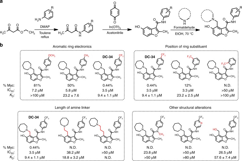
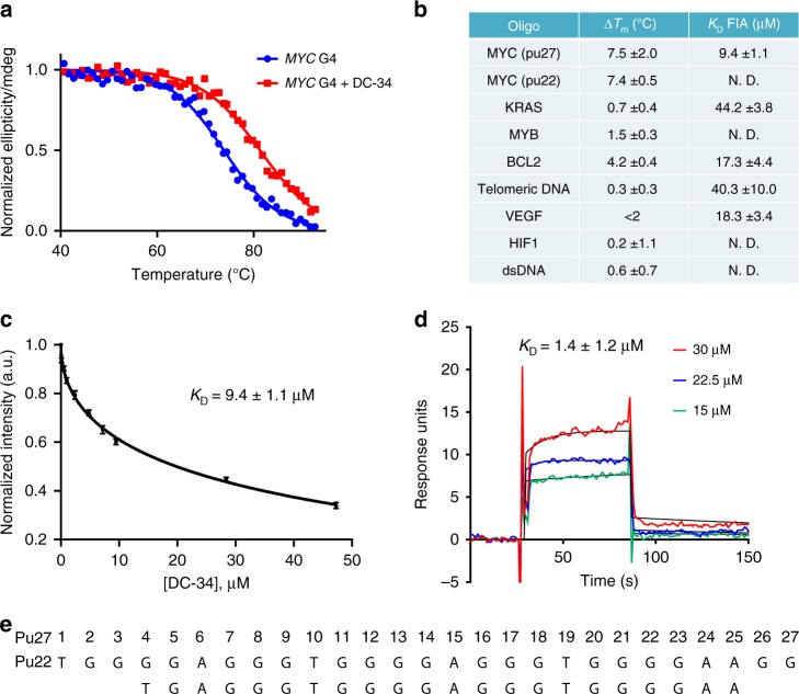
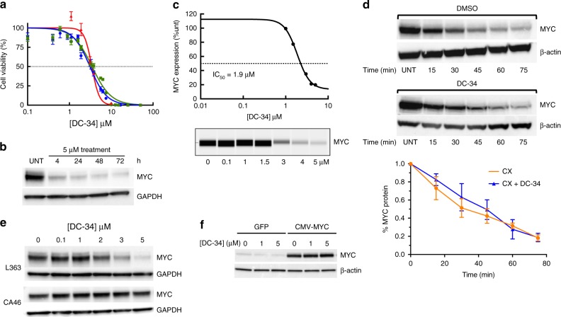
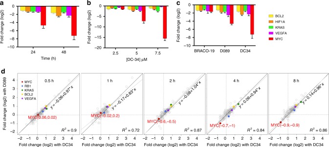
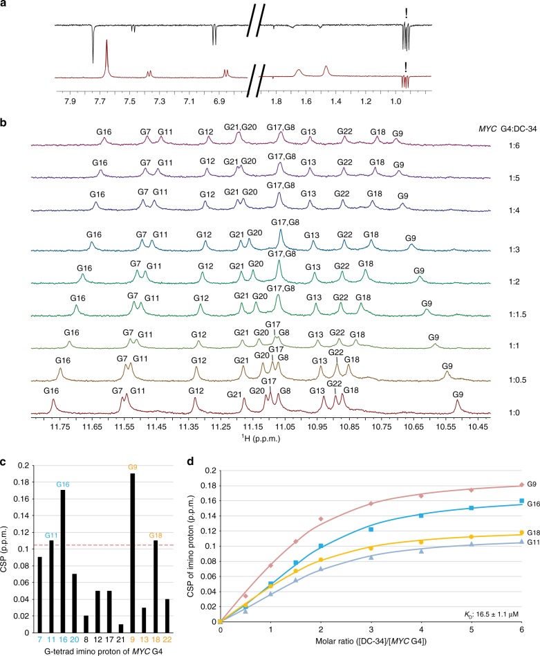
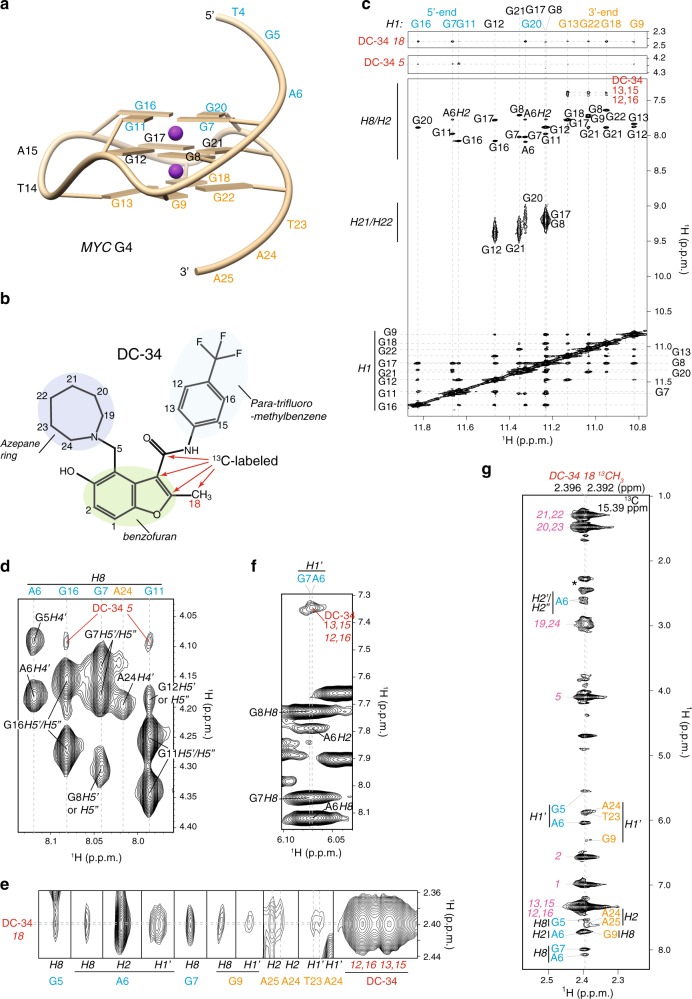
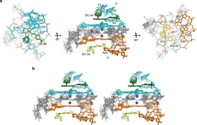
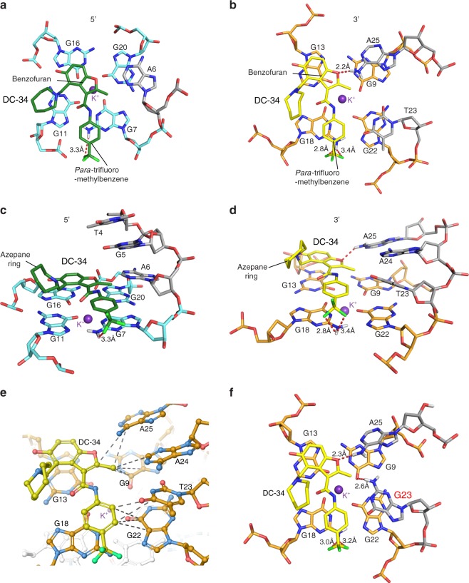
G-quadruplexes (G4s) are noncanonical DNA structures that frequently occur in the promoter regions of oncogenes, such as MYC, and regulate gene expression. Although G4s are attractive therapeutic targets, ligands capable of discriminating between different G4 structures are rare. Here, we describe DC-34, a small molecule that potently downregulates MYC transcription in cancer cells by a G4-dependent mechanism. Inhibition by DC-34 is significantly greater for MYC than other G4-driven genes. We use chemical, biophysical, biological, and structural studies to demonstrate a molecular rationale for the recognition of the MYC G4. We solve the structure of the MYC G4 in complex with DC-34 by NMR spectroscopy and illustrate specific contacts responsible for affinity and selectivity. Modification of DC-34 reveals features required for G4 affinity, biological activity, and validates the derived NMR structure. This work advances the design of quadruplex-interacting small molecules to control gene expression in therapeutic areas such as cancer.


Conflict of interest statement

The authors declare no competing interests.

Figures

Fig. 1
Fig. 1
Structure–activity relationship of DC-34. a Synthesis scheme of MYC G4-binding scaffold. b Table of selected analogs, with the parent compound on the left. %Myc indicates to the percent of MYC protein expressed at a 10 µM dose in L363 MM cells, IC50 is the half maximum concentration for cytotoxicity in L363 MM cells at 48 h, and KD determined by a fluorescence intensity assay. N. D. indicates that value was not determined
Fig. 2
Fig. 2
Biophysical analysis of the DC-34/MYC G4 interaction. a CD thermal melt of the MYC G4 Pu22 (10 µM) in the absence (blue) and presence (red) of DC-34 (40 µM). b Table of changes in thermal melting temperatures in the presence of four equivalents of DC-34 (measured by circular dichroism) and KD values of DC-34 against a panel of G4-forming sequences and dsDNA. c Fluorescence intensity curves of 5′ Alexa Fluor labeled Pu22 MYC upon titration of DC-34. KD = 9.4 ± 1.1 µM. d Sensorgrams corresponding to 15 to 30 µM injections of DC-34 over a 5′ biotin labeled MYC sensor chip. KD = 1.4 ± 1.2 µM. e Sequence of MYC Pu 27 and Pu22
Fig. 3
Fig. 3
DC-34 silencs MYC expression in cancer cells. a Inhibition of L363 MM cell proliferation at 24 (IC50 = 3.4 µM) (red), 48 (IC50 = 3.4 µM) (green) and 72 h (IC50 = 3.1 µM) (blue). b Inhibition of MYC protein translation with 5 μM of DC-34 is sustained over time in L363 cells. c The half maximal inhibitory concentration (IC50) of DC-34 with respect to MYC protein inhibition as determined by Peggy protein expression. d A Western blot of MYC protein during a representative cycloheximide-chase degradation experiment with L363 multiple myeloma cells. Cells were treated with 10 µg/mL of cycloheximide (CX) in the absence or presence of DC-34 (5 µM) and MYC protein expression was assessed at indicated time points. β-actin was used as loading control. e MYC protein levels are inhibited as a function of the dose of DC-34 in L363 cells; only the highest dose of DC-34 affected MYC in the more resistant CA46 Burkitt’s lymphoma cells. f Western blot analysis of 293T cells transiently transfected with either GFP or CMV-MYC plasmid (the CMV promoter lacks a MYC G4) (right) and dosed with different concentrations of DC-34. All western blots were exposed for <1 min
Fig. 4
Fig. 4
Effects of DC-34 on gene expression. a qPCR analysis of the indicated G4 containing genes following 5 μM treatment in L363 cells. b qPCR analysis of G4-associated genes at 48 h as a function of [DC-34]. c qPCR analysis of G4-associated genes after 48 h treatment with Braco-19, a pan-G4 binder, D089, a MYC G4 analog, and DC-34 at the indicated doses in L363 cells. Data in ac are the average log2 values for ΔΔCt of three replicates in L363 cells. d Effects of D089 (15 µM) and DC-34 (5 µM) on expression of genes (Nanostring) at the indicated time points. G4 controlled genes including MYC (red) are highlighted in color. Fitted linear regression lines (solid) for gene expression and identity lines (dashed) for drugs are indicated
Fig. 5
Fig. 5
Shifting of imino protons during a titration experiment indicates DC-34 binding to each end of MYC G4. a WaterLOGSY NMR spectra of DC-34 and N-methyl-l-valine (non-binding control, peaks indicated with !) in the absence (top) and presence (bottom) of MYC G4. b Expanded 1D 1H NMR spectra illustrating the imino region during titration of DC-34 into MYC G4. Molar ratios of MYC G4:DC-34 are as indicated at 1:0, 1:0.5, 1:1, 1:1.5, 1:2, 1:3, 1:4, 1:5, and 1:6. The G-tetrad imino protons are labeled in each spectrum. c Plot of chemical shift perturbation (CSP) comparing MYC G4 G-tetrad imino protons alone and with sixfold molar excess DC-34. The red dashed line indicates one standard deviation above the average value. d CSP values for G9 (red), G11 (purple), G16 (blue), and G18 (orange) H1 plotted against molar ratio of DC-34 to MYC G4. An overall KD value of 16.5 ± 1.1 µM was obtained by fitting the data to a non-cooperative binding mode with the software package Bindfit v0.5
Fig. 6
Fig. 6
Intermolecular NOE interactions indicate distinct binding modes for DC-34 at the 5′ and 3′ end of MYC G4. a Representative G-quadruplex structure for the MYC G4 with nucleic acid number indicated. The lowest energy structure displayed in Fig. 7 was used to make this figure. b Annotation of DC-34 indicating numbering scheme and 13C-labeling. cf Expanded regions of a 2D NOESY spectrum acquired on MYC G4 with twofold molar excess unlabeled DC-34 at pH 6.4 in buffer A (25 mM Tris-d11 and 50 mM KCl) with 90% H2O/10% DMSO-d6. H1–H1, H21/H22–H1, and H8/H2–H1 NOE interactions within MYC G4 are labeled (c, bottom panel), as are intermolecular NOE interactions between MYC G4 H1 and DC-34 methyl (c, top panel) or methanediyl (c, middle panel) groups. g Selected region from a 1H, 13C half-filtered NOESY experiment acquired with MYC G4 and twofold molar excess DC-34 with selective 13C-labeling as indicated by red arrows in b. Intermolecular NOE interactions between the DC-34 methyl group and MYC G4 protons are displayed and labeled as are intramolecular NOE interactions involving 12C bound DC-34 protons. A breakthrough intramolecular signal from the DC-34 methyl group is indicated by a black asterisk. Labeling of NOEs in cg is color coded with DC-34 in red (intermolecular) or pink (intramolecular) and MYC G4 in black, light blue (for the 5′ G-tetrad and flanking residues), or orange (for the 3′ G-tetrad and flanking residues)
Fig. 7
Fig. 7
Structure of the DC-34/MYC G4 complex indicates an additional stacked layer at each end and rearrangement of the flanking residues. a The 15 lowest energy structures of the DC-34/MYC G4 complex are displayed with a top (left panel), side (middle panel), and bottom (right panel) view relative to the cylindrical axis of the DNA. Residues of MYC G4 from the 5′ G-tetrad and flanking residues are highlighted in light blue, whereas those from the 3′ end are indicated in orange. The two DC-34 molecules are colored green and yellow, and the two potassium ions displayed as purple spheres. b Stereo view of the 15 lowest energy structures of the DC-34/MYC G4 complex
Fig. 8
Fig. 8
Specific contacts formed at each end of the MYC G4 provide a rationale for DC-34 as the preferred compound from the benzofuran-containing molecules screened. ad Expanded views of the DC-34/MYC G4 complex to display interactions at the 5′ (a, c) and 3′ (b, d, e) ends of the MYC G4. The color scheme follows Fig. 7 but with flanking residues gray and oxygen, nitrogen and fluorine in red, navy, and light green. Hydrogen bonds between MYC G4 and DC-34 are indicated by dotted red lines. In e hydrophobic contacts to flanking residues are indicated by gray-dashed lines. f Expanded view of an energy minimized model structure of DC-34/MYC G4 with wild-type guanine substituted for T23

References

    1. Burge S, Parkinson GN, Hazel P, Todd AK, Neidle S. Quadruplex DNA: sequence, topology and structure. Nucleic Acids Res. 2006;34:5402–5415. doi: 10.1093/nar/gkl655. - DOI - PMC - PubMed
    1. Bacolla A, Wells RD. Non-B DNA conformations, genomic rearrangements, and human disease. J. Biol. Chem. 2004;279:47411–47414. doi: 10.1074/jbc.R400028200. - DOI - PubMed
    1. Neidle S. Quadruplex nucleic acids as novel therapeutic targets. J. Med. Chem. 2016;59:5987–6011. doi: 10.1021/acs.jmedchem.5b01835. - DOI - PubMed
    1. Neidle Stephen. Quadruplex nucleic acids as targets for anticancer therapeutics. Nature Reviews Chemistry. 2017;1(5):0041. doi: 10.1038/s41570-017-0041. - DOI
    1. Thomas JR, Hergenrother PJ. Targeting RNA with small molecules. Chem. Rev. 2008;108:1171–1224. doi: 10.1021/cr0681546. - DOI - PubMed

Publication types

MeSH terms

Substances