. 2018 Oct 11;5(1):45.
doi: 10.1186/s40634-018-0157-1.

Force measurement metrics for simulated elbow arthroscopy training

Collaborators, Affiliations

Force measurement metrics for simulated elbow arthroscopy training

Nick F J Hilgersom et al. J Exp Orthop. .

Abstract

Background: Elbow arthroscopy is a difficult surgical technique. Objective metrics can be used to improve safe and effective training in elbow arthroscopy. Force exerted on the elbow tissue during arthroscopy can be a measure of safe tissue manipulation. The purpose of this study was to determine the force magnitude and force direction used by experts during arthroscopic elbow navigation in cadaveric specimens and assess their applicability in elbow arthroscopy training.

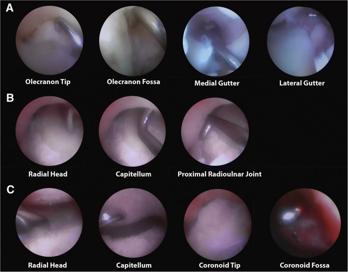
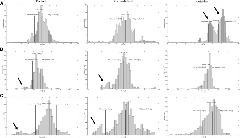
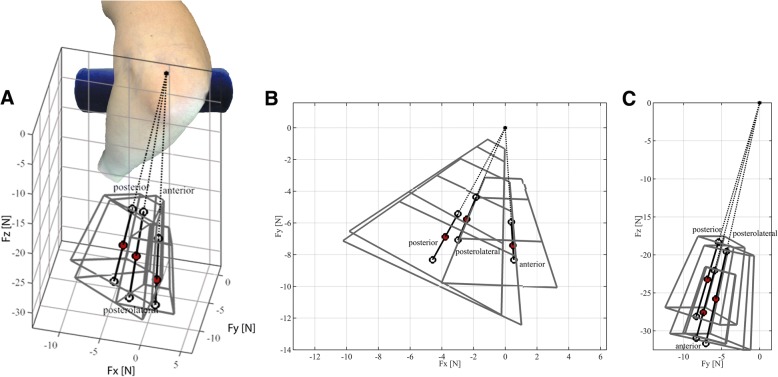
Methods: Two cadaveric elbows were mounted on a Force Measurement Table (FMT) that allowed 3-dimensional measurements (x-, y-, and z-plane) of the forces exerted on the elbow. Five experts in elbow arthroscopy performed arthroscopic navigation once in each of two cadaveric elbows, navigating through the posterior, posterolateral and anterior compartment in a standardized fashion with visualization of three to four anatomic landmarks per compartment. The total absolute force (Fabs) and force direction exerted (α and β) on the elbow during arthroscopy were recorded. α being the angle in the horizontal plane and β being the angle in the vertical plane. The 10th-90th percentiles of the data were used to set threshold levels for training.

Results: The median Fabs was 24 N (19 N - 30 N), 27 N (20 N - 33 N) and 29 N (23 N - 32 N) for the posterior, posterolateral and anterior compartment, respectively. The median α was - 29° (- 55° - 5°), - 23° (- 56° - -1°) and 4° (- 22° - -18°) for the posterior, posterolateral and anterior compartment, respectively. The median β was - 71° (- 80° - -65°), - 76° (- 86° - -69°) and - 75° (- 81° - -71°) for the posterior, posterolateral and anterior compartment, respectively.

Conclusion: Expert data on force magnitude and force direction exerted on the elbow during arthroscopic navigation in cadaveric specimens were collected. The proposed maximum allowable force of 30 N (smallest 90th percentile of Fabs) exerted on the elbow tissue, and the 10th-90th percentile range of the force directions (α and β) for each compartment may be used to provide objective feedback during arthroscopic skills training.

Keywords: Arthroscopy; Cadaver; Education; Elbow; Experts; Navigational forces; Skills assessment.


Conflict of interest statement

Ethics approval

The cadaveric specimens used in this study were derived from bodies that entered the department of anatomy, University of Utrecht, through a donation program. From these persons written consent was obtained during life that allowed the use of their entire bodies for educational and research purposes.

Consent for publication

Not applicable.

Competing interests

The authors declare that they have no competing interests.

Publisher’s Note

Springer Nature remains neutral with regard to jurisdictional claims in published maps and institutional affiliations.

Figures

Fig. 1
Fig. 1
Force Measurement Table. The FMT with custom-made stand with vice attached is shown. The FMT consists of the three squared frames and bending beams with Hall effect sensors and magnets in the x-, y- and z-plane of the FMT. The design of the FMT allows continuous recordings of the forces exerted on the cadaver specimen attached to the vice in three loading directions
Fig. 2
Fig. 2
Arthroscopic views of predefined landmarks per compartment. a Posterior compartment. b Posterolateral compartment. c Anterior compartment
Fig. 3
Fig. 3
Example of force measurement in time of one navigation task performed by one expert. This example shows force measurement in time of one navigation task performed by one expert in the posterior compartment. In the upper graph the individual force components as well as the overall combined force Fabs are illustrated. In the lower graph the stars indicate the moments of touching and displaying the assigned landmark. In this 2D representation, the first Area (A) represents a location were elbow flexing occurs indicated by an oppositely directed change in Force. The following area’s (B) represent instrument bone/tissue interaction with force fluctuations in similar direction
Fig. 4
Fig. 4
Histograms presenting the data points for Fabs, alfa and beta of each compartment. Histograms presenting the median, 10th and 90th percentile for frequency of total absolute force (Fabs), horizontal angle (α) and vertical angle (β) data points in the posterior, posterolateral and anterior compartment of both cadaver elbows. a Histogram showing Fabs. The two black arrows point to two separate peaks in frequency of Fabs, around 23 N and 30 N in the anterior compartment. b Histogram showing α. α is positive to the right, and negative to the left. The two black arrows point to a smaller peak around − 90° for α and β in the posterior and posterolateral compartment. c Histogram showing β. β is positive upward, and negative downward. The two black arrows point to a smaller peak around − 90° for α and β in the posterior and posterolateral compartment
Fig. 5
Fig. 5
Median force direction in the vertical plane (β) for all compartments combined. Schematic representation of the median direction of Fabs in the vertical plane for all compartments combined represented by β. The blue lines represent the lowest and highest values of the 10th and 90th percentiles of β. An elbow x-ray has been superimposed over the cadaveric elbow to further clarify the correlation between the overall median force direction and elbow joint
Fig. 6
Fig. 6
Safe force zone (magnitude and direction) for all compartments. The red dots indicate the median value of Fabs, α and β for each compartment. The thick black lines with white dots at their respective ends represent the 10th and 90th percentile value of Fabs in the median force direction. The grey boxes surrounding the thick black lines indicate the combined boundaries of the 10th -90th percentiles for Fabs, α and β. The origin is taken at the same position for each compartment. a 3D graphical representation. For reference an elbow in the lateral decubitus position is added. So the forces are directed towards the surgeon, b Top view, c Sagittal view

References

    1. Aggarwal R, Moorthy K, Darzi A. Laparoscopic skills training and assessment. Br J Surg. 2004;91:1549–1558. doi: 10.1002/bjs.4816. - DOI - PubMed
    1. Aim F, Lonjon G, Hannouche D, Nizard R. Effectiveness of virtual reality training in Orthopaedic surgery. Arthroscopy. 2016;32:224–232. doi: 10.1016/j.arthro.2015.07.023. - DOI - PubMed
    1. Camp CL, Krych AJ, Stuart MJ, Regnier TD, Mills KM, Turner NS. Improving resident performance in knee arthroscopy: a prospective value assessment of simulators and cadaveric skills laboratories. J Bone Joint Surg Am. 2016;98:220–225. doi: 10.2106/JBJS.O.00440. - DOI - PubMed
    1. Claessen F, Kachooei AR, Kolovich GP, Buijze GA, Oh LS, van den Bekerom MPJ, Doornberg JN. Portal placement in elbow arthroscopy by novice surgeons: cadaver study. Knee Surg Sports Traumatol Arthrosc. 2017;25:2247–2254. doi: 10.1007/s00167-016-4186-y. - DOI - PubMed
    1. Elfeddali R, Schreuder MH, Eygendaal D. Arthroscopic elbow surgery, is it safe? J Shoulder Elb Surg. 2013;22:647–652. doi: 10.1016/j.jse.2013.01.032. - DOI - PubMed

LinkOut - more resources