Review
. 2019 Feb;36(2):179-187.
doi: 10.1007/s10815-018-1326-y. Epub 2018 Oct 12.

The role of microRNAs in human embryo implantation: a review

Affiliations
Review

The role of microRNAs in human embryo implantation: a review

Anthea B M Paul et al. J Assist Reprod Genet. 2019 Feb.

Abstract

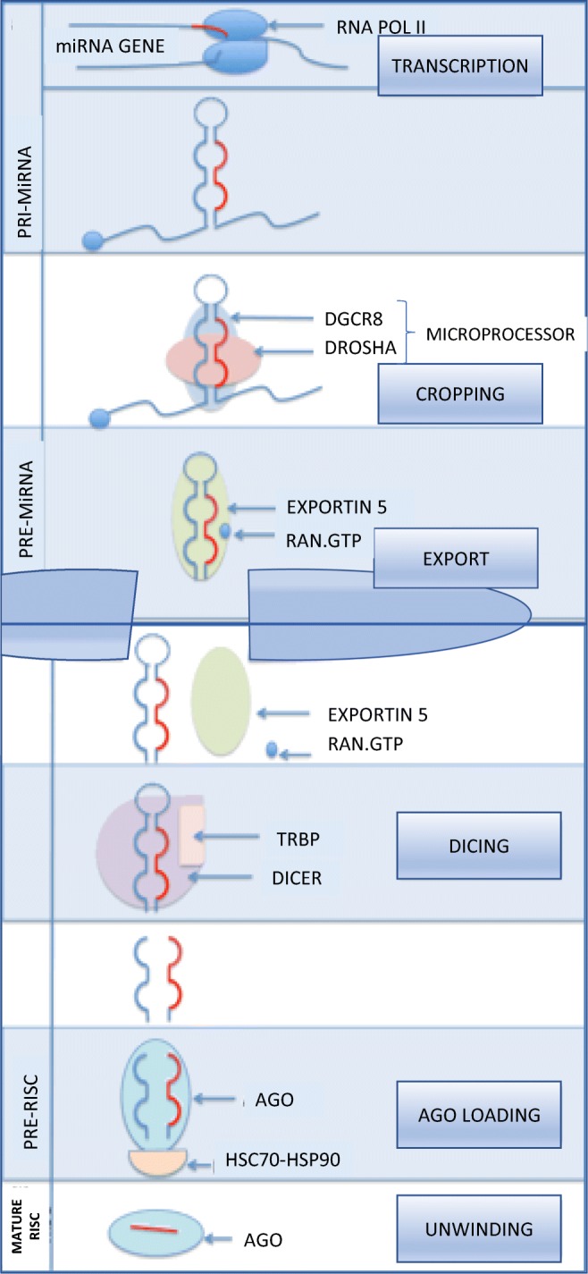
MicroRNAs (miRNAs) are emerging as important in human embryo implantation, and we present here a review of the literature from a clinical perspective. Implantation involves complex interactions between the blastocyst and endometrium. miRNAs have been shown to be differentially expressed in implanted compared with non-implanted blastocysts and euploid compared with aneuploid blastocysts. Further, miRNAs are differentially expressed in proliferative compared with decidualized endometrium, and in receptive compared with pre-receptive endometrium. miRNAs are also differentially expressed in endometrium of women who failed implantation, and in endometrium of women with recurrent implantation failure. Due to the complexity of miRNA signaling, studies have suffered from inconsistency in reproducibility of results. However, miRNAs show potential as biomarkers in the pursuit of more reliable prediction of embryo implantation.

Keywords: Endometrium; Human embryo; Implantation; MicroRNA; Pregnancy; Recurrent implantation failure.


Conflict of interest statement

The authors declare that they have no conflict of interest

Figures

Fig. 1
Fig. 1
The number of references found for each year of publication on the PubMed database using the keyword ‘microRNA’. In 2017, this number was 12,682. Image by author
Fig. 2
Fig. 2
A schematic diagram of signaling between blastocyst and receptive maternal endometrium. Microvilli from the blastocyst aid in attachment to the endometrial pinopodes. Further, ErbB4 on the blastocyst surface adheres to transmembrane HB-EGF (modulated by Lif) on the receptive endometrium. Integrin avB3 is attached to the maternal endometrium via osteopontin. l-selectin molecules on the blastocyst attach to selectin oligosaccharide ligand on the maternal surface. Image by author, adapted from Davidson et al. 2016 [10]
Fig. 3
Fig. 3
A schematic diagram of a attachment and b implantation between the blastocyst and receptive maternal endometrium. ErbB4 on the blastocyst surface adheres to transmembrane HB-EGF (modulated by Lif) along with l-selectin ligands (sLE) expressed by the maternal endometrium epithelium to l-selectin receptors on the blastocyst. The other key signaling pathways for attachment and implantation are also shown. AA, arachidonic acid; BMP2, bone morphogenetic protein 2; Cox2, cyclooxygenase-2; ENaC, epithelium sodium channel; ErbB4; epidermal growth factor receptor 1/4; gp130, glycoprotein 130; HB-EGF, heparin-binding epidermal growth factor-like growth factor; Hoxa10/11, homeobox A10/11; KLF5, Kruppel-like factor 5; LIF, leukemia inhibitory factor; LIFR, LIF receptor; LPA3, lysophosphatidic acid receptor 3; PG, prostaglandin; PPAR-δ; peroxisome proliferators–activating receptor δ; Wnt4, wingless-type MMTV integration site family members 4. Image by author, adapted from Cha et al. [15]
Fig. 4
Fig. 4
Nuclear events in miRNA canonical biogenesis. Schematic diagram of miRNA gene transcription by RNA polymerase II to form pre-miRNA. This is processed by the microprocessor complex of Drosha and DGCR8 (DiGeorge Syndrome Critical Region Gene 8) and cropped to form pre-miRNA. The pre-miRNA is exported from the nucleus via exportin 5 in complex with RAN.GTP. In the cytoplasm, after Dicer-processing RNA duplex is loaded onto AGO (Argonaute proteins) stabilized by a heatshock protein (HSC70-HSP90). The remaining “passenger” strand is discarded, and the mature miRNA strand is loaded onto the AGO protein. Image by author, adapted from Ha and Kim [38]

References

    1. Paria BC, Huet-Hudson YM, Dey SK. Blastocyst’s state of activity determines the “window” of implantation in the receptive mouse uterus. Proc Natl Acad Sci U S A. 1993;90(21):10159–10162. doi: 10.1073/pnas.90.21.10159. - DOI - PMC - PubMed
    1. Liu W, Niu Z, Li Q, Pang RTK, Chiu PCN, Yeung WSB. MicroRNA and embryo implantation. American Journal of Reproductive Immunology. 2016. - PubMed
    1. Evans J, Salamonsen LA, Winship A, Menkhorst E, Nie G, Gargett CE, Dimitriadis E. Fertile ground: human endometrial programming and lessons in health and disease. Nat Rev Endocrinol. 2016;12:654–667. doi: 10.1038/nrendo.2016.116. - DOI - PubMed
    1. Salamonsen LA, Evans J, Nguyen HPT, Edgell TA. The microenvironment of human implantation: Determinant of reproductive success. American Journal of Reproductive Immunology. 2016. - PubMed
    1. Liang J, Wang S, Wang Z. Role of microRNAs in embryo implantation. Reprod Biol Endocrinol. 2017, 15; - PMC - PubMed

LinkOut - more resources