. 2019 Jan;87(1):23-33.
doi: 10.1002/prot.25613. Epub 2018 Nov 8.

Increased sequence hydrophobicity reduces conformational specificity: A mutational case study of the Arc repressor protein

Affiliations

Increased sequence hydrophobicity reduces conformational specificity: A mutational case study of the Arc repressor protein

Katie L Stewart et al. Proteins. 2019 Jan.

Abstract

The amino-acid sequences of soluble, globular proteins must have hydrophobic residues to form a stable core, but excess sequence hydrophobicity can lead to loss of native state conformational specificity and aggregation. Previous studies of polar-to-hydrophobic mutations in the β-sheet of the Arc repressor dimer showed that a single substitution at position 11 (N11L) leads to population of an alternate dimeric fold in which the β-sheet is replaced by helix. Two additional hydrophobic mutations at positions 9 and 13 (Q9V and R13V) lead to population of a differently folded octamer along with both dimeric folds. Here we conduct a comprehensive study of the sequence determinants of this progressive loss of fold specificity. We find that the alternate dimer-fold specifically results from the N11L substitution and is not promoted by other hydrophobic substitutions in the β-sheet. We also find that three highly hydrophobic substitutions at positions 9, 11, and 13 are necessary and sufficient for oligomer formation, but the oligomer size depends on the identity of the hydrophobic residue in question. The hydrophobic substitutions increase thermal stability, illustrating how increased hydrophobicity can increase folding stability even as it degrades conformational specificity. The oligomeric variants are predicted to be aggregation-prone but may be hindered from doing so by proline residues that flank the β-sheet region. Loss of conformational specificity due to increased hydrophobicity can manifest itself at any level of structure, depending upon the specific mutations and the context in which they occur.

Keywords: conformational specificity; protein conformation; protein folding; sequence hydrophobicity; sequence-structure relationship; structural degeneracy.


Figures

Figure 1:
Figure 1:. Polar-to-hydrophobic substitutions in the β-strand region of Arc repressor decrease folding specificity.
A) Wild-type Arc (PDB ID 1ARR) and “switch” Arc (PDB ID 1NLA) homodimers, illustrating their distinct folding patterns. Residues 9–13 are β-sheet in wild-type Arc and helix in the alternate “switch” fold. B) Residues 9, 11 and 13 form the solvent-exposed surface of the sheet. C) Ball and stick diagrams schematically depicting structure and hydrophobicity for residues 9–14. Wild-type Arc adopts a homodimeric β-sheet structure in which hydrophobic residues (black balls) face the interior of the protein and polar residues (white balls) face solvent. Arc-N11L exists in equilibrium between the wild-type fold and the “switch” dimer fold in which the β-sheet is replaced by helices. Arc-N11L lowers conformational specificity by allowing multiple viable hydrophobic core arrangements: the introduced leucine is on the solvent-exposed surface of the wild-type fold but is buried in the hydrophobic core of the “switch” dimer. Two additional substitutions (Q9V and R13V) give the S-VLV variant and render the entire sequence of this region hydrophobic. S-VLV adopts both dimer folds, and also forms an octameric species. The structure adopted by residues 9–14 in the octamer is unknown (as indicated by dashed lines) but the global fold of the octamer is considerably less helical, and therefore different from either dimer fold. S-VLV therefore has at least three distinct folds. Our working model is that residues 9, 11 and 13 form a hydrophobic interface in the octamer.
Figure 2:
Figure 2:. One or two polar-to-hydrophobic substitutions in the β-strand are insufficient to convert Arc repressor dimer to a higher-order oligomer.
(A) Superdex 75 size exclusion chromatograms of Arc single and double polar-to-hydrophobic substitutions in SB250 buffer at 300 µM protein concentration and ambient temperature, following 4 h of a heat annealing treatment at 80 °C. See the text for an explanation of shorthand nomenclature for variants. Hydrophobic substitutions are indicated in bold. See also Figure S1 for column calibration. (B) Far ultraviolet circular dichroism spectra of the same variants, without a heat annealing treatment, in SB250 at 25 µM protein concentration in a 1 mm path length cuvette at 20 °C.
Figure 3:
Figure 3:. Three highly hydrophobic substitutions in the β-strand are necessary for population of the Arc repressor oligomer.
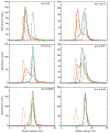
Each panel shows a set of Superdex 75 size exclusion chromatograms of Arc triple polar-to-hydrophobic substitutions in SB250 buffer at 300 µM protein concentration and ambient temperature, either without heat annealing treatment (green), or following 2 h (yellow) or 4 h (red) of annealing at 80 °C. For comparison, dashed lines show chromatograms for heated Arc-S-VLV oligomer (orange) and wild-type Arc dimer (blue). See also Figure S1 for column calibration.
Figure 4:
Figure 4:. Effect of dimer-stabilizing P8A/P8L substitutions on the oligomerization of S-VLV.
(A) Superdex 75 size exclusion chromatograms of S-VLV-P8A (purple) and S-VLV-P8L (red), in SB250 buffer at 150 µM protein concentration, before (dashed lines) and after (solid lines) a heat annealing treatment for 4 h at 80 °C. Chromatograms of heat annealed S-VLV octamer and wild-type Arc dimer in SB250 buffer are shown for comparison (brown). See also Figure S1 for column calibration. (B) Thermal denaturation curves for S-VLV P8A and S-VLV P8L in SB250 at 25 µM protein concentration in a 2 mm path length cuvette, monitored by circular dichroism at 222 nm.
Figure 5:
Figure 5:. Substitution of both P8 and P15 causes some higher-order aggregation in Arc-S-VLV, but does not affect dimer formation in a wild-type background.
(A) Analytical size exclusion of unheated P8A/P15A (blue) at 380 µM in SB250 buffer (blue). Traces for Arc-S-VLV and wild-type Arc (brown) are shown for comparison. See also Figure S1 for column calibration. (B) Far ultraviolet circular dichroism spectra at 50 µM protein concentration in SB250 buffer, in a 1 mm path length cuvette at 20 °C. (C) Thermal denaturation of P8A/P15A at 50 µM concentration in SB250 buffer, collected from 20–100 °C with a 2 °C step size in a 2 mm path length cuvette. (D) Thermal denaturation of Arc-S-VLV P8A/P15A (no prior heat treatment) at 25 µM concentration in SB250 buffer, collected from 20–100 °C with a 2 °C step size in a 2 mm path length cuvette.
Figure 6:
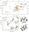
Figure 6:. Double hydrophobic substitution variants Arc-QLV and Arc-VLR adopt both the wild-type and helical dimer folds, while Arc-VNV resembles wild-type Arc.
(A) 15N-1H HSQC spectrum of uniform 15N-labeled variants at 87 μM protein concentration in 50 mM MES (pH 5.5), 50 mM KCl at 25 °C, compared to spectra of wild-type Arc and “switch Arc” (N11L/L12N), a variant known to exclusively adopt the helical dimer fold. In the lower left quadrant of the spectrum, the Trp 14 Ɛ1 resonance of S-VNV resembles that of wild-type Arc, while this resonance is invisible for S-QLV and S-VLR, putatively due to exchange between very different resonance positions in the wild-type and helical dimer folds. (B) Chemical shifts for the amide groups of A26, V33 and M42 in S-QLV and S-VLR are intermediate between the values observed for wild-type and switch Arc, while values for S-VNV are close to those of wild-type Arc. (C) Ribbon diagram of Arc repressor dimer structure with residues analyzed in panel B highlighted in color. These residues are in the helices of Arc and 10–20 Å away from the center of Arc’s β-sheet, but show some sensitivity to the dimer switch.

References

    1. Lattman EE, Rose GD. Protein folding--what’s the question? Proc Natl Acad Sci U S A 1993;90(2):439–441. - PMC - PubMed
    1. Bolon DN, Grant RA, Baker TA, Sauer RT. Specificity versus stability in computational protein design. Proc Natl Acad Sci U S A 2005;102(36):12724–12729. - PMC - PubMed
    1. Hoffmann J, Wrabl JO, Hilser VJ. The role of negative selection in protein evolution revealed through the energetics of the native state ensemble. Proteins. 2016;84(4):435–447. - PMC - PubMed
    1. Dill KA. Dominant Forces in Protein Folding. Biochemistry-Us 1990;29(31):7133–7155. - PubMed
    1. Kauzmann W Some Factors in the Interpretation of Protein Denaturation. Adv Protein Chem 1959;14:1–63. - PubMed

Publication types

Substances

LinkOut - more resources