. 2019 Feb;156(2):147-163.
doi: 10.1111/imm.13011. Epub 2018 Nov 8.

Different phenotypes of non-classical monocytes associated with systemic inflammation, endothelial alteration and hepatic compromise in patients with dengue

Affiliations

Different phenotypes of non-classical monocytes associated with systemic inflammation, endothelial alteration and hepatic compromise in patients with dengue

Juan S Naranjo-Gómez et al. Immunology. 2019 Feb.

Abstract

Although dengue can progress to severe stages, the exact causes of this phenomenon are unknown; however, the possibility of monocyte participation is acknowledged. It has been suggested that monocyte subsets (classical, intermediate and non-classical) play differential roles in dengue immunopathology. Therefore, we determined the count of monocyte subsets and obtained the clinical information of patients with dengue. We noted a significant decrease in the count of non-classical monocytes in patients compared with controls. With this finding, we focused on studying the phenotype of non-classical monocytes in the present study. An increase in activation and differentiation markers, such as CD64, CD86, the percentage of tumor necrosis factor-α+ cells and exposure of phosphatidylserine, were recorded in the non-classical monocytes of patients compared with controls. Moreover, a significant decrease in the expression of CX3CR1 with a corresponding increase in the expressions of CCR2, CCR5, CD11b and CD54 was detected in the non-classical monocytes of patients in comparison with that of the controls. Significant increases in the frequency of microparticles from endothelium and in the concentrations of interleukin-6 (IL-6), IL-8 and IL-10 were noted in the plasma of patients. These findings demonstrate that in patients with dengue, non-classical monocytes are activated, exhibiting a phenotype associated with more differentiation, produces tumor necrosis factor-α and has a profile of less endothelial surveillance closer to the cellular migration. These changes were associated with hepatic compromise, endothelial alteration and high concentration of circulating cytokines. Hence, alterations of non-classical monocytes seem to be associated with the immunopathology of dengue infection.

Keywords: dengue; monocyte subsets; monocytes; non-classical monocytes; severe dengue.


Figures

Figure 1
Figure 1
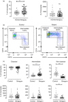
Decrease in the relative and absolute count of non‐classical monocytes in patients with dengue. (a) Relative (frequency, left panel) and absolute (right panel) counts of total monocytes (CD45+, CD14HLA‐DR+) in 63 patients with dengue and in 45 healthy controls. (b) Representative dot plots to define the monocyte subsets. The data from a dengue patient (right panel) and healthy control (left panel) are shown. (c) Frequency (top) and absolute counts (down) of the three monocyte subsets in 63 patients with dengue and in 45 healthy controls. The median is shown as a central tendency measure. Comparisons between groups were performed using the Mann–Whitney U‐test, *P ≤ 0·05, **P ≤ 0·01 and ***P ≤ 0·001.
Figure 2
Figure 2
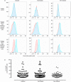
Similar percentage of positive cells to viral E protein in monocyte subsets of patients with dengue. (a) Representative histograms of the control cells (in red color, corresponding to staining without primary antibody) and the cells stained with the anti‐viral E protein primary antibody (in blue color) in the three monocyte subsets. The data from a healthy control and two dengue patients are shown (one with low frequency and one with high frequency of positive cells). (b) The frequency of positive cells to viral E protein in the three monocyte subsets of 58 patients with dengue. The median is shown as a central tendency measure. Comparisons among the groups were performed using the Kruskal–Wallis test and the Dunn's post hoc test.
Figure 3
Figure 3
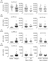
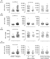
Low expression of CD16 and high expression of CD64, CCR2 and CCR5 in non‐classical monocytes of patients with dengue. The mean fluorescence intensity (MFI) of (a) CD16 and (b) CD64 FcR, (c) CCR2 and (d) CCR5 chemokine receptors in non‐classical monocytes of 63 patients with dengue and in 45 healthy controls. The results of patients divided according to the state of illness and the type of infection are shown in the middle and right panels, respectively. The median is shown as the central tendency measure. Comparisons between two groups were performed using the Mann–Whitney U‐test, *P ≤ 0·05, **P ≤ 0·01 and ***P ≤ 0·001.
Figure 4
Figure 4
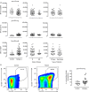
High expression of CX3CR1, CD11b and CD54 in non‐classical monocytes and the increase in the percentage of endothelium‐derived microparticles in patients with dengue. (a) The mean fluorescence intensity (MFI) of CX3CR1, CD11b and CD54 in non‐classical monocytes from 63 patients with dengue and in 45 healthy controls are shown. The results of the patients are divided according to the state of illness and the type of infection are shown in the middle and right panels, respectively. (b) Representative pseudocolor diagram of the HLA‐DR+ CD105+ microparticles analysis. The data from a dengue patient (middle panel) and healthy control (left panel) are shown. The frequency of the HLA‐DR+ CD105+ microparticles in 25 patients with dengue and in 20 healthy controls (right panel). The median is shown as a central tendency measure. Comparisons between groups were performed using the Mann–Whitney U‐test, *P ≤ 0·05, **P ≤ 0·01 and ***P ≤ 0·001.
Figure 5
Figure 5
High expression of CD14, CD68, CD86 and HLA‐DR in non‐classical monocytes of patients with dengue. The mean fluorescence intensity (MFI) of (a) CD14, (b) total CD68 (intra and extracellular), (c) CD86 and (d) HLA‐DR in non‐classical monocyte subset from 63 patients with dengue and from 45 healthy controls. The results of the patients divided according to the state of illness and the type of infection are shown in the middle and right panels, respectively. The median is shown as the central tendency measure. Comparisons between groups were performed using the Mann–Whitney U‐test, *P ≤ 0·05, **P ≤ 0·01 and ***P ≤ 0·001.
Figure 6
Figure 6
High expression of CD69 and CD68 (extracellular) and the frequency of tumor necrosis factor‐α‐positive (TNF‐α +) and Annexin‐V+ cells in the non‐classical monocyte subset of patients with dengue. (a) The mean fluorescence intensity (MFI) of CD69, CD68 (extracellular) and the frequency of TNF‐α + and Annexin‐V+ cells in the non‐classical subset of 15–25 patients with dengue and 10–20 healthy controls. (b) Analysis of the correlation between MFI of CD69 on non‐classical monocytes and the number of platelets/μl, and the percentage of TNF‐α + cells in non‐classical monocytes and the blood levels of bilirubin in 23 patients with dengue. The median is shown as a central tendency measure. Comparisons between groups were performed using the Mann–Whitney U‐test *P ≤ 0·05, **P ≤ 0·01 and ***P ≤ 0·001. Correlation analyses were performed by the Spearman rank coefficients at 95% confidence intervals.
Figure 7
Figure 7
High levels of circulating interleukin‐6 (IL‐6), IL‐8 and IL‐10 in patients with dengue. The plasmatic concentration of (a) IL‐6, (b) IL‐8 and (c) IL‐10 in 63 patients with dengue and in 45 healthy controls. The results of the patients divided according to the state and days of illness are shown in the middle and right panels, respectively. The median is shown as a central tendency measure. Comparisons between the two groups were performed using the Mann–Whitney U‐test. Other comparisons among the groups were performed using the Kruskal–Wallis test and the Dunn's post hoc test, *P ≤ 0·05, **P ≤ 0·01 and ***P ≤ 0·001.
Figure 8
Figure 8
Association between paraclinical and non‐classical monocyte variables in patients with dengue. Principal component analysis showing the correlations among paraclinical variables aspartate aminotransferase (AST), alanine aminotransferase (ALT), C‐reactive protein (CRP) in red lines; interleukin‐6 (IL‐6), IL‐8, IL‐10 in orange; the percentage of microparticles in green line; the number of total monocytes, the number of non‐classical monocytes, the percentage of tumor necrosis factor‐α‐positive (TNF‐α +) and annexin‐V+ in non‐classical monocytes and the mean fluorescence intensity (MFI) of CCR5, CCR2, CD11b, CD69, CD68, HLA‐DR, CD86, CD14, CD54 and CX3CR1 in non‐classical monocytes of dengue patient in blue lines.

References

    1. Halstead SB. Dengue. Lancet 2007; 370:1644–52. - PubMed
    1. WHO . Dengue: Guidelines for Diagnosis, Treatment, Prevention and Control. New Edition. Geneva: WHO; Guidelines Approved by the Guidelines Review Committee, 2009. - PubMed
    1. Guzman MG, Harris E. Dengue. Lancet 2015; 385:453–65. - PubMed
    1. Kwissa M, Nakaya HI, Onlamoon N, Wrammert J, Villinger F, Perng GC et al Dengue virus infection induces expansion of a CD14+CD16+ monocyte population that stimulates plasmablast differentiation. Cell Host Microbe 2014; 16:115–27. - PMC - PubMed
    1. Wong KL, Chen W, Balakrishnan T, Toh YX, Fink K, Wong SC. Susceptibility and response of human blood monocyte subsets to primary dengue virus infection. PLoS ONE 2012; 7:e36435. - PMC - PubMed

Publication types

MeSH terms