. 2018 Oct 13;15(1):69.
doi: 10.1186/s12977-018-0452-z.

Host SAMHD1 protein restricts endogenous reverse transcription of HIV-1 in nondividing macrophages

Affiliations

Host SAMHD1 protein restricts endogenous reverse transcription of HIV-1 in nondividing macrophages

Bijan Mahboubi et al. Retrovirology. .

Abstract

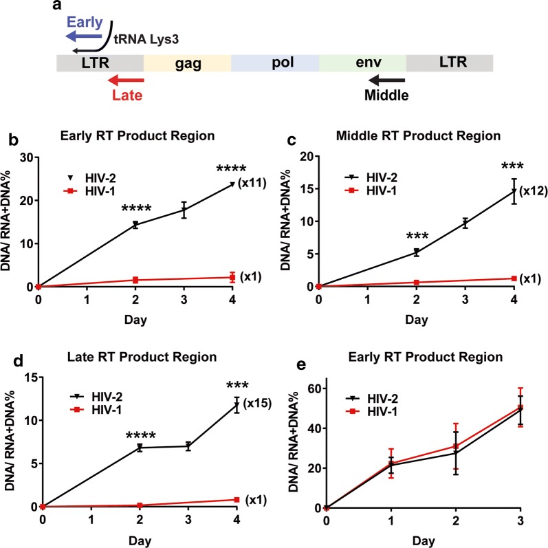
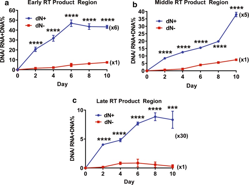
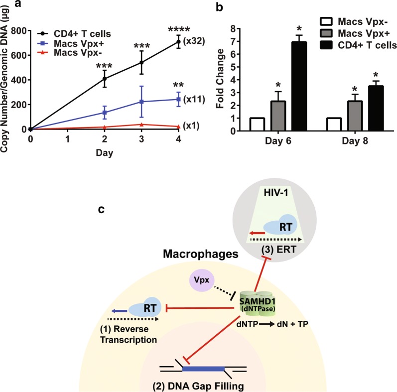
Background: SAM domain and HD domain containing protein 1 (SAMHD1) is a host anti-HIV-1 restriction factor known to suppress viral reverse transcription in nondividing myeloid cells by its dNTP triphosphorylase activity that depletes cellular dNTPs. However, HIV-2 and some SIV strains rapidly replicate in macrophages due to their accessory protein, viral protein X (Vpx), which proteosomally degrades SAMHD1 and elevates dNTP levels. Endogenous reverse transcription (ERT) of retroviruses is the extra-cellular reverse transcription step that partially synthesizes proviral DNAs within cell-free viral particles before the viruses infect new cells. ERT activity utilizes dNTPs co-packaged during budding from the virus-producing cells, and high ERT activity is known to enhance HIV-1 infectivity in nondividing cells. Here, since Vpx elevates cellular dNTP levels in macrophages, we hypothesize that HIV-2 should contain higher ERT activity than HIV-1 in macrophages, and that the Vpx-mediated dNTP elevation should enhance both ERT activity and infectivity of HIV-1 particles produced in macrophages.

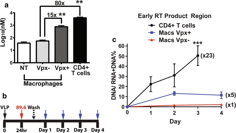
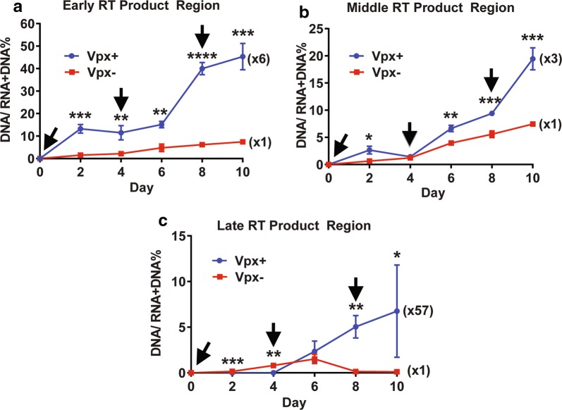
Results: Here, we demonstrate that HIV-2 produced from human primary monocyte derived macrophages displays higher ERT activity than HIV-1 produced from macrophages. Also, HIV-1 particles produced from macrophages treated with virus like particles (VLPs) containing Vpx, Vpx (+), displayed large increases of ERT activity with the enhanced copy numbers of early, middle and late reverse transcription products within the viral particles, compared to the viruses produced from macrophages treated with Vpx (-) VLPs. Furthermore, upon the infection with an equal p24 amount to fresh macrophages, the viruses produced from the Vpx (+) VLP treated macrophages demonstrated higher infectivity than the viruses from the Vpx (-) VLP treated macrophages.

Conclusions: This finding identifies the viral ERT step as an additional step of HIV-1 replication cycle that SAMHD1 restricts in nondividing myeloid target cells.

Keywords: Endogenous reverse transcription; HIV-1; HIV-2; Macrophages; Reverse transcription; SAMHD1; Vpx; dNTPs.


Figures

Fig. 1
Fig. 1
ERT activity comparison between HIV-1 and HIV-2 produced from human primary monocyte-derived macrophages and activated CD4 + T cells. a Three RT product regions (early, middle and late) of HIV-1 and HIV-2 genomes used for measuring viral endogenous reverse transcription (ERT) activity. bd ERT activities of HIV-1 89.6 and HIV-2 Rod10 produced from macrophages. Human primary monocyte derived macrophages from 5 healthy donors were infected with HIV-1 89.6 (red line) and HIV-2 Rod10 (black line) in triplicates, and the remaining uninfected viruses were extensively washed at 9 h post infection. The media containing the produced viruses were collected at days 2, 3 and 4 post infection, and the total viral nucleic acids of the produced viral particles were extracted for the Q-RT PCR (for RNA + DNA) and Q-PCR (for DNA only) for the early (b), middle (c), and late regions (d) of the viral genomes. ERT activity assay was determined in triplicates by the ratio between DNA copy number and (RNA + DNA) copy number in the produced viral particles. Fold differences between ERT activities of HIV-1 89.6 (1 ×) and HIV-2 Rod10 were calculated. e ERT activity of HIV-1 89.6 and HIV-2 Rod10 produced from activated CD4 + T cells isolated from the same donors. ERT assay was conducted for the early region with the viruses produced up to 3 days. The data are the mean of three independent experiments with qPCR performed in duplicate, and error bars represent the standard error of the mean. *P < 0.05; **P < 0.01; ***P < 0.001; ****P < 0.0001
Fig. 2
Fig. 2
ERT activity comparison of HIV-1 89.6 produced from macrophages treated with Vpx (−) and Vpx (+) virus like particles. a Comparison of cellular dATP concentrations among Vpx (−) virus like particles (VLPs) treated macrophages, Vpx (+) VLP treated macrophages, and activated CD4 + T cells. Human primary monocyte derived macrophages from 5 healthy donors were treated with Vpx (−) and Vpx (+) VLPs as well as without VLP treatment (NT) for 24 h, and cellular dNTPs were extracted from the cells for the RT-based dNTP assay. The dNTP concentrations were calculated based on their cell volumes. The dNTP concentration from activated CD4 + T cells were also determined, and the fold differences of the dNTP concentrations were calculated. Other three dNTP concentrations are shown in Additional file 1: Figure S1A. The VLP treatment and dNTP assay were conducted in triplicates. b Protocol for virus collection produced from macrophages for ERT assay. Human primary monocyte-derived macrophages were pre-treated with Vpx (−) and Vpx (+) VLPs for 24 h, and then infected with HIV-1 89.6 in triplicates. The remaining uninfected viruses were washed at 9 h post infection, and the viruses produced from these cells were collected for the ERT activity measurement at every 24 h for 4 days. The SAMHD1 degradation in these VLP treated macrophages were confirmed by western blots (Additional file 1: Figure S1B). c ERT activity of HIV-1 89.6 produced from macrophages treated with Vpx (−) (red line) and Vpx (+) (blue line) VLPs. ERT activity of the produced viruses were determined as described in Fig. 1. The ERT activity of HIV-1 89.6 from activated CD4 + T cells (black line) was used for the comparison. The data are the mean of three independent experiments with qPCR or dNTP assay performed in duplicate, and error bars represent the standard error of the mean. *P < 0.05; **P < 0.01; ***P < 0.001
Fig. 3
Fig. 3
ERT activity comparison between HIV-1 89.6 viruses produced during long-term culture of macrophages with multiple treatments of Vpx (−) and Vpx (+) VLPs. Primary macrophages were pre-treated with Vpx (−) or Vpx (+) VLPs for 24 h, and then infected with an equal p24 level of dual tropic HIV-1 89.6 in triplicates. The remaining uninfected viruses were washed at 9 h post infection. The infected macrophages were cultured for 10 days post infection. During the 10-day culture, the cells were repeatedly treated with Vpx (−) or Vpx (+) VLPs in every 4 days (see black arrows). The produced viruses were harvested at every 2 days, and total viral nucleic acids were extracted and used for measuring the ERT activity of the viral particles produced from Vpx (−) (red line) and Vpx (+) (blue line) VLP treated macrophages for a early, b middle, and c late RT products. The data are the mean of three independent experiments with qPCR performed in duplicate, and error bars represent the standard error of the mean. *P < 0.05; **P < 0.01; ***P < 0.001
Fig. 4
Fig. 4
ERT activity comparison between HIV-1 89.6 viruses produced from primary macrophages with and without dN treatment. Primary macrophages were pre-treated with dNs (2.5 mM) for 3 h, and then infected the treated macrophages with an equal p24 level of dual tropic HIV-1 89.6 in triplicates. The remaining uninfected viruses were washed at 9 h post infection. The infected macrophages were cultured for 10 days post infection. The produced viruses were harvested at every 2 days, and total viral nucleic acids were extracted and used for measuring the ERT activity of the viral particles produced from dN (−) (red line) and dN (+) (blue line) treated macrophages for a early, b middle, and c late RT products. The data are the mean of three independent experiments with qPCR performed in duplicate, and error bars represent the standard error of the mean. ***P < 0.001; ****P < 0.0001
Fig. 5
Fig. 5
Reverse transcription kinetics and Infectivity comparisons of HIV-1 89.6 produced from macrophages treated with Vpx (−) VLPs and Vpx (+) VLPs, and activated CD4+ T cells, and three steps of HIV-1 replication cycle restricted by host SAMHD1 protein. Fresh macrophages from healthy donors were infected in triplicates with the HIV-1 89.6 viruses collected at day 8 from Vpx (−) VLP treated macrophages, Vpx (+) VLP treated macrophages (from Fig. 3) or collected at day 3 from the activated CD4+ T cells (see Fig. 2). a Reverse transcription kinetics of these three viruses were determined by measuring the copy numbers of 2LTR circle DNAs with the cellular genomic DNAs isolated from the infected cells. b Infectivity of the three different viruses was determined by measuring the viral RNA copy numbers (early RT products) at days 6 and 8 post infection. c Three steps of HIV-1 replication cycle restricted by host SAMHD1 in macrophages. SAMHD1 suppression against the three steps of HIV-1 cycle that consume dNTPs is marked as red lines. HIV-1 particles: grey, macrophage cytoplasm: yellow, and macrophages nucleus: pink. Reverse transcribed products by RT are marked as thick arrows. Red arrow: ERT product within the core (green) of produced viral particle, Blue arrow: reverse transcribed product in cytoplasm post infection. SAMHD1 tetramer depletes cellular dNTPs by hydrolyzing dNTP to dN and triphosphate (TP). Vpx (purple) counteracts SAMHD1. The data are the mean of three independent experiments with qPCR performed in duplicate, and error bars represent the standard error of the mean. *P < 0.05; **P < 0.01; ***P < 0.001; ****P < 0.0001

References

    1. Weinberg JB, Matthews TJ, Cullen BR, Malim MH. Productive human immunodeficiency virus type 1 (HIV-1) infection of nonproliferating human monocytes. J Exp Med. 1991;174(6):1477–1482. doi: 10.1084/jem.174.6.1477. - DOI - PMC - PubMed
    1. Lewis PF, Emerman M. Passage through mitosis is required for oncoretroviruses but not for the human immunodeficiency virus. J Virol. 1994;68(1):510–516. - PMC - PubMed
    1. O’Brien WA. HIV-1 entry and reverse transcription in macrophages. J Leukoc Biol. 1994;56(3):273–277. doi: 10.1002/jlb.56.3.273. - DOI - PubMed
    1. O’Brien WA, Namazi A, Kalhor H, Mao SH, Zack JA, Chen IS. Kinetics of human immunodeficiency virus type 1 reverse transcription in blood mononuclear phagocytes are slowed by limitations of nucleotide precursors. J Virol. 1994;68(2):1258–1263. - PMC - PubMed
    1. Mace K, Duc Dodon M, Gazzolo L. Restriction of HIV-1 replication in promonocytic cells: a role for IFN-alpha. Virology. 1989;168(2):399–405. doi: 10.1016/0042-6822(89)90282-1. - DOI - PubMed

Publication types

MeSH terms

Substances