. 2018 Nov 21;100(4):926-939.e3.
doi: 10.1016/j.neuron.2018.09.029. Epub 2018 Oct 11.

Somatostatin Interneurons Facilitate Hippocampal-Prefrontal Synchrony and Prefrontal Spatial Encoding

Affiliations

Somatostatin Interneurons Facilitate Hippocampal-Prefrontal Synchrony and Prefrontal Spatial Encoding

Atheir I Abbas et al. Neuron. .

Abstract

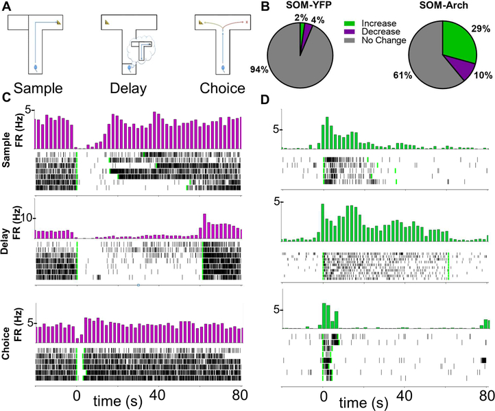
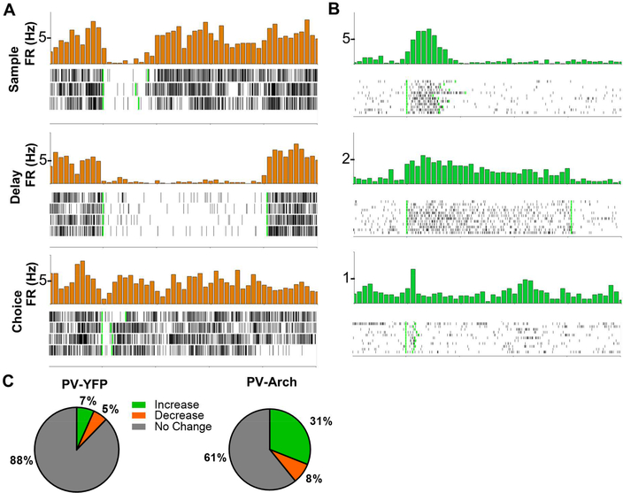
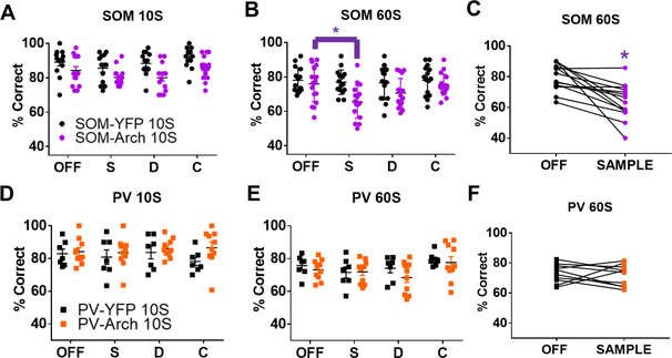
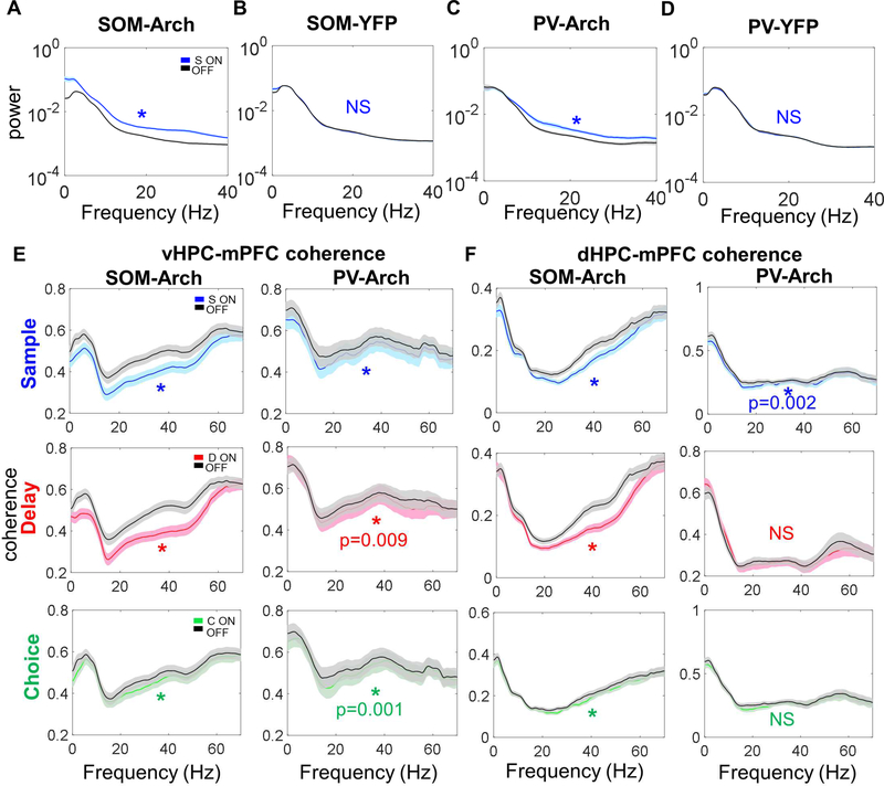
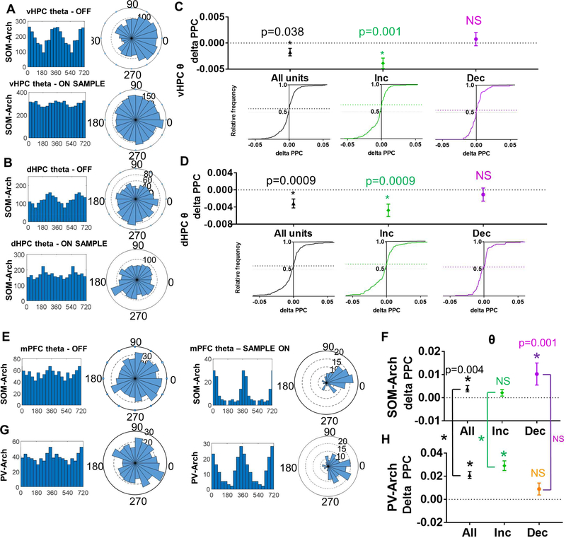
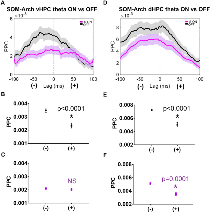
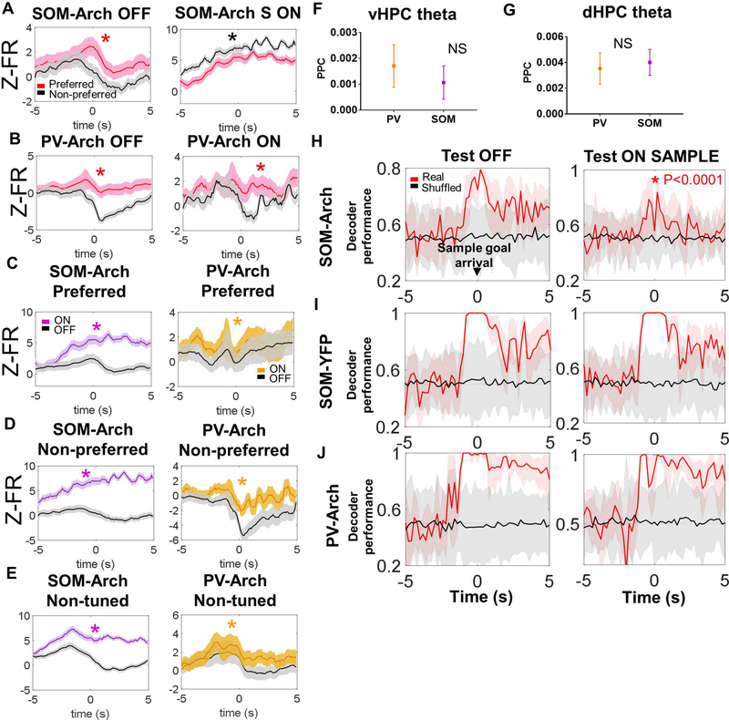
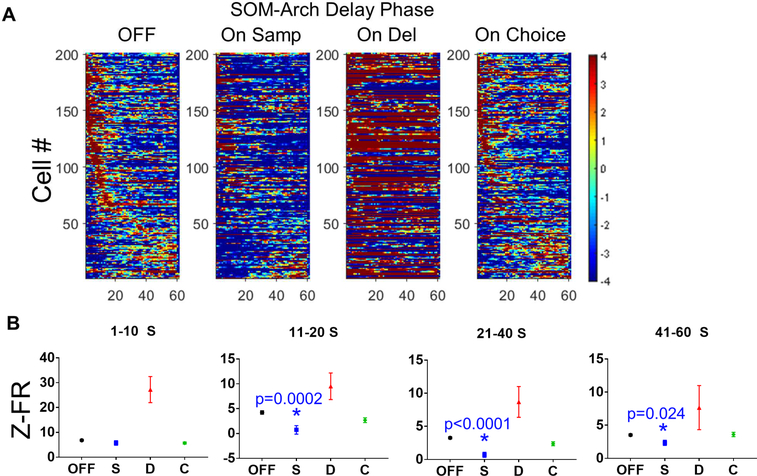
Decreased hippocampal-prefrontal synchrony may mediate cognitive deficits in schizophrenia, but it remains unclear which cells orchestrate this long-range synchrony. Parvalbumin (PV)- and somatostatin (SOM)-expressing interneurons show histological abnormalities in individuals with schizophrenia and are hypothesized to regulate oscillatory synchrony within the prefrontal cortex. To examine the relationship between interneuron function, long-range hippocampal-prefrontal synchrony, and cognition, we optogenetically inhibited SOM and PV neurons in the medial prefrontal cortex (mPFC) of mice performing a spatial working memory task while simultaneously recording neural activity in the mPFC and the hippocampus (HPC). We found that inhibiting SOM, but not PV, interneurons during the encoding phase of the task impaired working memory accuracy. This behavioral impairment was associated with decreased hippocampal-prefrontal synchrony and impaired spatial encoding in mPFC neurons. These findings suggest that interneuron dysfunction may contribute to cognitive deficits associated with schizophrenia by disrupting long-range synchrony between the HPC and PFC.

Keywords: encoding; hippocampal-prefrontal; inhibitory neuron; interneuron; oscillations; parvalbumin; prefrontal cortex; somatostatin; synchrony; theta; working memory.


Conflict of interest statement

Declaration of Interests

The authors declare no competing interests.

Figures

Figure 1:
Figure 1:. Archaerhodopsin Inhibition of SOM Interneurons Decreases Interneuron Firing and Increases Pyramidal Cell Firing
A.) Schematic for T-maze delayed-non-match-to-sample test. Neural recordings are obtained during this spatial working memory task. B.) Proportions of cells with a significant increase or decrease in firing rate in response to sample phase green light as determined by bootstrapping (p<0.01) in SOM-YFP (N =394 cells) and SOM-Arch (N = 382 cells) animals. C.) Time triggered average (green light turns on at time = 0, marked by first vertical green line; green light turns off at second vertical green line) and raster plot of a cell inhibited by Archaerhodopsin during sample, delay, and choice phases. D.) Time triggered average and raster plot of a cell that is disinhibited during SOM interneuron inhibition in sample, delay, and choice phases.
Figure 2:
Figure 2:. Archaerhodopsin Inhibition of PV Interneurons Decreases Interneuron Firing and Increases Pyramidal Cell Firing
A.) Time triggered average (green light turns on at time = 0, marked by first vertical green line; green light turns off at second vertical green line) and raster plot of a cell inhibited by Archaerhodopsin during the sample, delay, and choice phases. B.) Time triggered average and raster plot of a cell disinhibited during PV interneuron inhibition during the sample, delay, and choice phases. C.) Proportions of cells with a significant increase or decrease in firing rate in response to sample phase green light as determined by bootstrapping in PV-YFP (N = 179 cells) or PV-Arch (N = 244 cells) animals.
Figure 3:
Figure 3:. Inhibiting SOM Interneurons During Working Memory Encoding Impairs Performance
Above, “OFF” refers to trials in which the laser was off. “S” or “Sample”, “D”, and “C” refer to trials in which the laser was on during the sample, delay, or choice phase, respectively. Inhibiting SOM interneurons during the DNMTS spatial working memory task does not significantly impair performance when the delay is A.) 10 seconds, but does significantly impair performance when the delay is B.) and C.) 60 seconds. Inhibiting PV interneurons during sample, delay, or choice phases of the task does not impair spatial working memory performance at D.) 10 second or E.) and F.) 60 second delay. See main text for summary of statistics.
Figure 4:
Figure 4:. Inhibiting SOM and PV Interneurons Leads to Decreased vHPC-mPFC Coherence and dHPC-mPFC Coherence
A.) Inhibiting SOM interneurons in SOM-Arch mice during the sample phase leads to a broadband increase in power (dark lines in this figure represent the mean, with the lighter shaded band representing the 95% confidence interval; for all comparisons in this figure, significance was determined by two-tailed Wilcoxon matched-pairs signed rank test Bonferroni corrected for multiple comparisons; unless otherwise indicated here and throughout, * indicates p<0.0001). B.) Green light during the sample phase in SOMYFP mice does not affect mPFC power. C.) Inhibiting PV interneurons in PV-Arch mice during the sample phase leads to a broadband increase in power. D.) Green light during the sample phase in PV-YFP mice does not affect mPFC power. E.) Left column, Inhibiting SOM interneurons during the sample, delay, and choice phases decreases vHPC-mPFC coherence over a wide range of frequencies. Right column, Inhibiting PV interneurons during sample, delay, and choice phases generally leads to a smaller decrease in vHPC-mPFC coherence than that seen with SOM inhibition. F.) Left column, Inhibiting SOM interneurons during the sample, delay, and choice phases decreases dHPC-mPFC coherence. Right column, Inhibiting PV interneurons during the sample, but not delay or choice phases, leads to a small decrease in dHPC-mPFC coherence.
Figure 5:
Figure 5:. Inhibiting SOM Interneurons During the Sample Phase Leads to Decreased Phase Locking of mPFC Units to vHPC and dHPC Theta and Decreased Phase Locking to Local mPFC Theta
A.) Histogram and rose plots illustrating a decrease in phase locking of mPFC single units to vHPC theta oscillations during SOM-inhibited trials (see bottom row) as compared to non-inhibited trials (see top row). B.) Histogram and rose plots illustrating a decrease in phase locking of mPFC single units to dHPC theta oscillations during SOM-inhibited trials (see bottom row) as compared to non-inhibited trials (see top row). C-D.) Cumulative frequency distribution of delta PPC (Pairwise Phase Consistency; Sample ON PPC – Sample OFF PPC) calculated for all single units (left column, black), units that increase in response to SOM inhibition (middle column, green), and units that decrease in response to SOM inhibition (right column, purple). When calculating phase locking of mPFC units to C.) vHPC theta and D.) dHPC theta, the frequency distribution skews towards negative delta PPC values for all units and increasers, but not for decreasers (data in C-H are represented as mean ± SEM; statistical comparisons in this figure between ON and OFF by Wilcoxon matched-pairs signed rank test Bonferroni corrected for multiple comparisons). E.) Histogram and rose plots illustrating an increase in phase locking of mPFC single units to mPFC theta oscillations during SOMinhibited trials (see right column) as compared to non-inhibited trials (see left columns). F.) SOM inhibition leads to a significant increase in phase locking of mPFC units to local mPFC theta oscillations as illustrated by delta PPC (PPC SAMPLE ON – PPC SAMPLE OFF) values that are significantly greater than zero. G.) Histogram and rose plots illustrating a decrease in phase locking of mPFC single units to dHPC theta oscillations during PV-inhibited trials (see bottom row) are compared to non-inhibited trials (see top row). H.) PV inhibition also leads to a significant increase in phase locking of mPFC units to local mPFC theta oscillations which is significantly greater than that seen in SOM-Arch mice when comparing all cells and increasers (comparisons signified by the vertical lines between F and H, * p<0.0001).
Figure 6:
Figure 6:. Sample Phase SOM Inhibition impairs vHPC to mPFC Directionality During Long Range Synchronization
A.) mPFC units exhibit higher phase locking to negative lag vHPC theta as compared to positive lag vHPC theta during the sample phase, but this directionality is abolished by SOM inhibition (dark lines in A and D represent the mean, with the lighter shaded band representing the 95% confidence interval). B.) During the sample phase in OFF trials, the mean PPC at negative lags is significantly greater than the mean PPC at positive lags (data in B-C and E-F are represented as mean ± SEM; statistical comparisons between mean PPC at negative vs positive lags is by Wilcoxon matched-pairs signed rank test Bonferroni corrected for multiple comparisons; * indicates p<0.0001). C.) During the sample phase in SOM-inhibited trials, there is no significant difference between the mean PPC to vHPC theta at negative vs positive lags. D.) mPFC units exhibit higher phase locking to negative lag dHPC theta as compared to positive lag dHPC theta during the sample phase, both during OFF trials and SOM inhibited trials. E.) and F.) During the sample phase in OFF and SOM inhibited trials, the mean PPC to dHPC theta at negative lags is significantly greater than the mean PPC to dHPC theta at positive lags.
Figure 7:
Figure 7:. Inhibition SOM but not PV Interneurons Impairs Spatial Encoding in the mPFC
A.) Z-scored firing rate is higher in the preferred arm of spatially tuned neurons during the sample phase around the time of sample goal arrival during laser off trials (left panel; (dark lines in A-E represent the mean, with the lighter shaded band representing the 95% confidence interval) but not during SOM inhibition (right panel; for this panel and others in this figure, asterisks indicate statistical comparisons that are significant by Wilcoxon matched-pairs signed rank test). B.) Z-scored firing rate is higher in the preferred arm of spatially tuned neurons during the sample phase around the time of sample goal arrival during laser off trials (left panel); this spatial tuning is not significantly affected by PV inhibition (right panel). C.-E) Firing rate is significantly increased during laser on (i.e., during SOM or PV inhibition) trials in the preferred and non-preferred arms of spatially tuned cells and in non-spatially tuned cells; this effect appears more robust during SOM inhibition as compared to PV inhibition. F.) and G.) There is no significant difference in phase locking of SOMs and PVs to vHPC theta or dHPC theta oscillations (data in F-G are represented as mean ± SEM). H.) Sample goal identity can be decoded from the mPFC population in SOM-Arch mice when the decoder is trained and tested on laser off trials (left panel; in H- J, solid lines – mean decoder accuracy, shaded areas – 95% confidence intervals), but not as well when the decoder is trained in laser off trials and tested in sample on trials (right panel, asterisk in graph indicates significance by two-way ANOVA followed by Bonferroni-corrected posthoc testing with p-value shown next to asterisk). I.) Sample goal identity can be decoded from the mPFC population in SOM-YFP mice when the decoder is trained and tested on laser off trials (left panel) and when the decoder is trained in laser off trials and tested in sample on trials (right panel). J.) Sample goal identity can also be decoded from the mPFC population in PV-Arch mice when the decoder is trained and tested in laser off trials (left panel) and when the decoder is trained in laser off trials and tested in sample on trials (right panel).
Figure 8:
Figure 8:. Inhibiting SOMs During the Sample Phase Disrupts Subsequent Delay-Elevated mPFC Activity
A.) Normalized firing rates for delay-elevated mPFC neurons during the delay phase for light OFF trials and On Sample, On Delay, and On Choice trials. Inhibiting SOMs during the sample phase (On Sample trials) appears to disrupt delay-elevated activity. It is difficult to visualize the transient elevations in firing rate when SOMs are inhibited during the delay period (On Delay) due to a large increase in the baseline firing rate for many of the neurons. Delay-elevated mPFC firing during On Choice trials, prior to SOM inhibition, appears to be preserved. B.) SOM inhibition during the sample phase significantly decreases firing rates in delay-elevated neurons during much of the delay period (data in B are represented as mean ± SEM; for cells whose peaks occur during 1–10 S, 11–20 S, and 21–40 S periods; statistical significance was determined by oneway ANOVA with Bonferroni-corrected post-hoc testing; asterisks indicate significance with p-value listed on graph).

References

    1. Akhlaghpour H, Wiskerke J, Choi JY, Taliaferro JP, Au J, and Witten IB (2016). Dissociated sequential activity and stimulus encoding in the dorsomedial striatum during spatial working memory. Elife 5. - PMC - PubMed
    1. Arts B, Jabben N, Krabbendam L, and van Os J (2008). Meta-analyses of cognitive functioning in euthymic bipolar patients and their first-degree relatives. Psychol Med 38, 771–785. - PubMed
    1. Atallah BV, Bruns W, Carandini M, and Scanziani M (2012). Parvalbuminexpressing interneurons linearly transform cortical responses to visual stimuli. Neuron 73, 159–170. - PMC - PubMed
    1. Bolkan SS, Stujenske JM, Parnaudeau S, Spellman TJ, Rauffenbart C, Abbas AI, Harris AZ, Gordon JA, and Kellendonk C (2017). Thalamic projections sustain prefrontal activity during working memory maintenance. Nat Neurosci 20, 987–996. - PMC - PubMed
    1. Brekke JS, Hoe M, Long J, and Green MF (2007). How neurocognition and social cognition influence functional change during community-based psychosocial rehabilitation for individuals with schizophrenia. Schizophr Bull 33, 1247–1256. - PMC - PubMed

Publication types

LinkOut - more resources