

Case Reports
doi: 10.4103/ijri.IJRI_469_17.
Mucinous cystadenoma of pancreas with honeycombing appearance: Radiological-Pathological correlation
Affiliations
- PMID: 30319210
- PMCID: PMC6176663
- DOI: 10.4103/ijri.IJRI_469_17
Item in Clipboard
Case Reports
Mucinous cystadenoma of pancreas with honeycombing appearance: Radiological-Pathological correlation
Indian J Radiol Imaging.
2018 Jul-Sep.
Abstract
Most mucinous cystadenomas of pancreas are solitary and multilocular with a few large compartments. Serous cystadenomas usually have a polycystic or microcystic (honeycomb) pattern consisting of collection of cysts (usually >6) that range from few millimetres up to 2 cm in size. Here we present a case of mucinous cystadenoma of pancreas showing an unusual appearance of honeycombing (which has not been described so far) using imaging studies such as endoscopic ultrasound and computed tomography with histopathological confirmation of the diagnosis.
Keywords: Cystic; Mucinous; honeycombing; pancreatic; tumors.
Conflict of interest statement
There are no conflicts of interest.
Figures

(A) CECT abdomen shows a well-defined cystic lesion in the head and neck of the pancreas (arrow) with enhancing septations within. (B) CECT abdomen demonstrating the mass effect of the cystic lesion causing downstream dilatation of main pancreatic duct (arrow)

Endoscopic ultrasound demonstrates a predominantly isoechoic lesion with few cystic portions noted giving ‘‘honeycomb’’ appearance (arrow)
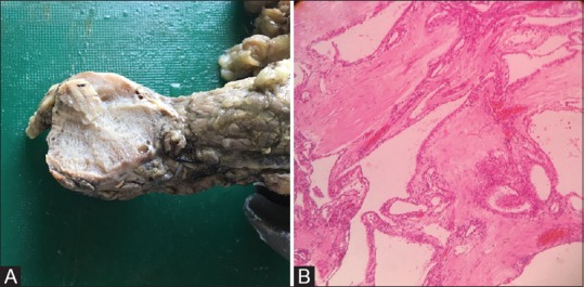
(A) Gross specimen shows a well-circumscribed pancreatic lesion which appears spongy and trabeculated with no secondary changes. (B) Microscopic examination shows an unencapsulated neoplasm in the pancreas composed of cysts of varying sizes lined by single layer of flattened to cuboidal epithelium with occasional papillary processes
References
-
- De Lima J, Javitt M, Mathur S. Residents’ teaching files: Mucinous cystic neoplasm of the pancreas. Radiographics. 1999;19:807–11. - PubMed
-
- Fernández-del Castillo C. Mucinous cystic neoplasms. J Gastrointest Surg. 2008;12:411–3. - PubMed
-
- Suzuki M, Fujita N, Onodera H, Kayaba Y, Suzuki S, Kagaya H, et al. Mucinous cystic neoplasm in a young male patient. J Gastroenterol. 2005;40:1070–4. - PubMed
-
- Tseng JF. Management of serous cystadenoma of the pancreas. J Gastrointest Surg. 2008;12:408–10. - PubMed
Publication types
LinkOut - more resources
Full Text Sources
Molecular Biology Databases