. 2017 Mar 24;27(12):1605352.
doi: 10.1002/adfm.201605352. Epub 2017 Jan 17.

Gold Nanocomposite Bioink for Printing 3D Cardiac Constructs

Affiliations

Gold Nanocomposite Bioink for Printing 3D Cardiac Constructs

Kai Zhu et al. Adv Funct Mater. .

Abstract

Bioprinting is the most convenient microfabrication method to create biomimetic three-dimensional (3D) cardiac tissue constructs, which can be used to regenerate damaged tissue and provide platforms for drug screening. However, existing bioinks, which are usually composed of polymeric biomaterials, are poorly conductive and delay efficient electrical coupling between adjacent cardiac cells. To solve this problem, we developed a gold nanorod (GNR) incorporated gelatin methacryloyl (GelMA)-based bioink for printing 3D functional cardiac tissue constructs. The GNR concentration was adjusted to create a proper microenvironment for the spreading and organization of cardiac cells. At optimized concentration of GNR, the nanocomposite bioink had a low viscosity, similar to pristine inks, which allowed for the easy integration of cells at high densities. As a result, rapid deposition of cell-laden fibers at a high resolution was possible, while reducing shear stress on the encapsulated cells. In the printed GNR constructs, cardiac cells showed improved cell adhesion and organization when compared to the constructs without GNRs. Furthermore, the incorporated GNRs bridged the electrically resistant pore walls of polymers, improved the cell-to-cell coupling, and promoted synchronized contraction of the bioprinted constructs. Given its advantageous properties, this gold nanocomposite bioink may find wide application in cardiac tissue engineering.

Keywords: Alginate; Bioprinting; Cardiac tissue engineering; Gelatin; Gold nanorods.


Conflict of interest statement

The authors declare no conflict of interests in this work.

Figures
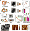
Figure 1
Figure 1. Preparation of G-GNR/GelMA hydrogel
(a) Schematic of coating GelMA molecules on GNRs. i) CTAB bilayer was found on GNRs by TEM before treatment. ii) GelMA-coated layer was found on GNRs by TEM after treatment. (b) UV-Vis spectra showed G-GNRs were the same as that of C-GNRs, thus indicating that G-GNRs were prepared without changing aspect ratio and forming aggregates. (c) Zeta potential of C-GNRs and G-GNRs. (d) Schematic of preparation for G-GNR/GelMA prepolymer solution with various concentrations of G-GNRs. TEM image showed the uniform distribution of G-GNRs in the solution. Darker color was observed with higher concentrations of G-GNRs in the solution. (e) Impedance and (f) Young’s modulus of G-GNR/GelMA hydrogel with various concentrations of G-GNRs. (g) No obvious G-GNR aggregation was observed on the G-GNR (0.1 mg/mL)/GelMA (7%) hydrogel matrix wall under SEM. (h) AFM images of the prisitne GelMA (7%) hydrogel and G-GNR (0.1 mg/mL)/GelMA (7%) hydrogel.
Figure 2
Figure 2. CMs on G-GNR/GelMA hydrogel
(a) Schematic of G-GNR/GelMA hydrogel constructs. (b) Optical images of CMs grown on the prisitne GelMA (7%) hydrogel and G-GNR (0.1 mg/mL)/GelMA (7%) hydrogel at day 1. (c) Quantification analysis showed that CMs on the G-GNR (0.1 mg/mL)/GelMA (7%) hydrogel had a better retention rate than those on pristine GelMA (7%) hydrogel at day 1 (* P < 0.05). (d) The G-GNR (0.1 mg/mL)/GelMA (7%) hydrogel showed a more uniform cell coverage on day 5 after cell seeding. Expression of sarcomeric α-actinin, Cx-43 (e) and troponin I (f) in CMs grown on pristine GelMA (7%) hydrogel and G-GNR (0.1 mg/mL)/GelMA (7%) hydrogel at day 7.
Figure 3
Figure 3. 3D bioprinting using G-GNR nanocomposite bioink
(a) Schematic of bioprinting process using the G-GNR nanocomposite bioink. The inset showed the bioprinted 30-layered construct. (b) The printing procedure on x, y and z axises. (c) Stacked layers (layer 1–4) using G-GNR nanocomposite bioink could be observed under microscope. (d) Constructs with different inner grid could be bioprinted (green beads were embedded in G-GNR nanocomposite bioink to exhibit the printed fibers). (e) Stable bioprinted constructs in culture medium up to day 5 and forced degradation of bioprinted construct using a collagenase solution at 37 °C. (f) The absorbance spectrum analysis of culture medium confirmed that the G-GNRs were kept into bioprinted constructs and released upon forced degradation of constructs. (g) Schematic of 3D embedded bioprinting using the G-GNR nanocomposite bioink. The inset showed the printed spiral structure in a support bath (2% gelatin, 11 mM CaCl2). (h) The spiral construct in the support bath after printing. (i) Bioprinted constructs could be obtained without damage and cultured in medium.
Figure 4
Figure 4. Cell viability in bioprinted constructs
(a) Schematic of shear stress effect on cells inside of the printing nozzle. (b) Rheological characterization of 7% GelMA prepolymer solution, GelMA/algiante bioink (7% GelMA, 2% alginate), and G-GNR nanocomposite bioink (0.1 mg/mL G-GNR, 7% GelMA, 2% alginate). (c) Computational simulation of velocity of the bioink in the nozzle while using the extrusion speed of 10 µL/min. (d) The shear stress profile of the G-GNR nanocomposite bioink was higher with the increase of extrusion speed. r is the radial coordinate, where the origin is located at the axis of the printing needle. R is the radius of the printing needle. (e, f) Live/dead assay of CFs within G-GNR nanocomposite bioink printed constructs after printing with different extrusion speed (* P < 0.05). (g) Live/dead assay of CFs within G-GNR nanocomposite bioink printed constructs under different UV exposure time (* P < 0.05). (h) Live/dead assay of CFs within different layer of G-GNR nanocomposite bioink printed constructs. (i) No significant difference of cell viability was observed between different layers (P > 0.05).
Figure 5
Figure 5. Bioprinted cardiac tissue construct
(a) Pseudo-3D brightfield micrograph showed cells were homogeneously distributed in bioprinted construct using G-GNR nanocomposite bionink (0.1 mg/mL G-GNR, 7% GelMA, 2% alginate). Fluorescence cell tracker-labeled CMs (red) and CFs (green) were observed in construct after printing. (b) Fluorescence F-actin and DAPI images of bioprinted cardiac cells within GelMA/alginate bioink and G-GNR nanocomposite bioink-printed constructs on day 5. (c) Fluorescence F-actin images of bioprinted cardiac cells within G-GNR nanocomposite bioink-printed constructs on day 12. (d) Presto blue assay showed that there was no significant differences in cell proliferation within GelMA/alginate bioink and G-GNR nanocomposite bioink-printed constructs. (e) The dry weight change of cell-laden G-GNR nanocomposite bioink-printed constructs during culture. (f) The morphology and inner grids of the G-GNR nanocomposite bioink-printed construct were maintained during culture. (g) Immunostaining of sarcomeric α-actinin (green), nuclei (blue), and Cx-43 (red) revealed that cardiac tissues (14-day culture) within GelMA/alginate bioink and G-GNR nanocomposite bioink-printed constructs were phenotypically different. (h) Quantification of Cx-43 expression by the CMs in the printed constructs, plotted as percentages of area coverage calculated from the fluorescence images (* P < 0.05). (i) Spontaneous beating rates of the GelMA/alginate bioink and G-GNR nanocomposite bioink-printed constructs (* P < 0.05).

References

    1. Murphy SV, Atala A. Nat. Biotechnol. 2014;32:773. - PubMed
    2. Ozbolata IT, Hospodiuk M. Biomaterials. 2016;76:321. - PubMed
    3. Kang HW, Lee SJ, Ko IK, Kengla C, Yoo JJ, Atala A. Nat. Biotechnol. 2016;34:312. - PubMed
    1. Kolesky DB, Truby RL, Gladman AS, Busbee TA, Homan KA, Lewis JA. Adv. Mater. 2014;26:3124. - PubMed
    1. Blaeser A, Campos DFD, Puster U, Richtering W, Stevens MM, Fischer H. Adv. Healthcare Mater. 2015;5:326. - PubMed
    1. Colosi C, Shin SR, Manoharan V, Massa S, Costantini M, Barbetta A, Dokmeci MR, Dentini M, Khademhosseini A. Adv. Mater. 2015;28:677. - PMC - PubMed
    1. Tamayol A, Najafabadi AH, Aliakbarian B, Arab-Tehrany E, Akbari M, Annabi N, Juncker D, Khademhosseini A. Adv. Healthcare Mater. 2015;4:2146. - PubMed
    2. Jia W, Gungor-Ozkerim PS, Zhang YS, Yue K, Zhu K, Liu W, Pi Q, Byambaa B, Dokmeci MR, Shin SR, Khademhosseini A. Biomaterials. 2016;106:58. - PMC - PubMed

LinkOut - more resources