. 2018 Sep 24:12:668.
doi: 10.3389/fnins.2018.00668. eCollection 2018.

CuII(atsm) Attenuates Neuroinflammation

Affiliations

CuII(atsm) Attenuates Neuroinflammation

Xin Yi Choo et al. Front Neurosci. .

Abstract

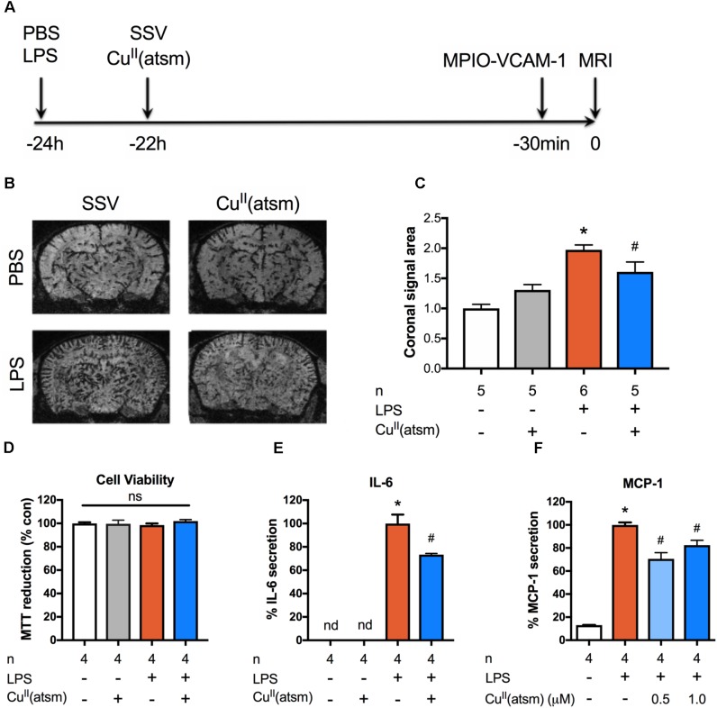
Background: Neuroinflammation and biometal dyshomeostasis are key pathological features of several neurodegenerative diseases, including Alzheimer's disease (AD). Inflammation and biometals are linked at the molecular level through regulation of metal buffering proteins such as the metallothioneins. Even though the molecular connections between metals and inflammation have been demonstrated, little information exists on the effect of copper modulation on brain inflammation. Methods: We demonstrate the immunomodulatory potential of the copper bis(thiosemicarbazone) complex CuII(atsm) in an neuroinflammatory model in vivo and describe its anti-inflammatory effects on microglia and astrocytes in vitro. Results: By using a sophisticated in vivo magnetic resonance imaging (MRI) approach, we report the efficacy of CuII(atsm) in reducing acute cerebrovascular inflammation caused by peripheral administration of bacterial lipopolysaccharide (LPS). CuII(atsm) also induced anti-inflammatory outcomes in primary microglia [significant reductions in nitric oxide (NO), monocyte chemoattractant protein 1 (MCP-1), and tumor necrosis factor (TNF)] and astrocytes [significantly reduced NO, MCP-1, and interleukin 6 (IL-6)] in vitro. These anti-inflammatory actions were associated with increased cellular copper levels and increased the neuroprotective protein metallothionein-1 (MT1) in microglia and astrocytes. Conclusion: The beneficial effects of CuII(atsm) on the neuroimmune system suggest copper complexes are potential therapeutics for the treatment of neuroinflammatory conditions.

Keywords: astrocyte; copper; inflammation; microglia; neurodegeneration.


Figures

FIGURE 1
FIGURE 1
Copper delivery reduces inflammation induced by administration of LPS. (A) Timeline demonstrating time points for administration of the indicated compounds to adult mice and in vivo imaging of inflammation by MRI. (B) Representative MRI images acquired 30 min after intravenous administration of MPIO-VCAM-1 (24 h after LPS and 22 h after CuII(atsm) or SSV) showing signal voids corresponding to MPIO binding. (C) Quantitative analysis of the volume of signal void in the brain induced by MPIO-VCAM-1 following treatment with LPS and CuII(atsm). (D–F) Cultured adult astrocytes were stimulated with 20 μg/ml LPS with or without 0.5 μM CuII(atsm) for 48 h and (D) MTT reduction, (E) IL-6 secretion, and (F) MCP-1 secretion were measured 48 h later. Sample sizes per group (n) as indicated. p < 0.05 compared to control, #p < 0.05 compared to LPS-treated group. ns, not significant. nd, not detected.
FIGURE 2
FIGURE 2
Copper delivery dampens microglial activation during inflammation. Primary microglia were stimulated with 15 ng/ml IFNγ and 10 ng/ml TNFα with or without 0.5 μM CuII(atsm). (A) MTT reduction was measured to determine the cell viability 24 h later. Primary microglia were stimulated with 15 ng/ml IFNγ and 10 ng/ml TNFα with or without 0.5 μM CuII(atsm) or 20 μM minocycline for 24 h. (B) Nitric oxide release into the media was measured 24 h later. (C) The secretion of MCP-1 into the media was measured by ELISA. qRT-PCR was used to measure microglial mRNA expression levels of (D) Ccl2, (E) Tnf, and (F) the indicated genes. (F) Presented as log fold-increase over control. Sample sizes per group (n) as indicated. p < 0.05 compared to control, #p < 0.05 compared to IFNγ/TNFα-treated group. ns, not significant.
FIGURE 3
FIGURE 3
Anti-inflammatory effects of copper delivery in astrocytes. Neonatal astrocytes were stimulated for 48 h with 15 ng/ml IFNγ and 10 ng/ml TNFα with or without 2μM CuII(atsm) for the final 24 h (A–H). (A) MTT reduction and (B) LDH release was measured to determine the cell viability and cell death, respectively, after treatment. (C) MCP-1 secretion and (D) nitric oxide release were measured after treatment. qRT-PCR was used to measure astrocyte mRNA expression levels of (E) Ccl2, (F) Nos2, (G) Tnf, and (H) the indicated genes. (H) Presented as log fold-increase over control. Sample sizes per group (n) as indicated, or 6–7/treatment group for (H). p < 0.05 compared to control, #p < 0.05 compared to IFNγ/TNFα-treated group. ns, not significant.
FIGURE 4
FIGURE 4
CuII(atsm) delivers copper into cells. (A,B) Neonatal astrocytes were treated for 48 h with 15 ng/ml IFNγ and 10 ng/ml TNFα with or without 2 μM CuII(atsm) for the final 24 h. (C,D) Microglia were treated for 24 h and with 15 ng/ml IFNγ and 10 ng/ml TNFα with or without 0.5 μM CuII(atsm). Cellular copper content of (A) astrocytes and (C) microglia was measured by ICP-MS. mRNA expression of Mt1 was measured by qRT-PCR in (B) astrocytes and (D) microglia. Sample sizes per group (n) as indicated. (E) Representative images of microglia analyzed for cellular copper content by XFM (reflected by elemental areal density). Scale bar 100 μm. (F) Quantification of XFM-determined copper levels per cell in microglia treated with 15 ng/ml IFNγ and 10 ng/ml TNFα with or without 0.5 μM CuII(atsm). Cells per treatment group (n) as indicated. p < 0.05 compared to control, #p < 0.05 compared to IFNγ/TNFα-treated group.

References

    1. Adlard P. A., Bica L., White A. R., Nurjono M., Filiz G., Crouch P. J., et al. (2011). Metal ionophore treatment restores dendritic spine density and synaptic protein levels in a mouse model of alzheimer’s disease. PLoS One 6:e17669. 10.1371/journal.pone.0017669 - DOI - PMC - PubMed
    1. Adlard P. A., West A. K., Vickers J. C. (1998). Increased density of metallothionein I/II-immunopositive cortical glial cells in the early stages of alzheimer’s disease. Neurobiol. Dis. 5 349–356. 10.1006/nbdi.1998.0203 - DOI - PubMed
    1. Agullo L., Garcia A., Hidalgo J. (1998). Metallothionein-I + II induction by zinc and copper in primary cultures of rat microglia. Neurochem. Int. 33 237–242. 10.1016/S0197-0186(98)00022-9 - DOI - PubMed
    1. Banks W. A., Robinson S. M. (2010). Minimal penetration of lipopolysaccharide across the murine blood-brain barrier. Brain Behav. Immun. 24 102–109. 10.1016/j.bbi.2009.09.001 - DOI - PMC - PubMed
    1. Bayer T. A., Schafer S., Simons A., Kemmling A., Kamer T., Tepest R., et al. (2003). Dietary cu stabilizes brain superoxide dismutase 1 activity and reduces amyloid abeta production in APP23 transgenic mice. Proc. Natl. Acad. Sci. U.S.A. 100 14187–14192. 10.1073/pnas.2332818100 - DOI - PMC - PubMed