Review
. 2018 Sep 27:9:570.
doi: 10.3389/fendo.2018.00570. eCollection 2018.

Regulation of Placental Development and Its Impact on Fetal Growth-New Insights From Mouse Models

Affiliations
Review

Regulation of Placental Development and Its Impact on Fetal Growth-New Insights From Mouse Models

Laura Woods et al. Front Endocrinol (Lausanne). .

Abstract

The placenta is the chief regulator of nutrient supply to the growing embryo during gestation. As such, adequate placental function is instrumental for developmental progression throughout intrauterine development. One of the most common complications during pregnancy is insufficient growth of the fetus, a problem termed intrauterine growth restriction (IUGR) that is most frequently rooted in a malfunctional placenta. Together with conventional gene targeting approaches, recent advances in screening mouse mutants for placental defects, combined with the ability to rapidly induce mutations in vitro and in vivo by CRISPR-Cas9 technology, has provided new insights into the contribution of the genome to normal placental development. Most importantly, these data have demonstrated that far more genes are required for normal placentation than previously appreciated. Here, we provide a summary of common types of placental defects in established mouse mutants, which will help us gain a better understanding of the genes impacting on human placentation. Based on a recent mouse mutant screen, we then provide examples on how these data can be mined to identify novel molecular hubs that may be critical for placental development. Given the close association between placental defects and abnormal cardiovascular and brain development, these functional nodes may also shed light onto the etiology of birth defects that co-occur with placental malformations. Taken together, recent insights into the regulation of mouse placental development have opened up new avenues for research that will promote the study of human pregnancy conditions, notably those based on defects in placentation that underlie the most common pregnancy pathologies such as IUGR and pre-eclampsia.

Keywords: DMDD; IUGR; fetal growth restriction; mouse models; placenta; trophoblast.


Figures
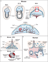
Figure 1
Figure 1
Overview of mouse placental development and similarities to human placenta. (A) Following implantation, the blastocyst's mural trophectoderm differentiates into trophoblast giant cells (TGCs), while trophectodermal cells that overlie the inner cell mass form the extra-embryonic ectoderm (ExE) and the ectoplacental cone (EPC). The future embryo originates from the blastocyst's inner cell mass that differentiates into the epiblast (EPI). The conceptus is embedded into the maternal decidua (Dec) which differentiates from the uterine endometrium. With gastrulation, the chorion (Ch) consisting of a trophoblast and an extra-embryonic mesodermal cell layer, the amnion (Am) and the allantois (All) are formed. Cells at the margins of the of the EPC differentiate into invasive, secondary TGCs, that remodel the maternal vasculature [highlighted by the inset, see (B)]. The allantois grows out and attaches to the chorion (chorio-allantoic fusion) around E8.5, a critical process for developmental progression. At E9.5, allantoic blood vessels start to invaginate into the chorionic ectoderm to initiate formation of the placental labyrinth (Lab). Trophoblast cells overlying this layer differentiate into future spongiotrophoblast (SpT) and glycogen cells (GCs). The mature mouse placenta is established around mid-gestation (~E10.5) and continues to grow in size and complexity. It consists of three main layers: the labyrinth (Lab), the junctional zone (JZ) made up of SpT, GCs and TGCs, and the maternal decidua (Dec). The labyrinth is the main site of nutrient and gas exchange [highlighted by the inset, see (C)]. Black bar indicates thickness of the labyrinth zone. (B) Magnified view of the tip of the EPC where invasive TGCs remodel maternal spiral arteries (SA) by eroding their smooth muscle lining and displacing their endothelial cell (EC) layer. (C) Close-up view of the interhaemal barrier, consisting (from maternal to fetal side) of a discontinuous layer of sinusoidal TGCs, two layers of syncytiotrophoblast (SynT-I and -II), and the endothelial cell layer of the fetal blood vessels. (D) Despite significant morphological differences, comparison with the human placenta reveals structural and/or functional similarities. Human placental villi are made up of a mesenchymal core that contains the fetal blood vessels, a layer of villous cytotrophoblast (vCTB) and one overlying layer of syncytiotrophoblast (ST) that is directly exposed to maternal blood. Thus, the haemochorial organization and the exchange barrier are similarly organized between mouse and human placentas. vCTBs are perhaps most analogous to the chorionic ectoderm in mice. In anchoring villi, cytotrophoblast cells form cytotrophoblast cell columns (CCCs) that invade into the maternal decidua (Dec). Cells at the base of the CCCs proliferate, pushing cells along the column where they progressively differentiate into invasive extravillous trophoblast (EVT). Trophoblast invasion occurs along two routes, interstitially into the decidual stroma, and along an endovascular route to replace the endothelial cell (EC) lining of maternal spiral arteries (SA). This spiral artery remodeling process is instrumental for healthy pregnancy progression, and is equally shared with the mouse where these processes occur between ~E7.5 and E10.5 as shown.
Figure 2
Figure 2
Schematic representation of JZ defects that can be associated with IUGR. (A) The junctional zone (JZ) is composed of three cell types: spongiotrophoblast cells (SpT), glycogen cells (GCs) and trophoblast giant cells (TGCs). The JZ provides energetic (glycogen), hormonal and physical support to ensure correct placentation and pregnancy progression. (B–D) Recurring JZ phenotypes entail (B) a reduced thickness of the JZ layer (indicated by the red bar) with fewer SpT and/or GCs, (C) an increased size of the JZ with more SpT and/or GCs and (D) a mislocalisation of GCs and SpT in the labyrinth. All of these phenotypes can be associated with IUGR, highlighting that JZ size alone is not indicative of placental efficiency, but that other parameters such as effect on cellular function, and precise cell localisation in relation to blood vessels and blood conduits, is important. Examples of mouse mutants or overexpression models in which these dysmorphologies are observed are given (fetal genotype is shown).
Figure 3
Figure 3
Schematic representation of labyrinth defects associated with IUGR. (A) Within a normal placenta, the labyrinth is the largest layer and is the site of all nutrient and gas exchange between the maternal and fetal blood circulations (black bar indicates thickness of the labyrinth layer). As such, failure in the establishment of this intricately organized layer is a direct cause of IUGR, or in more severe cases of intrauterine lethality. Defects in the development of the labyrinth often originate from defective or insufficient invagination of allantoic blood vessels into the chorionic ectoderm (see Figure 1, E9.5) and the subsequent branching morphogenesis that has to occur to form this exchange surface. (B) Such defects can lead to a small labyrinth layer (indicated by the red bar, often coupled with an overall reduced size of the placenta) or (C) a reduced complexity of the vascular organization within the labyrinth, in which there are fewer, disorganized and/or inappropriately dilated fetal blood vessels and maternal conduits. Principally, these structural defects lead to a reduction of the surface area available for transport, which hence cause placental insufficiency. (D) Impaired nutrient transfer may also occur because of a thickened or dysfunctional interhaemal barrier (IHM). (E) The transport surface may be diminished due to overt lesions in the labyrinth layer, that frequently entail thrombotic or necrotic patches or the accumulation of fibrotic tissue. (F) An unusual trophoblast differentiation defect is observed in some mouse mutants with the formation of multinucleate trophoblast giant cells (mTGCs). These mTGCs likely have their origin in inappropriate fusion and syncytialisation of labyrinth trophoblast cells, disrupting the intricate labyrinthine architecture. Examples of mouse mutants or overexpression models in which these dysmorphologies are observed are given (fetal genotype is shown).
Figure 4
Figure 4
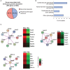
Data mining approaches to identify molecular networks of potential importance in placental development. (A) Pie chart of genes extracted from the Mouse Genome Informatics database (www.informatics.jax.org) that are associated with embryonic growth retardation (MP: 0003984; 505 genes in total) and that are either scored as having an abnormal placenta (MP:0002086 or MP:0004264) or an unknown placental phenotype. (B) Breakdown of the proportion of mutants with unknown placental phenotype that also exhibit defects in heart and/or brain development. Since placental defects are often co-associated with cardiovascular and brain abnormalities (56), this selection will enrich for genes that have a function in the placenta. (C) Molecular network analysis using String (https://string-db.org/) to identify physical and/or functional gene interactions for selected examples, and expression levels of the identified complex components in trophoblast stem cells (TSCs) compared to embryonic stem cells (ESCs). The network components are enriched in the trophoblast compartment, arguing for a potential function in placental development.

References

    1. Militello M, Pappalardo EM, Ermito S, Dinatale A, Cavaliere A, Carrara S. Obstetric management of IUGR. J Prenat Med. (2009) 3:6–9. - PMC - PubMed
    1. Yung HW, Hemberger M, Watson ED, Senner CE, Jones CP, Kaufman RJ, et al. . Endoplasmic reticulum stress disrupts placental morphogenesis: implications for human intrauterine growth restriction. J. Pathol. (2012) 228:554–64. 10.1002/path.4068 - DOI - PMC - PubMed
    1. Vijayaselvi R, Cherian AG. Risk assessment of intrauterine growth restriction. Curr Med Issues (2017) 15:262–6. 10.4103/cmi.cmi_76_17 - DOI
    1. Gardosi J, Madurasinghe V, Williams M, Malik A, Francis A. Maternal and fetal risk factors for stillbirth: population based study. BMJ (2013) 346:f108. 10.1136/bmj.f108 - DOI - PMC - PubMed
    1. von Beckerath AK, Kollmann M, Rotky-Fast C, Karpf E, Lang U, Klaritsch P. Perinatal complications and long-term neurodevelopmental outcome of infants with intrauterine growth restriction. Am J Obstet Gynecol. (2013) 208:130.e1–6. 10.1016/j.ajog.2012.11.014 - DOI - PubMed