Review
. 2018 Sep 26:9:2174.
doi: 10.3389/fimmu.2018.02174. eCollection 2018.

Cell-Mediated Immune Responses and Immunopathogenesis of Human Tick-Borne Encephalitis Virus-Infection

Affiliations
Review

Cell-Mediated Immune Responses and Immunopathogenesis of Human Tick-Borne Encephalitis Virus-Infection

Kim Blom et al. Front Immunol. .

Abstract

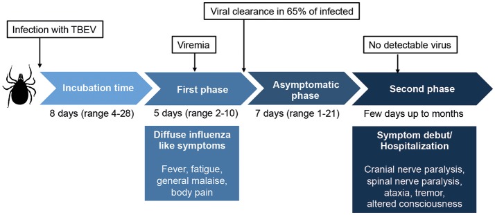
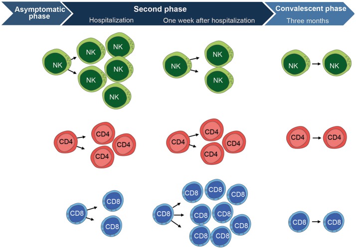
Tick-borne encephalitis virus (TBEV) is a flavivirus that belongs to the Flaviviridae family. TBEV is transmitted to humans primarily from infected ticks. The virus causes tick-borne encephalitis (TBE), an acute viral disease that affects the central nervous system (CNS). Infection can lead to acute neurological symptoms of significant severity due to meningitis or meningo(myelo)encephalitis. TBE can cause long-term suffering and has been recognized as an increasing public health problem. TBEV-affected areas currently include large parts of central and northern Europe as well as northern Asia. Infection with TBEV triggers a humoral as well as a cell-mediated immune response. In contrast to the well-characterized humoral antibody-mediated response, the cell-mediated immune responses elicited to natural TBEV-infection have been poorly characterized until recently. Here, we review recent progress in our understanding of the cell-mediated immune response to human TBEV-infection. A particular emphasis is devoted to studies of the response mediated by natural killer (NK) cells and CD8 T cells. The studies described include results revealing the temporal dynamics of the T cell- as well as NK cell-responses in relation to disease state and functional characterization of these cells. Additionally, we discuss specific immunopathological aspects of TBEV-infection in the CNS.

Keywords: NK cells; T cells; cell-mediated immunity; flavivirus; tick-borne encephalitis; tick-borne encephalitis virus; viral immunopathogenesis.


Figures

Figure 1
Figure 1
Overview of the classic biphasic disease-pattern of human TBEV infection. The viremic first phase includes influenza-like symptoms and occurs around 1 week after virus transmission. It is estimated that 65–70% of infected individuals clear the virus after this phase, but for one third of the patients, an asymptomatic disease phase follows before the second phase of disease begins. In this phase, symptoms of meningitis or encephalitis occur, including fever, headache, tremor, nystagmus, altered state of consciousness, cranial nerve paralysis, and spinal nerve paralysis. Classically, no virus is detected in sera or plasma in the second phase of disease. Around 30% of patients that enter the second phase of disease will suffer from long lasting sequeale, with a decreased quality of life. Figure compiled from Lindquist and Vapalahti (3), Taba et al. (4), and Haglund and Gunther (6).
Figure 2
Figure 2
Overview of the cell-mediated immune response to TBEV-infection. CD56dim NK cells (mainly highly differentiated CD57+ cells) are highly activated at the time of hospitalization during the second phase of disease. They express significantly higher levels of Ki67, CD38, and produce less cytokines in response to target cells as compared to the convalescent phase. NK cells then become fully normalized, comparable to healthy control levels, at the convalescent phase as assessed 3 months after hospitalization. CD4 T cells show a similar pattern as the CD56dim NK cells. They are activated at hospitalization and express significantly higher levels of Ki67, CD38, HLA-DR, and Granzyme B as compared to the convalescent phase. CD4 T cells have retracted to normal healthy control levels at the convalescent phase. CD8 T cells show a different pattern and peak in activation at 1 week after hospitalization. They express significantly higher levels of Ki67, CD38, HLA-DR, Granzyme B, Perforin, PD-1, T-bet, and Eomesodermin as compared CD8 T cells at the convalescent phase. The CD8 T cell activation subsequently return to normal healthy control levels at the convalescent phase, just as is the case for CD4 T cells and NK cells. Figure compiled from Blom et al. (58) and Blom et al. (59).

References

    1. Calisher CH. Antigenic classification and taxonomy of flaviviruses (family Flaviviridae) emphasizing a universal system for the taxonomy of viruses causing tick-borne encephalitis. Acta Virol. (1988) 32:469–78. - PubMed
    1. Heinz FX, Collett MS, Purcell RH, Gould EA, Howard CR, Houghton M, et al. Family Flaviviridae. San Diego, CA: Academic Press; (2000).
    1. Lindquist L, Vapalahti O. Tick-borne encephalitis. Lancet (2008) 371:1861–71. 10.1016/S0140-6736(08)60800-4 - DOI - PubMed
    1. Taba P, Schmutzhard E, Forsberg P, Lutsar I, Ljostad U, Mygland A, et al. . EAN consensus review on prevention, diagnosis and management of tick-borne encephalitis. Eur J Neurol. (2017) 24:1214–e61. 10.1111/ene.13356 - DOI - PubMed
    1. Kohl I, Kozuch O, Eleckova E, Labuda M, Zaludko J. Family outbreak of alimentary tick-borne encephalitis in Slovakia associated with a natural focus of infection. Eur J Epidemiol. (1996) 12:373–5. 10.1007/BF00145300 - DOI - PubMed

Publication types

MeSH terms