. 2018 Sep 30;3(9):11633-11642.
doi: 10.1021/acsomega.7b01533. Epub 2018 Sep 24.

Preparation of Tyrosylprotein Sulfotransferases for In Vitro One-Pot Enzymatic Synthesis of Sulfated Proteins/Peptides

Affiliations

Preparation of Tyrosylprotein Sulfotransferases for In Vitro One-Pot Enzymatic Synthesis of Sulfated Proteins/Peptides

Chen-Chu Wang et al. ACS Omega. .

Abstract

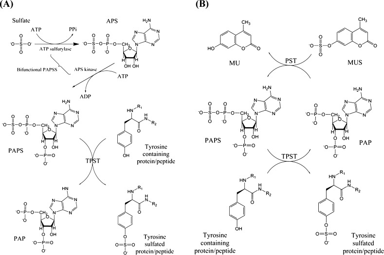
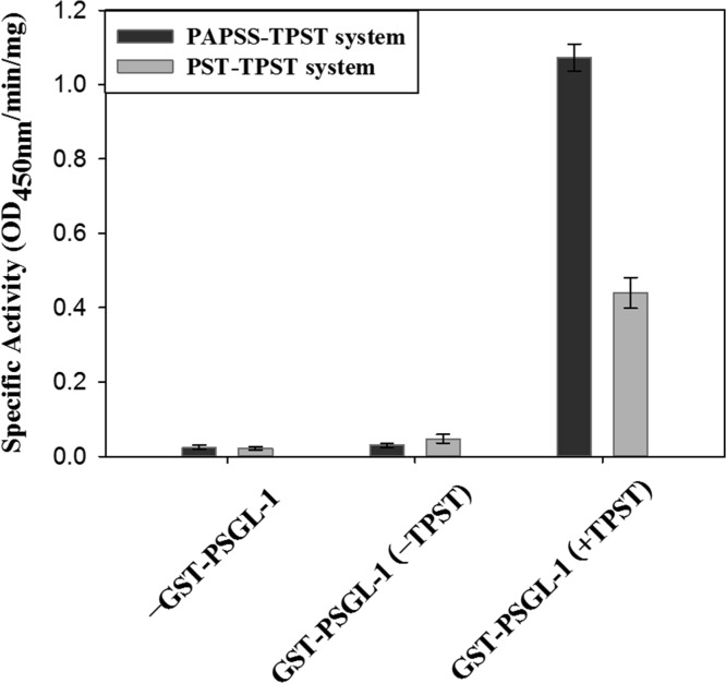
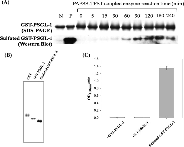
Protein tyrosine sulfation (PTS), catalyzed by membrane-anchored tyrosylprotein sulfotransferase (TPST), is one of the most common post-translational modifications of secretory and transmembrane proteins. PTS, a key modulator of extracellular protein-protein interactions, accounts for various important biological activities, namely, virus entry, inflammation, coagulation, and sterility. The preparation and characterization of TPST is fundamental for understanding the synthesis of tyrosine-sulfated proteins and for studying PTS in biology. A sulfated protein was prepared using a TPST-coupled protein sulfation system that involves the generation of the active sulfate 3'-phosphoadenosine-5'-phosphosulfate (PAPS) through either PAPS synthetase (PAPSS) or phenol sulfotransferase. The preparation of sulfated proteins was confirmed through radiometric or immunochemical assays. In this study, enzymatically active Drosophila melanogaster TPST (DmTPST) and human TPSTs (hTPST1 and hTPST2) were expressed in Escherichia coli BL21(DE3) host cells and purified to homogeneity in high yield. Our results revealed that recombinant DmTPST was particularly useful considering its catalytic efficiency and ease of preparation in large quantities. This study provides tools for high-efficiency, one-step synthesis of sulfated proteins and peptides that are useful for further deciphering the mechanisms, functions, and future applications of PTS.


Conflict of interest statement

The authors declare no competing financial interest.

Figures

Figure 1
Figure 1
Sequence analysis of TPSTs from humans and D. melanogaster. The pairwise sequence alignment was performed by ClustalW (http://www.ebi.ac.uk/Tools/clustalw2/index.html) and sorted and shaded by the BOXSHADE web server (http://www.ch.embnet.org/software/BOX_form.html). The black background indicates identical amino acids, and the gray one indicates conserved substitutions. The residue colored in red is the predicted transmembrane domain calculated by PSIPRED (http://bioinf.cs.ucl.ac.uk/psipred/psiform.html) and ranged from residues 6 to 28 for both human TPST1 and TPST2 and from 12 to 28 for D. melanogaster TPST. The pairwise sequence identities of these TPSTs were 57, 56, and 65% for DmTPST–hTPST1, DmTPST–hTPST2, and hTPST1–hTPST2, respectively. In the absence of the transmembrane region, the sequence identity of the catalysis domain increased to approximately 75%.
Figure 2
Figure 2
Plasmid construction, expression, and purification of recombinant TPSTs. (A) Schematic of recombinant TPSTs in fusion proteins. DmTPST and hTPST1 were expressed as NusA at the N-terminal fused protein containing two his-tags. hTPST2 was also expressed with NusA as only one his-tag at the N terminus. The calculated molecular weight of the TPST fusion protein was approximately 96 kDa. (B) SDS–PAGE of recombinant TPST before and after purification for DmTPST, hTPST1, hTPST2, and their crude extracts. Lane “C-” indicates crude extracts. Lane M is the standard protein molecular weight marker. The arrowheads indicate TPSTs.
Figure 3
Figure 3
Preparation of sulfated protein by two types of coupled enzyme systems. (A) PAPSS–TPST coupled PTS. Activated sulfate in PAPS was first obtained from inorganic sulfate and ATP catalyzed by PAPSS. The sulfated protein was prepared from PAPS and a protein substrate catalyzed by TPST. Radioactive sulfate can be used to produce radioactive-labeled sulfated proteins. (B) Phenol sulfotransferase–TPST coupled PTS. PAPS is regenerated from PAP and MUS catalyzed by phenol sulfotransferase to produce MU that yields fluorescence.
Figure 4
Figure 4
In situ determination of two PTS schemes by ELISA. GST–PSGL-1 was coated on an ELISA plate, which was subsequently blocked with milk. The immobilized GST–PSGL-1 was then treated with the PAPSS–TPST or phenol sulfotransferase–TSPT system. Control experiments in the absence of each critical ingredient, GST–PSGL-1, and TPST were conducted to confirm that the PTS reaction on the target protein proceeded as expected. The sulfated proteins/peptides were recognized by anti-sulfotyrosine antibody, as described in the Materials and Methods section. The specific activity was determined according to the results of the HRP-produced signal per minute at OD450nm, and the DmTPST reaction time and amount were 60 min and 10 μg, respectively. Each data point was obtained from three independent measurements, and the error bar indicates standard deviation (SD).
Figure 5
Figure 5
PTS under long incubation time. PTS was determined by ELISA, as described in Figure 4, which was conducted from 5 to 150 min by using either the PAPSS–TPST or phenol sulfotransferase–TPST system. Three recombinant enzymes, DmTPST (circle dot), hTPST1 (triangle dot), and hTPST2 (square dot), were examined. The reaction condition was the same as that described in Figure 4, except for the reaction time. PTS by the PAPSS–TPST system is shown as filled dots and that by the phenol sulfotransferase–TPST system is shown as open dots. The total activity was determined according to the HRP-produced signal per minute at OD450nm, and the TPST reaction amount was 10 μg. The mole of sulfated products was calculated by plotting the standard curve of sulfated GST–PSGL-1 in terms of the HRP-produced signal per minute at OD450nm. Each data point was obtained from three independent measurements, and the error bar indicates SD.
Figure 6
Figure 6
Detection of radioactive sulfated proteins/peptides. The sulfation of PSGL-1 was conducted using the scheme of the PAPSS–TPST system (Figure 1A). The 35S-labeled substrate yielded radioactive sulfate from PAPS synthesized in situ from 35S-containing inorganic sulfate. Lanes “–substrate (+TPST),” “PSGL-1 (−TPST),” “GST (−TPST),” and “GST–PSGL-1 (−TPST)” were the negative controls, indicating controlled reactions in the absence of one such component from the complete reaction mixture. Lanes “PSGL-1 (+TPST),” “GST (+TPST),” and “GST–PSGL-1 (+TPST)” contained a complete reaction mixture, as described in the Materials and Methods section for the sulfation of the substrate. The arrowheads indicate the spot of [35S] sulfated proteins/peptides. The bottom spots indicate unreacted [35S] sulfate and [35S] PAPS.
Figure 7
Figure 7
Identification and confirmation of sulfated product. (A) In vitro synthesis of sulfated GST–PSGL-1 with the PAPSS–TPST system by DmTPST. The amount of 5 μg GST–PSGL-1 was loaded in each well. SDS–PAGE of total GST–PSGL-1 stained with Coomassie blue is shown in the upper panel as an internal control. Western blotting for sulfated GST–PSGL-1 was probed by the anti-sulfotyrosine monoclonal antibody (lower panel). Lanes N and P indicate GST–PSGL-1 and sulfated GST–PSGL-1 as negative and positive controls, respectively. The sulfated GST–PSGL-1 (lane P) was obtained with the PAPSS–TPST system reacted for 240 min and was purified using the GSTrap sepharose column. The detailed procedure is reported in the Materials and Methods section. (B) Native PAGE of GST, GST–PSGL-1, and purified sulfated GST–PSGL-1 obtained with the PAPSS–TPST system after reaction for 240 min. Electrophoresis was performed under 8% PAGE in pH 8.0. (C) The sulfated products were confirmed by ELISA with blank, GST–PSGL-1, and purified sulfated GST–PSGL-1. Furthermore, 1 μg of sulfated GST–PSGL-1 was coated on an ELISA plate. The Y-axis represents HRP-produced signal per minute at OD 450nm. Each data point was obtained from three independent measurements, and the error bar indicates SD.

Similar articles

Cited by

References

    1. Bettelheim F. R. Tyrosine-O-sulfate in a peptide from fibrinogen. J. Am. Chem. Soc. 1954, 76, 2838–2839. 10.1021/ja01639a073. - DOI
    1. Moore K. L. The biology and enzymology of protein tyrosine O-sulfation. J. Biol. Chem. 2003, 278, 24243–24246. 10.1074/jbc.r300008200. - DOI - PubMed
    1. Mishiro E.; Sakakibara Y.; Liu M.-C.; Suiko M. Differential enzymatic characteristics and tissue-specific expression of human TPST-1 and TPST-2. J. Biochem. 2006, 140, 731–737. 10.1093/jb/mvj206. - DOI - PubMed
    1. Nishimura M.; Naito S. Tissue-specific mRNA expression profiles of human carbohydrate sulfotransferase and tyrosylprotein sulfotransferase. Biol. Pharm. Bull. 2007, 30, 821–825. 10.1248/bpb.30.821. - DOI - PubMed
    1. Baeuerle P. A.; Huttner W. B. Tyrosine sulfation of yolk proteins 1, 2, and 3 in Drosophila melanogaster. J. Biol. Chem. 1985, 260, 6434–6439. - PubMed

LinkOut - more resources