Case Reports
. 2018 Oct 2;102(1):62.
doi: 10.5334/jbsr.1587.

Retro-Odontoid Pseudotumor in a Patient with Atlanto-Occipital Assimilation

Affiliations
Case Reports

Retro-Odontoid Pseudotumor in a Patient with Atlanto-Occipital Assimilation

Arvy Buttiens et al. J Belg Soc Radiol. .

Abstract

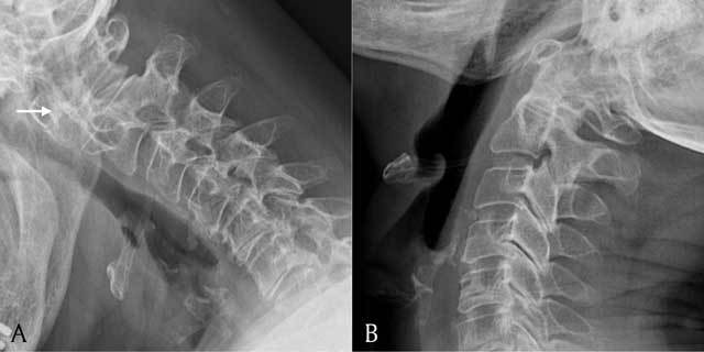
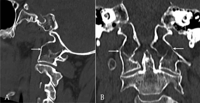
A retro-odontoid pseudotumor is an uncommon non-neoplastic mass. They are mostly associated with rheumatoid arthritis and atlanto-axial subluxation. The pathogenesis is degeneration of the transverse ligament due to chronic mechanical stress. In this case report, an atlanto-occipital assimilation altered the biomechanics of the cervical spine, causing chronic mechanical stress on the transverse ligament and subsequently the development of a retro-odontoid pseudotumor. This is in accordance with previous studies that have attributed the development of retro-odontoid pseudotumor to a loss of mobility of the cervical spine, in cases without associated rheumatoid arthritis or atlanto-axial subluxation.

Keywords: Odontoid process; atlanto-axial subluxation; pseudotumor; rheumatoid arthritis; transverse ligament.


Figures

Figure 1
Figure 1
Axial T2-weighted image. The retro-odontoid pseudotumor is seen as a hyperintense mass (black arrow) which extrudes through the transverse ligament and compresses the myelum (white arrow).
Figure 2
Figure 2
(A) Sagittal T1-weighted image. The retro-odontoid pseudotumor is seen as a mass (black arrow) extruding through the transverse ligament and compressing the myelum (white arrow). It has an isointense signal compared to the myelum. (B) Sagittal T2-weighted image. The retro-odontoid pseudotumor has a hyperintense signal (black arrow) and compresses the myelum (white arrow).
Figure 3
Figure 3
A = lateral view of the cervical spine in flexion; B = lateral view of the cervical spine in extension. Plain radiograph showing a lateral view of the cervical spine in flexion and extension. Note widening of the atlantodental interval in flexion (white arrow).
Figure 4
Figure 4
Sagittal (A) and coronal (B) reconstructions of a CT scan of the cervical spine shows assimilation of the massa lateralis of C1 and the occipital condyle (white arrows).

Similar articles

Cited by

References

    1. Yu, SH, Choi, HJ, Cho, WH, Cha, SH and Han, IH. Retro-odontoid pseudotumor without atlantoaxial subluxation or rheumatic arthritis. Korean J Neurotrauma. 2016; 12: 180–184. DOI: 10.13004/kjnt.2016.12.2.180 - DOI - PMC - PubMed
    1. Goel, A and Dange, N. Immediate postoperative regression of retroodontoid pannus after lateral mass reconstruction in a patient with rheumatoid disease of the craniovertebral junction. Case report. J Neurosurg Spine. 2008; 9: 273–276. DOI: 10.3171/SPI/2008/9/9/273 - DOI - PubMed
    1. Sze, G, Brant-Zawadzki, MN, Wilson, CR, Norman, D and Newton, TH. Pseudotumor of the craniovertebral junction associated with chronic subluxation: MR imaging studies. Radiology. 1986; 161: 391–394. DOI: 10.1148/radiology.161.2.3763907 - DOI - PubMed
    1. Kakutani, K, Doita, M, Yoshikawa, M, et al. C1 laminectomy for retro-odontoid pseudotumor without atlantoaxial subluxation: Review of seven consecutive cases. Eur Spine J. 2013; 22: 1119–1126. DOI: 10.1007/s00586-013-2681-0 - DOI - PMC - PubMed
    1. Yoshida, K, Hanyu, T and Takahashi, HE. Progression of rheumatoid arthritis of the cervical spine: Radiographic and clinical evaluation. J Orthop Sci. 1999; 4: 399–406. DOI: 10.1007/s007760050122 - DOI - PubMed

Publication types

LinkOut - more resources