. 2018 Dec;42(6):3404-3414.
doi: 10.3892/ijmm.2018.3919. Epub 2018 Oct 9.

Cytokine-induced alterations of BAMBI mediate the reciprocal regulation of human Th17/Treg cells in response to cigarette smoke extract

Affiliations

Cytokine-induced alterations of BAMBI mediate the reciprocal regulation of human Th17/Treg cells in response to cigarette smoke extract

Hong-Ju Liu et al. Int J Mol Med. 2018 Dec.

Abstract

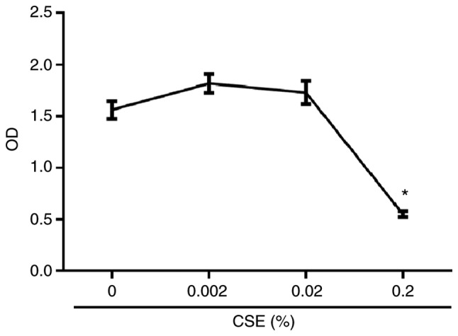
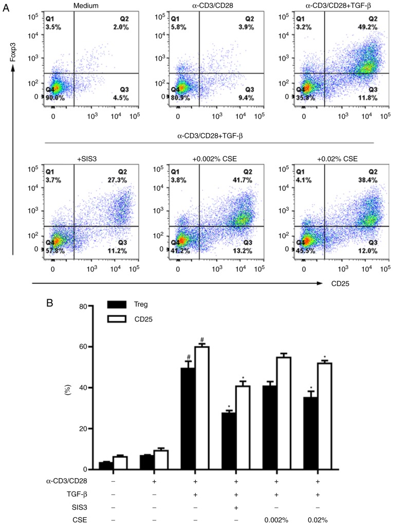
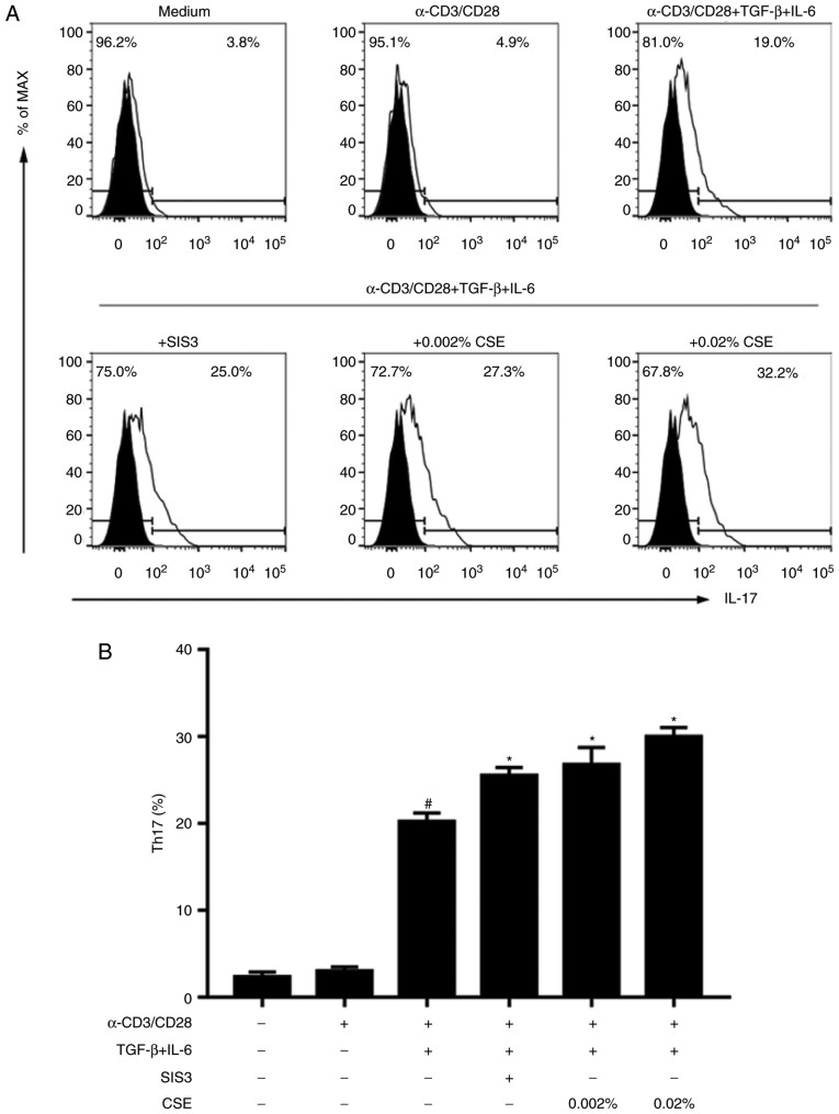
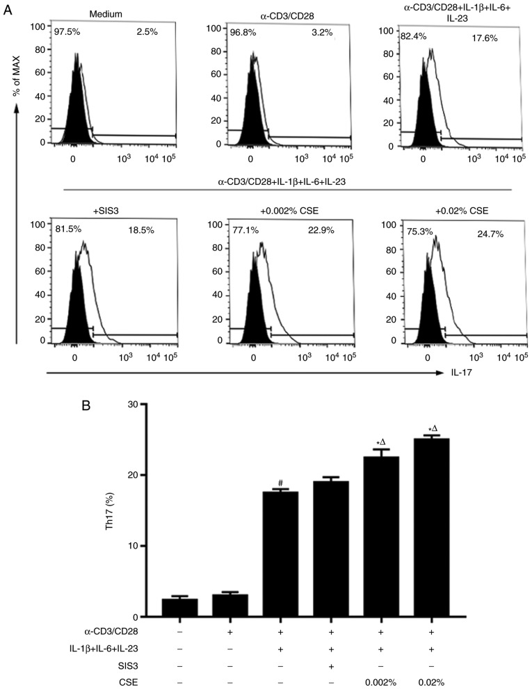
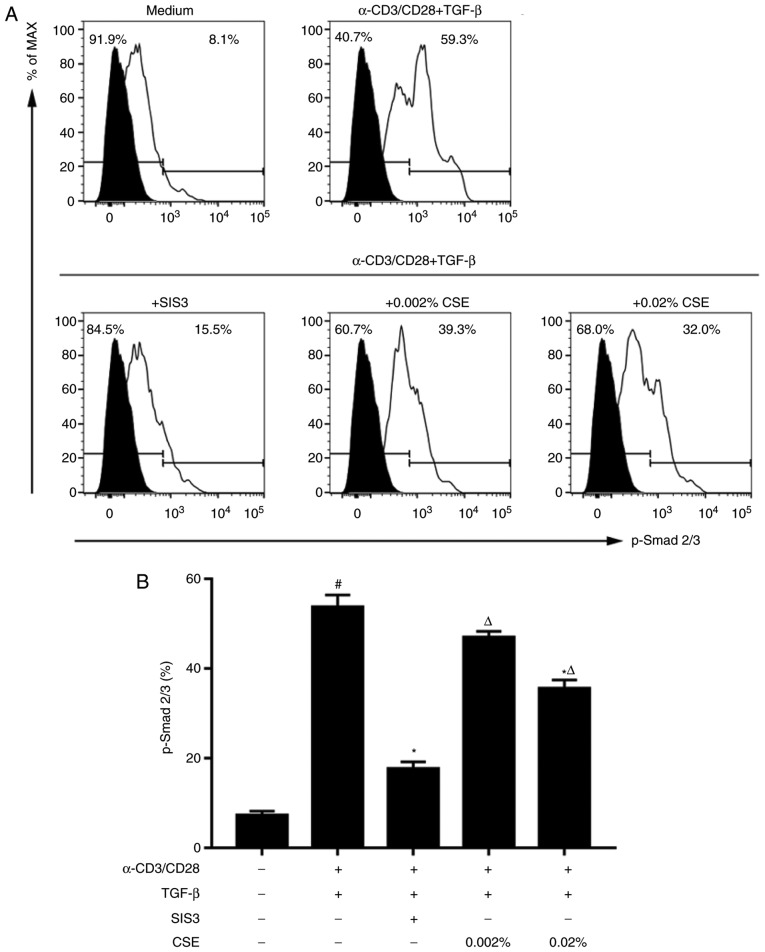
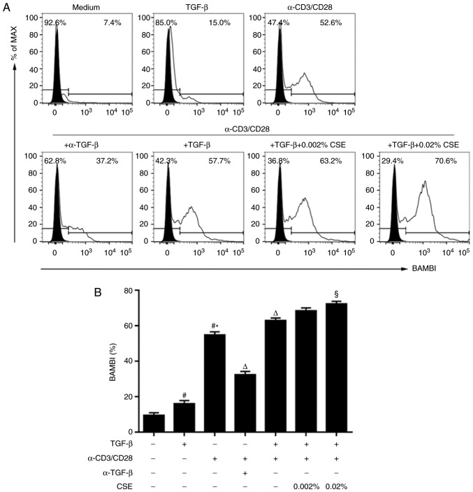
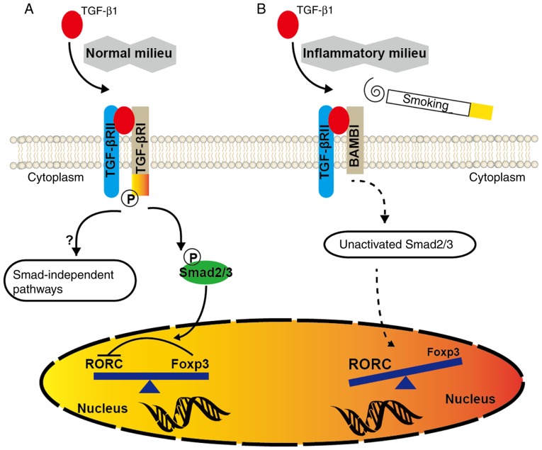
In CD4+ T helper (Th) cells, transforming growth factor β (TGF‑β) is indispensable for the induction of both regulatory T (Treg) and interleukin‑17‑producing effector T helper (Th17) cells. Although BMP and activin membrane‑bound inhibitor (BAMBI) is part of a rheostat‑like mechanism for the regulation of TGF‑β signalling and autoimmune arthritis in mouse models, the underlying activity of BAMBI on the human Th17/Treg cell axis, particularly during exposure to cigarette smoke, remains to be elucidated. The present study aimed to further characterize BAMBI expression in human CD4+ cells, as well as immune imbalance during activation and cigarette smoke exposure. Results from the present study indicated that exposure to cigarette smoke extract partially suppressed Treg differentiation and promoted Th17 cell generation under stimulation by anti‑CD3/28 antibodies and TGF‑β1. Additionally, exposure to cigarette smoke induced an inhibition of phosphorylated‑Smad2/Smad3, which may have arisen from a concomitant enhancement of BAMBI expression. In conclusion, human BAMBI may function as a molecular switch to control TGF‑β signalling strength and the Th17/Treg cell balance, which may be used not only as a biomarker but also as a target of new treatment strategies for maintaining immune tolerance and for the treatment of smoking‑induced immune disorders.


Figures

Figure 1
Figure 1
Effects of CSE on cell viability. Naive CD4+ T cells isolated from peripheral blood were stimulated with plate-bound α-CD3 and α-CD28 monoclonal antibodies in the presence of CSE at 0, 0.002, 0.02 and 0.2% for 5 days. Cell viability was examined by Cell Counting Kit-8 at an absorbance of 450 nm. The data are presented as the mean ± standard error of the mean (n=4), and are representative of three independent experiments; *P<0.05 vs. 0% CSE. CSE, cigarette smoke extract; OD, optical density.
Figure 2
Figure 2
Effects of CSE on Treg differentiation. (A and B) Naive CD4+ T cells isolated from peripheral blood were cultured in complete medium and stimulated with plate-bound α-CD3 and α-CD28 monoclonal antibodies under the indicated conditions for 5 days. (A) Cells were co-stained for CD25 and FOXP3 expression and measured by flow cytometry; representative pseudocolour dot plots gated on CD4+ T cells are shown. (B) Summary data of CD25+ FOXP3+ Tregs and CD25+ T cells in each condition, from (A) Data are presented as the mean ± standard error of the mean (n=4), and are representative of three independent experiments; #P<0.05 vs. Untreated control or α-CD3/28; *P<0.05 vs. respective α-CD3/28 + TGF-β1. CSE, cigarette smoke extract; FOXP3, forkhead box P3; TGF-β1, transforming growth factor β1; Treg, regulatory T cell; SIS3, a Smad3‑specific inhibitor.
Figure 3
Figure 3
Effects of CSE on Th17 cell differentiation. (A and B) Naive CD4+ T cells isolated from peripheral blood were cultured in complete medium and stimulated with plate-bound α-CD3 and α-CD28 monoclonal antibodies under the indicated conditions for 5 days. (A) Th17 cell counts were determined by flow cytometry, and representative histograms gated on lymphocytes are presented. (B) Summary data of Th17 cells in each condition from (A) Data are presented as the mean ± standard error of the mean (n=4), and are representative of three independent experiments; #P<0.05 vs. Untreated or α-CD3/28; *P<0.05 vs. α-CD3/28 + TGF-β1 + IL-6. CSE, cigarette smoke extract; IL, interleukin; TGF-β1, transforming growth factor β1; Th17, IL-17-producing T cells; SIS3, a Smad3‑specific inhibitor.
Figure 4
Figure 4
Effects of cigarette smoke extract (CSE) on Th17 cell differentiation. (A and B) Naive CD4+ T cells isolated from peripheral blood were cultured in complete medium and stimulated with plate-bound α-CD3 and α-CD28 monoclonal antibodies under the indicated conditions for 5 days. (A) Th17 cell counts were determined by flow cytometry, and representative histograms gated on lymphocytes are presented. (B) Summary data of Th17 cells in each condition from (A). Data are presented as the mean ± standard error of the mean (n=4), and are representative of three independent experiments; #P<0.05 vs. Untreated or α-CD3/28; *P<0.05 vs. α-CD3/28 + IL-1β/IL-6/IL-23; ΔP<0.05 vs. α-CD3/28 + IL-1β/IL-6/IL-23 + SIS3. CSE, cigarette smoke extract; IL, interleukin; Th17, IL‑17‑producing T cells; SIS3, a Smad3‑specific inhibitor.
Figure 5
Figure 5
Effects of CSE on Smad2/Smad3 phosphorylation. (A and B) Naive CD4+ T cells isolated from peripheral blood were cultured in complete medium and stimulated with plate-bound α-CD3 and α-CD28 monoclonal antibodies under the indicated conditions for 5 days. (A) Phosphorylation levels of Smad2/Smad3 were determined by flow cytometry, and representative histograms gated on CD4+ T cells are shown. (B) Summary data of Smad2/Smad3 phosphorylation in each condition from (A). Data are presented as the mean ± standard error of the mean (n=4), and are representative of three independent experiments; #P<0.05 vs. Untreated control; *P<0.05 vs. α-CD3/28 + TGF-β1. ΔP<0.05 vs. α-CD3/28 + TGF-β1 + SIS3. CSE, cigarette smoke extract; p, phosphorylated; TGF-β1, transforming growth factor β1; SIS3, a Smad3‑specific inhibitor.
Figure 6
Figure 6
BAMBI expression increases during activation and CSE exposure. (A and B) Naive CD4+ T cells isolated from peripheral blood were cultured in complete medium and stimulated with plate-bound α-CD3 and α-CD28 monoclonal antibodies under the indicated conditions for 5 days. (A) Expression levels of BAMBI were determined by flow cytometry, and representative histograms gated on CD4+ T cells are provided. (B) Summary data of BAMBI expression in each condition from (A). Data are presented as the mean ± standard error of the mean (n=4), and are representative of three independent experiments; #P<0.05 vs. Untreated control; *P<0.05 vs. TGF-β1; ΔP<0.05 vs. α-CD3/28; §P<0.05 vs. α-CD3/28 + TGF-β1. BAMBI, BMP and activin membrane-bound inhibitor; CSE, cigarette smoke extract; TGF-β1, transforming growth factor β1.
Figure 7
Figure 7
Schematic representation of the TGF-β/BAMBI pathway. (A) In the presence of TGF-β1, TCR-activated CD4+ T cells express both RORC and FOXP3. Enough Tregs keep effector T cells in check since RORC (a Th17‑specific transcription factor) activity is antagonized by FOXP3. (B) However, upon inflammatory insult or CSE exposure, BAMBI overexpression by the activated immune system in susceptible individuals will suppress the generation of TGF-β-induced Tregs and evoke pro‑inflammatory responses dominated by Th17 cells. BAMBI, BMP and activin membrane‑bound inhibitor; FOXP3, forkhead box P3; TCR, T cell receptor; TGF-β, transforming growth factor β; RORC, retinoic acid-related orphan receptor C.

Similar articles

Cited by

References

    1. Nicholson LB, Kuchroo VK. Manipulation of the Th1/Th2 balance in autoimmune disease. Curr Opin Immunol. 1996;8:837–842. doi: 10.1016/S0952-7915(96)80013-6. - DOI - PubMed
    1. Tran EH, Prince EN, Owens T. IFN-gamma shapes immune invasion of the central nervous system via regulation of chemokines. J Immunol. 2000;164:2759–2768. doi: 10.4049/jimmunol.164.5.2759. - DOI - PubMed
    1. Korn T, Bettelli E, Oukka M, Kuchroo VK. IL-17 and Th17 cells. Annu Rev Immunol. 2009;27:485–517. doi: 10.1146/annurev.immunol.021908.132710. - DOI - PubMed
    1. Sakaguchi S, Yamaguchi T, Nomura T, Ono M. Regulatory T cells and immune tolerance. Cell. 2008;133:775–787. doi: 10.1016/j.cell.2008.05.009. - DOI - PubMed
    1. Postigo J, Iglesias M, Alvarez P, Jesús Augustin J, Buelta L, Merino J, Merino R. Bone morphogenetic protein and activin membrane-bound inhibitor, a transforming growth factor β rheostat that controls murine treg Cell/Th17 cell differentiation and the development of autoimmune arthritis by reducing interleukin-2 signaling. Arthritis Rheumatol. 2016;68:1551–1562. doi: 10.1002/art.39557. - DOI - PubMed