The possible association between AQP9 in the intestinal epithelium and acute liver injury‑induced intestinal epithelium damage
- PMID: 30320400
- PMCID: PMC6236304
- DOI: 10.3892/mmr.2018.9542
The possible association between AQP9 in the intestinal epithelium and acute liver injury‑induced intestinal epithelium damage
Abstract
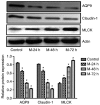
The present study aimed to investigate the expression and function of aquaporin (AQP)9 in the intestinal tract of acute liver injury rat models. A total of 20 Sprague Dawley rats were randomly divided into four groups: Normal control (NC) group and acute liver injury groups (24, 48 and 72 h). Acute liver injury rat models were established using D‑amino galactose, and the serum levels of alanine aminotransferase (ALT), aspartate aminotransferase (AST), total bilirubin (Tbil) and albumin were determined using an automatic biochemical analyzer. Proteins levels of myosin light chain kinase (MLCK) in rat intestinal mucosa were investigated via immunohistochemistry. Pathological features were observed using hematoxylin and eosin (H&E) staining. MLCK, AQP9 and claudin‑1 protein expression levels were detected via western blotting. Levels of ALT and AST in acute liver injury rats were revealed to steadily increase between 24 and 48 h time intervals, reaching a peak level at 48 h. Furthermore, TBil levels increased significantly until 72 h. Levels of ALT were revealed to significantly increase until the 48 h time interval, and then steadily decreased until the 72 h time interval. The acute liver injury 72 h group exhibited the greatest levels of MLCK expression among the three acute liver injury groups; however, all three acute liver injury groups exhibited enhanced levels of MLCK expression compared with the NC group. Protein levels of AQP9 and claudin‑1 were enhanced in the NC group compared with the three acute liver injury groups. H&E staining demonstrated that terminal ileum mucosal layer tissues obtained from the acute liver injury rats exhibited visible neutrophil infiltration. Furthermore, the results revealed that levels of tumor necrosis factor‑α, interleukin (IL)‑6 and IL‑10 serum cytokines were significantly increased in the acute liver injury groups. In addition, AQP9 protein expression was suppressed in acute liver injury rats, which induced pathological alterations in terminal ileum tissues may be associated with changes of claudin‑1 and MLCK protein levels.
Keywords: aquaporin 9; light junction; intestinal epithelium damage; acute liver injury; myosin light chain kinase; claudin-1.
Figures


Similar articles
-
[Effect of acupoint thread-embedding on tight junction of intestinal mucosa epithelium in rats with ulcerative colitis].Zhongguo Zhen Jiu. 2021 Aug 12;41(8):899-905. doi: 10.13703/j.0255-2930.20200629-k0003. Zhongguo Zhen Jiu. 2021. PMID: 34369702 Chinese.
-
[Myosin light chain kinase involved in change of intestinal mucosal barrier function in nonalcoholic steatohepatitis mice model].Zhonghua Nei Ke Za Zhi. 2015 May;54(5):434-8. Zhonghua Nei Ke Za Zhi. 2015. PMID: 26080824 Chinese.
-
Phosphorylated‑myosin light chain mediates the destruction of small intestinal epithelial tight junctions in mice with acute liver failure.Mol Med Rep. 2021 May;23(5):392. doi: 10.3892/mmr.2021.12031. Epub 2021 Mar 24. Mol Med Rep. 2021. PMID: 33760163 Free PMC article.
-
[Effect of curcumin on intestinal mucosal mechanical barrier in rats with non-alcoholic fatty liver disease].Zhonghua Gan Zang Bing Za Zhi. 2017 Feb 20;25(2):134-138. doi: 10.3760/cma.j.issn.1007-3418.2017.02.011. Zhonghua Gan Zang Bing Za Zhi. 2017. PMID: 28297801 Chinese.
-
[Correlation between the liver injury and the expression of interleukin-10 in severe acute pancreatitis at high altitude: a rat experimental result].Zhonghua Wei Zhong Bing Ji Jiu Yi Xue. 2018 Nov;30(11):1077-1082. doi: 10.3760/cma.j.issn.2095-4352.2018.011.013. Zhonghua Wei Zhong Bing Ji Jiu Yi Xue. 2018. PMID: 30541649 Chinese.
Cited by
-
Clinical value and molecular mechanism of AQGPs in different tumors.Med Oncol. 2022 Aug 16;39(11):174. doi: 10.1007/s12032-022-01766-0. Med Oncol. 2022. PMID: 35972604 Free PMC article. Review.
-
Screening gene signatures for clinical response subtypes of lung transplantation.Mol Genet Genomics. 2022 Sep;297(5):1301-1313. doi: 10.1007/s00438-022-01918-x. Epub 2022 Jul 3. Mol Genet Genomics. 2022. PMID: 35780439
-
Licorice Ameliorates Cisplatin-Induced Hepatotoxicity Through Antiapoptosis, Antioxidative Stress, Anti-Inflammation, and Acceleration of Metabolism.Front Pharmacol. 2020 Nov 10;11:563750. doi: 10.3389/fphar.2020.563750. eCollection 2020. Front Pharmacol. 2020. PMID: 33240085 Free PMC article.
References
MeSH terms
Substances
LinkOut - more resources
Full Text Sources

