. 2019 Feb;33(2):2858-2869.
doi: 10.1096/fj.201800529RR. Epub 2018 Oct 15.

Metformin increases glucose uptake and acts renoprotectively by reducing SHIP2 activity

Affiliations

Metformin increases glucose uptake and acts renoprotectively by reducing SHIP2 activity

Zydrune Polianskyte-Prause et al. FASEB J. 2019 Feb.

Abstract

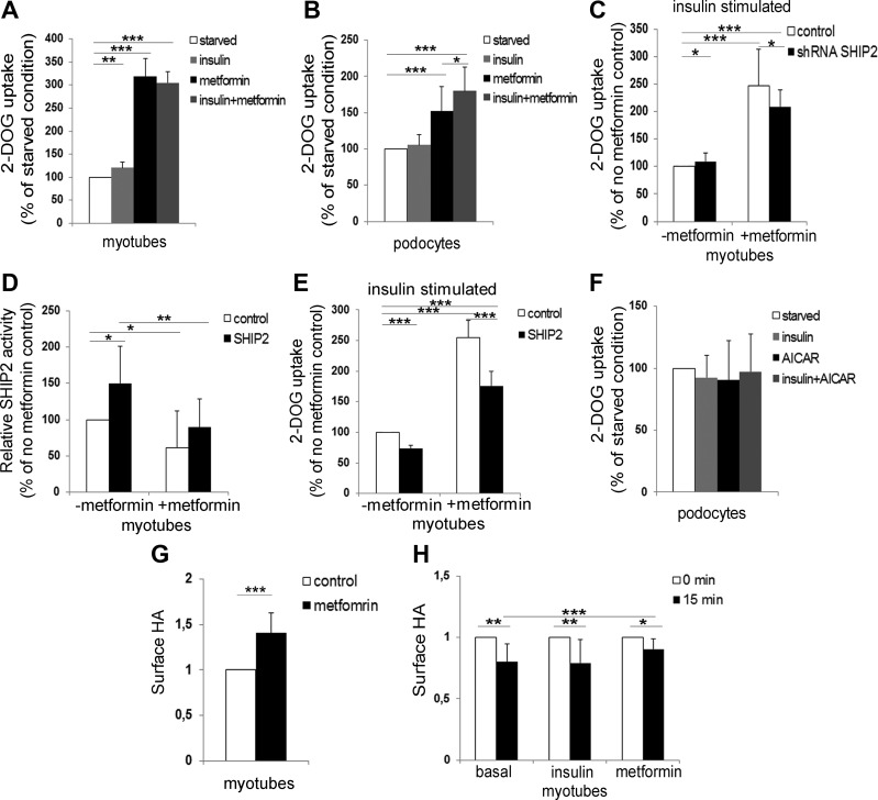
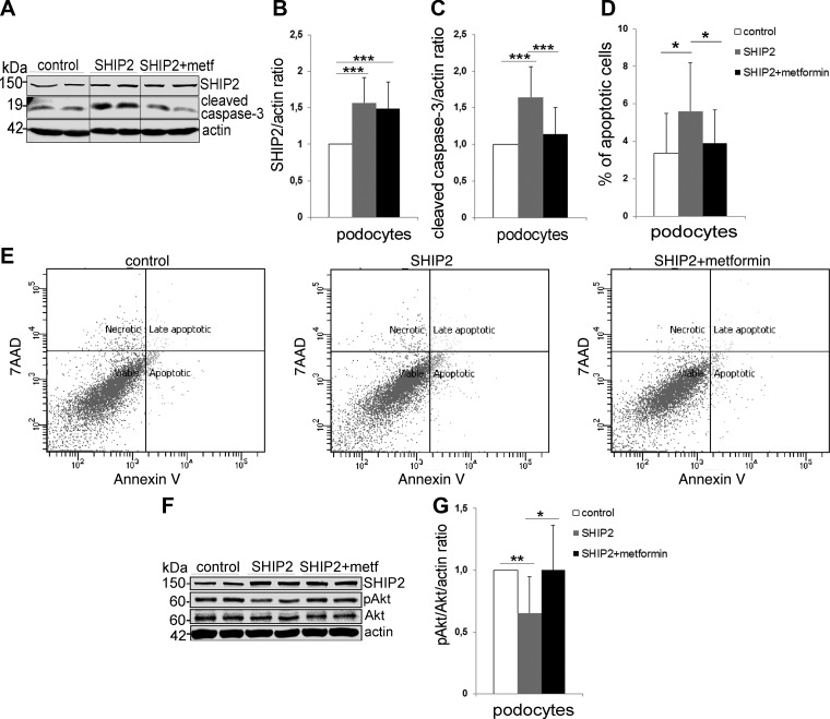
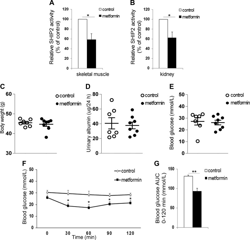
Metformin, the first-line drug to treat type 2 diabetes (T2D), inhibits mitochondrial glycerolphosphate dehydrogenase in the liver to suppress gluconeogenesis. However, the direct target and the underlying mechanisms by which metformin increases glucose uptake in peripheral tissues remain uncharacterized. Lipid phosphatase Src homology 2 domain-containing inositol-5-phosphatase 2 (SHIP2) is upregulated in diabetic rodent models and suppresses insulin signaling by reducing Akt activation, leading to insulin resistance and diminished glucose uptake. Here, we demonstrate that metformin directly binds to and reduces the catalytic activity of the recombinant SHIP2 phosphatase domain in vitro. Metformin inhibits SHIP2 in cultured cells and in skeletal muscle and kidney of db/db mice. In SHIP2-overexpressing myotubes, metformin ameliorates reduced glucose uptake by slowing down glucose transporter 4 endocytosis. SHIP2 overexpression reduces Akt activity and enhances podocyte apoptosis, and both are restored to normal levels by metformin. SHIP2 activity is elevated in glomeruli of patients with T2D receiving nonmetformin medication, but not in patients receiving metformin, compared with people without diabetes. Furthermore, podocyte loss in kidneys of metformin-treated T2D patients is reduced compared with patients receiving nonmetformin medication. Our data unravel a novel molecular mechanism by which metformin enhances glucose uptake and acts renoprotectively by reducing SHIP2 activity.-Polianskyte-Prause, Z., Tolvanen, T. A., Lindfors, S., Dumont, V., Van, M., Wang, H., Dash, S. N., Berg, M., Naams, J.-B., Hautala, L. C., Nisen, H., Mirtti, T., Groop, P.-H., Wähälä, K., Tienari, J., Lehtonen, S. Metformin increases glucose uptake and acts renoprotectively by reducing SHIP2 activity.

Keywords: diabetic kidney disease; insulin resistance; lipid phosphatase; podocyte; type 2 diabetes.


Conflict of interest statement

The authors thank M. Saleem (Southmead Hospital, University of Bristol, Bristol United Kingdom) for kindly providing immortalized human podocytes, S. Cushman (National Institutes of Health, Bethesda, MD, USA) for the HA-GLUT4-GFP construct, and C. A. Mitchell (Monash University, Clayton, VIC, Australia) and G. Krystal (Terry Fox Laboratory, Vancouver, BC, Canada) for human SHIP2 (NP_001558) and SHIP1 (NP_005532) cDNA constructs, respectively. Leena Saikko, High Throughput Biomedicine Unit, Biomedicum Flow Cytometry Unit, Biochemical Analysis Core for Experimental Research, and Genome Biology Unit (University of Helsinki) are acknowledged for excellent technical assistance. This work was supported by the European Research Council (Grant 242820; to S.L.), Academy of Finland (Grants 131255, 218021, and 255551 to S.L.; Grant 134379 to P.-H.G.), Jane and Aatos Erkko Foundation (to S.L.), European Foundation for the Study of Diabetes/Boehringer Ingelheim Research Programme (to S.L.), Sigrid Jusélius Foundation (to S.L.), Päivikki and Sakari Sohlberg Foundation (to S.L.), Diabetes Research Foundation (to S.L.), Business Finland (1964/31/2017 to S.L.), Faculty of Medicine, University of Helsinki (to S.L.), Helsinki University Hospital (to J.T.), Doctoral Programme in Biomedicine (to T.A.T. and V.D.), Folkhälsan Research Foundation (to P.-H.G.), and Novo Nordisk Foundation (to P.-H.G.). P.-H.G. has received research grants from Eli Lilly and Roche, and is an advisory board member for AbbVie, AstraZeneca, Boehringer-Ingelheim, Cebix, Eli Lilly, Janssen, Medscape, Musculoskeletal Diseases (MSD), and Novartis. P.-H.G. has received lecture honoraria from AstraZeneca, Boehringer-Ingelheim, Eli Lilly, Genzyme, MSD, Novartis, Novo Nordisk, and Sanofi. However, none of the above are relevant to the article. The remaining authors declare no conflicts of interest.

Figures

Figure 1
Figure 1
Metformin reduces the catalytic activity of SHIP2. A) Predicted binding orientation of metformin to the active site of SHIP2. Hydrogen bond is shown in green. Hydrophobic residues are shown in gray, positive-charged residues in blue, and negative-charged residues in red. B) Metformin (10 µM) reduces the activity of purified His-tagged SHIP2 phosphatase domain (100 ng). C, D) Metformin (1 mM, 20–24 h) reduces the activity of SHIP2 immunoprecipitated from the lysates of L6 myotubes (C) and podocytes (D). E, F) Metformin and AS1949490 at 20 µM concentration (20–24 h) reduce the activity of SHIP2 immunoprecipitated from the lysates of L6 myotubes (E) and podocytes (F). The catalytic activity of SHIP2 was measured by manual malachite green phosphate assay. Data are presented as the mean ± sd of 3–5 independent experiments . **P ≤ 0.01, ***P ≤ 0.001 (Student’s t test).
Figure 2
Figure 2
Knockdown and overexpression of SHIP2 decrease metformin-induced glucose uptake, and metformin slows down the endocytosis of GLUT4. A, B) Metformin increases glucose uptake in myotubes and podocytes. Serum-starved L6-GLUT4 myotubes (A) and podocytes (B), which were additionally starved of insulin-transferrin-selenium for 72 h, were treated with metformin (2 mM; 20–24 h) and/or stimulated with insulin (100 or 200 nM, respectively; 15 min), followed by glucose uptake assay. C) Knockdown of SHIP2 reduces metformin-induced glucose uptake. L6-GLUT4 myotubes were infected with SHIP2 or control shRNAs, treated with metformin (2 mM, 20–24 h) under insulin (100 nM, 15 min)-stimulated conditions, followed by glucose uptake assay. D) SHIP2 overexpression increases SHIP2 activity, and metformin restores it back to the level of the control. L6 myotubes overexpressing SHIP2 or empty vector (control) were incubated with metformin (1 mM; 20–24 h), and the phosphatase activity of immunoprecipitated SHIP2 was measured by malachite green phosphate assay. E) SHIP2 overexpression abrogates the effect of metformin to induce glucose uptake. L6-GLUT4 myotubes overexpressing SHIP2 were treated and glucose uptake performed as described in C. F) AICAR (1 mM; 20–24 h), alone or together with insulin (200 nM, 15 min), does not increase glucose uptake in podocytes. G) Quantification of the On-Cell Western signal for HA revealed that metformin increases the HA-GLUT4-GFP level at the PM. L6-GLUT4 myotubes were incubated under basal conditions and treated with metformin (2 mM; 20–24 h), followed by On-Cell Western assay. H) Metformin slows down endocytosis of HA-GLUT4-GFP in L6-GLUT4 cells. Time point 0 min indicates the amount of HA on the cell surface after metformin (2 mM; 20–24 h) or insulin (100 nM; 15 min) treatment and is set to value 1. Time point 15 min indicates the amount of HA detected on the cells after 15 min internalization. Data are presented as the mean ± sd of 3–4 independent experiments. *P ≤ 0.05, **P ≤ 0.01, ***P ≤ 0.001 (Student’s t test).
Figure 3
Figure 3
Metformin restores prosurvival Akt signaling and protects podocytes from SHIP2 overexpression-induced apoptosis. A) SHIP2 overexpression increases the expression of cleaved caspase-3 in podocytes, and metformin (2 mM, 48 h) restores it back to the level in the control (empty vector). Cell lysates were subjected to immunoblot analysis with anti-SHIP2, anti-cleaved caspase-3, and anti-actin IgGs. Representative protein bands are from the same immunoblot. B, C) Quantification of SHIP2 and cleaved caspase-3 levels in A, normalized to actin. D) SHIP2 overexpression increases the proportion of apoptotic cells in podocytes, and metformin (2 mM, 48 h) restores it back to the level in the control (empty vector). Apoptosis was measured by flow cytometry using Annexin V-FITC and 7-aminoactinomycin D (7AAD) double staining. E) Representative images of 5 independent experiments, each with 3 replicates, showing flow cytometry analysis of control (empty vector) or SHIP2-overexpressing podocytes treated or not with metformin. F) SHIP2 overexpression decreases insulin-induced pAkt in podocytes, and metformin restores it back to the level in the control (empty vector). Podocytes overexpressing SHIP2 or empty vector (control) were incubated with metformin (2 mM; 20–24 h) and stimulated with insulin (200 nM; 15 min). Cell lysates were subjected to immunoblot analysis with anti-SHIP2, anti-Akt, anti-pAkt, and anti-actin IgGs. G) Quantification of pAkt levels in F presented as pAkt/Akt after normalizing to actin. Data are presented as the mean ± sd of 3–5 independent experiments. *P ≤ 0.05, **P ≤ 0.01, ***P ≤ 0.001 (Student’s t test).
Figure 4
Figure 4
Metformin decreases the catalytic activity of SHIP2 in db/db mice. A, B) Metformin decreases the catalytic activity of SHIP2 in skeletal muscle (A) and kidney (B) of db/db mice. SHIP2 was immunoprecipitated from tissue lysates and its catalytic activity measured by malachite green phosphate assay (n = 6–8). CE) Twelve d metformin treatment has no effect on body weight (C), urinary albumin excretion (D), and fasting blood glucose (E) in db/db mice (n = 7–8). F, G) Metformin improves insulin sensitivity of db/db mice. Insulin tolerance test was performed and blood glucose measured at the indicated times after insulin injection, and the area under the curve (AUC) was calculated (n = 5–6). Data are presented as the mean ± sem. *P ≤ 0.05, **P ≤ 0.01 (Student’s t test).
Figure 5
Figure 5
SHIP2 activity and podocyte loss are reduced in kidneys of human patients with T2D receiving metformin. A) SHIP2 activity is increased in kidneys of patients with T2D receiving nonmetformin medication compared with people without T2D. SHIP2 activity in patients with T2D receiving metformin medication is similar to people without T2D. Kidney samples were lysed, and the enzymatic activity of immunoprecipitated SHIP2 was analyzed by malachite green phosphate assay (n = 4–7). B) SHIP2 expression level in glomeruli in D was quantified with the HistoQuant (3DHistech) program. Ten randomly chosen glomeruli were analyzed from each patient (n = 6–7). C) SHIP2 expression level in kidney cortex was quantified with 3DHistech (n = 5–7). D) SHIP2 staining in human glomeruli of patients with T2D receiving metformin or nonmetformin medication and people without T2D. Paraffin sections of nephrectomy samples were processed for immunoperoxidase staining and labeled with anti-SHIP2 IgG. E) WT1 staining in human glomeruli of patients with T2D receiving metformin or nonmetformin medication and people without T2D. Paraffin sections of nephrectomy samples were processed for immunoperoxidase staining and labeled with anti-WT1 IgG. F) Podocyte loss is decreased in kidneys of patients with T2D receiving metformin medication compared with patients receiving nonmetformin medication. Podocyte number per glomeruli was quantified by counting WT1-positive cells. Ten randomly chosen glomeruli were analyzed from each patient (n = 9–30). Original scale bars, 50 μm. Data are presented as means ± sem. *P ≤ 0.05, **P ≤ 0.01, ***P ≤ 0.001; ANOVA (B–F); Student’s t test (A).

References

    1. Foretz, M., Guigas, B., Bertrand, L., Pollak, M., Viollet, B. (2014) Metformin: from mechanisms of action to therapies. Cell Metab. 20, 953–966 10.1016/j.cmet.2014.09.018 - DOI - PubMed
    1. Hundal, R. S., Krssak, M., Dufour, S., Laurent, D., Lebon, V., Chandramouli, V., Inzucchi, S. E., Schumann, W. C., Petersen, K. F., Landau, B. R., Shulman, G. I. (2000) Mechanism by which metformin reduces glucose production in type 2 diabetes. Diabetes 49, 2063–2069 10.2337/diabetes.49.12.2063 - DOI - PMC - PubMed
    1. Féry, F., Plat, L., Balasse, E. O. (1997) Effects of metformin on the pathways of glucose utilization after oral glucose in non-insulin-dependent diabetes mellitus patients. Metabolism 46, 227–233 10.1016/S0026-0495(97)90307-3 - DOI - PubMed
    1. Aatsinki, S. M., Buler, M., Salomäki, H., Koulu, M., Pavek, P., Hakkola, J. (2014) Metformin induces PGC-1α expression and selectively affects hepatic PGC-1α functions. Br. J. Pharmacol. 171, 2351–2363 10.1111/bph.12585 - DOI - PMC - PubMed
    1. Petersen, M. C., Vatner, D. F., Shulman, G. I. (2017) Regulation of hepatic glucose metabolism in health and disease. Nat. Rev. Endocrinol. 13, 572–587 10.1038/nrendo.2017.80 - DOI - PMC - PubMed

Publication types

MeSH terms

Substances