. 2019 Feb 15;74(3):315-324.
doi: 10.1093/gerona/gly237.

Marmoset as a Model to Study Kidney Changes Associated With Aging

Affiliations

Marmoset as a Model to Study Kidney Changes Associated With Aging

Hak Joo Lee et al. J Gerontol A Biol Sci Med Sci. .

Erratum in

Abstract

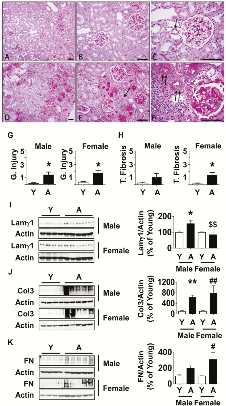
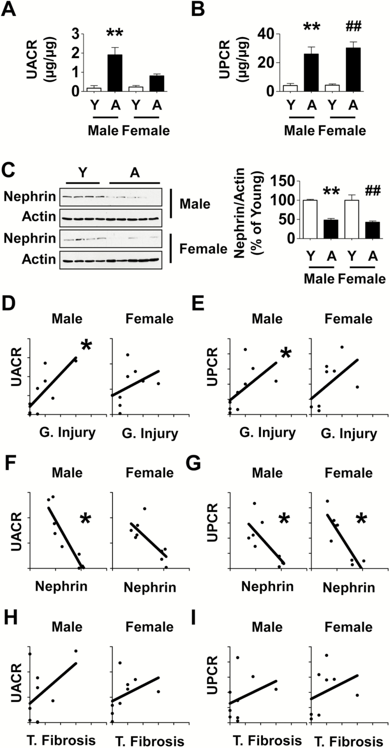
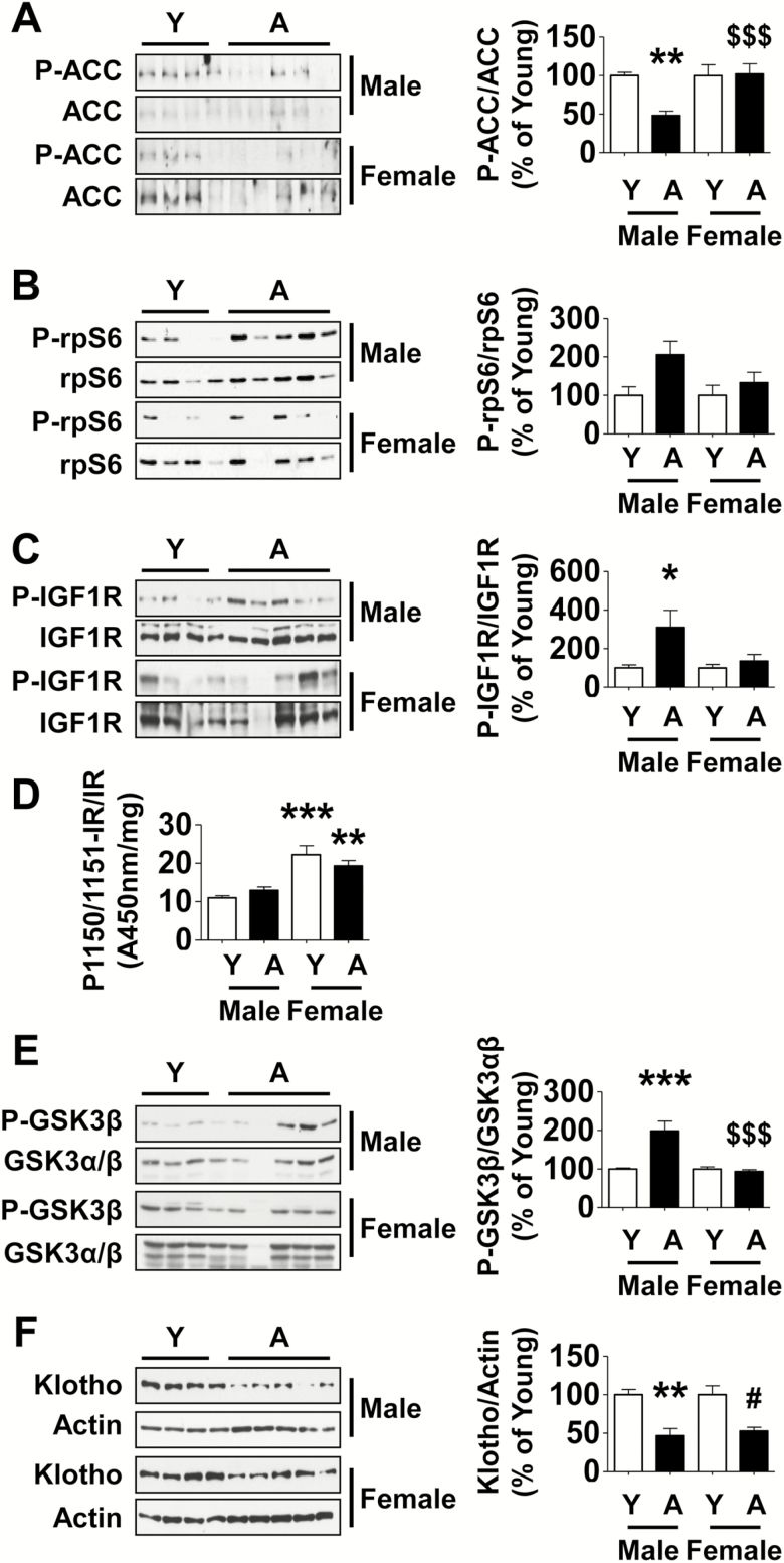
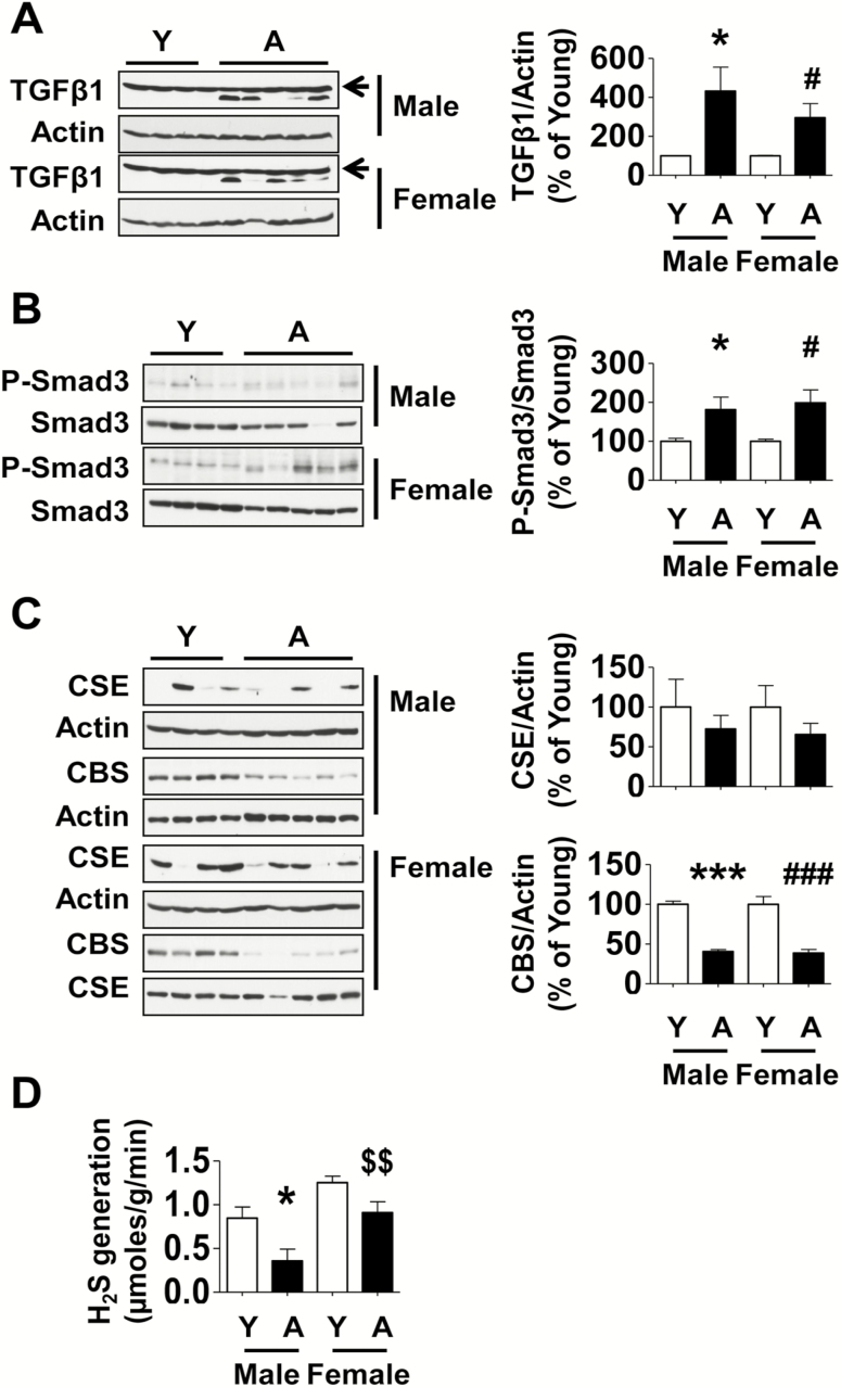
We evaluated whether the marmoset, a nonhuman primate, can serve as a good model to study aging-related changes in the kidney by employing healthy young and aged marmosets of both sexes. Aging was associated with glomerulosclerosis, interstitial fibrosis, and arteriolosclerosis in both sexes; correspondingly, the content of matrix proteins was increased. Functionally, aging resulted in an increase in urinary albumin and protein excretion. There was a robust correlation between markers of fibrosis and functional changes. We explored signaling pathways as potential mechanistic events. Aging in males, but not in females, was associated with reduced renal cortical activity of AMP-activated protein kinase (AMPK) and a trend toward activation of mechanistic target of rapamycin complex 1 (mTORC1); upstream of AMPK and mTORC1, Akt and IGF-1 receptor were activated. In both sexes, aging promoted kidney activation of transforming growth factor β-1 signaling pathway. While the expression of cystathionine β-synthase (CBS), an enzyme involved hydrogen sulfide (H2S) synthesis, was reduced in both aged males and females, decreased H2S generation was seen in only males. Our studies show that the marmoset is a valid model to study kidney aging; some of the signaling pathways involved in renal senescence differ between male and female marmosets.

Keywords: Extracellular matrix; Fibrosis; Glomerulus; Primates; Signaling.


Figures

Figure 1.
Figure 1.
Kidney histology and extracellular matrix protein expression in aged marmosets. (AC) By PAS stain, in young marmosets glomeruli displayed normal cellularity without segmental or global accentuation of mesangial matrix. Tubules were closely packed without atrophy. Blood vessels (arrow in C) were thin walled and lumens were patent. (DF) In aged marmosets, glomeruli showed either segmental (part of an individual glomerulus) or total (whole of an individual glomerulus) sclerosis. Parietal glomerular basement membrane showed thickening and wrinkling. Tubules showed significant atrophy, loss, thickening of tubular basement membrane and many contained casts. There were rare areas of interstitial nephritis composed of mononuclear cells (arrow in E). Blood vessels showed narrowing of lumen and severe medial hypertrophy and sclerosis (double arrows in F). (G and H) Semi-quantitative scoring of indices of glomerular injury and tubular fibrosis are shown (Y = young, A = aged). Data are shown as mean ± SEM, n = 4 young male and n = 4 young female marmosets, n = 5 aged male and n = 5 aged female marmosets (*p < .05 by t-test). Renal cortical lysates were employed to perform immunoblotting using antibodies against (I) laminin γ1, (J) collagen III, and (K) fibronectin. Actin expression was used to assess loading (Y = young, A = aged). Data are shown as mean ± SEM, n = 4 young male and n = 4 young female marmosets, n = 5 aged male and n = 5 aged female marmosets (*p < .05, **p < .01 vs young male; #p < .05, ##p < .01 vs young female, $$p < .01 vs aged male by two-way ANOVA).
Figure 2.
Figure 2.
Aging is associated with changes in kidney function in marmosets and they correlate with histological changes. (A and B) Urinary albumin to creatinine ratio (UACR) and urinary protein to creatinine ratio (UPCR) data in aged marmosets and young marmosets are shown. (C) Immunoblotting of kidney cortical lysates showed reduction in the expression of nephrin in aged marmosets (Y = young, A = aged). Data in A–C are shown as mean ± SEM, n = 4 young male and n = 3–4 young female marmosets, n = 5 aged male and n = 5 aged female marmosets (**p < .01 vs young male; ##p < .01 vs young female by two-way ANOVA). Correlation between glomerular injury (G. Injury) and tubular fibrosis (T. Fibrosis) indices and UACR, UPCR was studied. (D and E) A high degree of correlation existed between glomerular injury and UACR (r = .78, *p = .0172), and UPCR (r = .80, *p = .0138) in aged males; a trend toward correlation between glomerular injury and UPCR (r = .68, p = .0694) but not UACR (r = .58, p = .1080) was seen in aged females. (F and G) A strong negative relationship was present between nephrin expression and UACR (r = −0.88, *p = .0031) and UPCR (r = −.70, *p = .0433) in aged males; in aged females, a negative correlation was present between nephrin and UPCR (r = −0.81, *p = .0218) and a trend with UACR (r = −0.65, p = .0666). (H and I) In aged males, tubular fibrosis did not have a significant relationship with UACR (r = .25, p = .5206) or UPCR (r = .38, p = .3125); in aged females a trend was seen with UACR (r = .67, p = .0589) but not with UPCR (r = .51, p = .1966).
Figure 3.
Figure 3.
Signaling pathways are dysregulated in the kidney cortex of aged male but not female marmosets. Renal cortical lysates were employed to perform immunoblotting using antibodies against (A) phospho-ACC, ACC, (B) phospho-rpS6, rpS6, (C) Tyr-1165/1166 phosphorylated IGF-1 receptor (P-IGF 1R), IGF 1R, (E) Ser-9 phosphorylated GSK 3β, GSK 3β, (F) Klotho and actin. (D) ELISA was performed to assess changes in Tyr-1150/1151 phosphorylation of the IR (Y = young, A = aged). Data are shown as mean ± SEM, n = 4 young male and n = 4 young female marmosets, n = 4–5 aged male and n = 4–5 aged female marmosets (*p < .05, **p < .01, ***p < .001 vs young male; #p < .05 vs young female, $$$p < .001 vs aged male by two-way ANOVA).
Figure 4.
Figure 4.
Changes in TGFβ and H2S pathways in the kidney cortex of aged marmosets. Equal amounts of renal cortex were employed in immunoblotting using antibodies against (A) TGFβ1 and actin (unchanged band above TGFβ1 is nonspecific [arrow]), (B) phospho-Smad3 and Smad3, and (C) CSE and CBS. (D) H2S generation was measured as described in Method section (Y = young, A = aged). Data are shown as mean ± SEM, n = 4 young male and n = 4 young female marmosets, n = 5 aged male and n = 5 aged female marmosets (*p < .05, ***p < .001 vs young male; #p < .05, ###p < .001 vs young female, $$p < .01 vs aged female by two-way ANOVA).

Similar articles

Cited by

References

    1. Choudhury D, Levi M. Kidney aging—inevitable or preventable?Nat Rev Nephrol. 2011;7:706–717. doi:10.1038/nrneph.2011.104 - DOI - PubMed
    1. Sataranatarajan K, Feliers D, Mariappan MM, et al. . Molecular events in matrix protein metabolism in the aging kidney. Aging Cell. 2012;11:1065–1073. doi:10.1111/acel.12008 - DOI - PMC - PubMed
    1. Maeda H, Gleiser CA, Masoro EJ, Murata I, McMahan CA, Yu BP. Nutritional influences on aging of Fischer 344 rats: II. Pathology. J Gerontol. 1985;40:671–688. - PubMed
    1. Ikeno Y, Hubbard GB, Lee S, et al. . Housing density does not influence the longevity effect of calorie restriction. J Gerontol A Biol Sci Med Sci. 2005;60:1510–1517. - PubMed
    1. Weindruch R, Masoro EJ. Concerns about rodent models for aging research. J Gerontol. 1991;46:B87–B88. - PubMed

Publication types