. 2019 Jun 1;11(6):496-508.
doi: 10.1093/jmcb/mjy056.

GSDMB promotes non-canonical pyroptosis by enhancing caspase-4 activity

Affiliations

GSDMB promotes non-canonical pyroptosis by enhancing caspase-4 activity

Qin Chen et al. J Mol Cell Biol. .

Abstract

Gasdermin B (GSDMB) has been reported to be associated with immune diseases in humans, but the detailed molecular mechanisms remain unsolved. The N-terminus of GSDMB by itself, unlike other gasdermin family proteins, does not induce cell death. Here, we show that GSDMB is highly expressed in the leukocytes of septic shock patients, which is associated with increased release of the gasdermin D (GSDMD) N-terminus. GSDMB expression and the accumulation of the N-terminal fragment of GSDMD are induced by the activation of the non-canonical pyroptosis pathway in a human monocyte cell line. The downregulation of GSDMB alleviates the cleavage of GSDMD and cell death. Consistently, the overexpression of GSDMB promotes GSDMD cleavage, accompanied by increased LDH release. We further found that GSDMB promotes caspase-4 activity, which is required for the cleavage of GSDMD in non-canonical pyroptosis, by directly binding to the CARD domain of caspase-4. Our study reveals a GSDMB-mediated novel regulatory mechanism for non-canonical pyroptosis and suggests a potential new strategy for the treatment of inflammatory diseases.

Keywords: GSDMB; GSDMD; pyroptosis; sepsis.


Figures

Figure 1
Figure 1
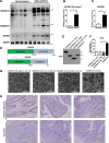
Sepsis and Crohn’s disease are accompanied with GSDMB upregulation. (A) We collected blood from sepsis patients and healthy individuals and examined the GSDMB protein expression as well as the GSDMD cleavage level in white blood cells using western blotting. GAPDH was used as the loading control. (B and C) The intensities of the GSDMD N-terminus (B) and GSDMB protein (C) bands were quantified using the ImageJ software program and normalized to that of GAPDH. (D) Graphic representation of GSDMB and GSDMD proteins. (EG) 293T cells were transfected with full-length GSDMB (1–417 aa), GSDMB N-terminus (1–236 aa), GSDMB C-terminus (237–417 aa), and GSDMD N-terminus (1–275 aa) plasmids. (E) Western blotting analysis of the protein expression levels in cells transfected with plasmids after 12 h of transfection. (F) LDH was released from 293T cells after 16 h of transfection. (G) The cell morphology of 293T cells after 16 h of transfection. Only the GSDMD N-terminus (1–275 aa) showed distinct cell death. (H) Human colon tissues from Crohn’s disease patients and colon cancer patients (para-carcinoma colon tissues) were examined via immunohistochemistry with an anti-GSDMB antibody (n = 6 subjects per group). Data are presented as mean ± SD. *P < 0.05 between groups as indicated.
Figure 2
Figure 2
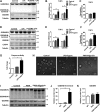
Activation of non-canonical pyroptosis promotes GSDMB expression in THP-1 cells. (AG) THP-1 cells were treated with LPS or transfected with LPS using FuGENE HD, and analyzed for protein levels of GSDMB, full-length GSDMD, and the GSDMD N-terminus in THP-1 cells after 8 h (A) or 16 h (D), expression profiles of GSDMB and GSDMD in THP-1 cells after 8 h (B) or 16 h (E), LDH release from THP-1 cells after 8 h (C) or 16 h (F), and the protein level of caspase-4 (D) and enzyme activity of caspase-4 (G) in THP-1 cells after 16 h of treatment. (H) The morphology of THP-1 cells in canonical and non-canonical pyroptosis. For non-canonical pyroptosis, images of THP-1 cells were taken after 16 h of transfection with LPS using FuGENE HD. For canonical pyroptosis, images of THP-1 cells were taken after PMA pretreatment for 24 h, followed by Nigericin treatment for 4 h. (IK) THP-1 cells were pre-treated with PMA and stimulated with Nigericin or without Nigericin. (I) Protein levels of GSDMB, full-length GSDMD, and GSDMD N-terminus in THP-1 cells were analyzed after 4 h of stimulation. The intensities of GSDMD N-terminus (J) and GSDMB protein (K) bands were quantified using the ImageJ software program and normalized to that of Tubulin. The data are presented as mean ± SD. *P < 0.05 between groups as indicated.
Figure 3
Figure 3
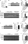
GSDMB expression is important for non-canonical pyroptosis in THP-1 cells. (AE) THP-1 cells were infected with lentiviruses (pLKO puro) driving the expression of shRNAs targeting GSDMB or control shRNAs and analyzed for GSDMB transcript levels (A) and GSDMB protein levels (B) in THP-1 cells upon GSDMB shRNA knockdown, as well as LDH released from GSDMB-knockdown THP-1 cells after 16 h of transfection with LPS (C). (D and E) The cleavage of GSDMD protein was measured in GSDMB-knockdown THP-1 cells after 16 h of transfection with LPS (D) and GSDMD N-terminus protein bands were quantitatively analyzed using the ImageJ software program (E). (FJ) THP-1 cells were infected with lentivirus (pTRIPZ puro) driving the expression of GSDMB or eGFP. (F and G) GSDMB protein levels were measured in THP-1 cells upon GSDMB overexpression and quantitatively analyzed. (H) LDH released from GSDMB-overexpressing THP-1 cells was measured after 16 h of transfection with LPS. (I and J) The cleavage of GSDMD protein was measured in GSDMB-overexpressing THP-1 cells after 16 h of transfection with LPS (I) and GSDMD N-terminus bands were quantitatively analyzed (J). The data are presented as mean ± SD. *P < 0.05 between groups as indicated.
Figure 4
Figure 4
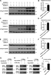
GSDMB enhances the enzyme activity of caspase-4 by binding to caspase-4. (A, C, E) Western blot analyses show the cleavage of GSDMD in 293T cells. (B, D, F) Graphs show GSDMD N-terminus levels normalized to GAPDH expression using the ImageJ software program. (A and B) 293T cells were co-transfected with plasmids encoding 3×FLAG-full-length GSDMD and either pcs2 (+) vector, pcs2 (+)-caspase-4, or pcs2 (+)-caspase-4 plus 3×FLAG-full-length GSDMB. (C and D) 293T cells were co-transfected with plasmids encoding 3×FLAG-full-length GSDMD and either pcs2 (+) vector, pcs2 (+)-caspase-11, or pcs2 (+)-caspase-11 plus 3×FLAG-full-length GSDMB. (E and F) 293T cells were co-transfected with plasmids encoding 3×FLAG-full-length GSDMD and either pcs2 (+) vector, pcs2 (+)-caspase-1, or pcs2 (+)-caspase-1 plus 3×FLAG-full-length GSDMB. (G) GSDMB did not interact with GSDMD. Co-IP analysis was performed in GSDMB-GFP- and FLAG-GSDMD-transfected 293T cells. (H and I) GSDMB interacted with caspase-4. Co-IP analysis was performed in caspase-4-Myc- and FLAG-GSDMB-transfected 293T cells. (J) The enzyme activity of caspase-4 in 293T cells. 293T cells were co-transfected with plasmids encoding pcs2 (+)-caspase-4 and either pcs2 (+) vector or pcs2 (+)-GSDMB. Graphs show mean ± SD of triplicate wells and represent three independent experiments. *P < 0.05 between groups as indicated.
Figure 5
Figure 5
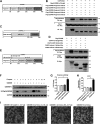
Full-length GSDMB promotes the enzyme activity of caspase-4 through the GSDMB N-terminus (1–83 aa) binding with the CARD domain of caspase-4. (A) Schematic of the GSDMB protein and the truncation fragments: fragment 1 (1–83 aa), fragment 2 (84–167 aa), fragment 3 (168–251 aa), fragment 4 (252–335 aa), and fragment 5 (336–417 aa). (B) 293T cells were transfected with the caspase-4-Myc expression plasmid and plasmids expressing the indicated FLAG-tagged GSDMB truncation form. Immunoprecipitation was performed using a monoclonal FLAG antibody, and samples were analyzed with a monoclonal Myc antibody via western blotting. (C) Graphic representation of the caspase-4 protein and the functional domains: CARD domain (1–80 aa), subunit 1 (81–270 aa), and subunit 2 (291–380 aa). (D) 293T cells were transfected with FLAG-GSDMB expression plasmid and plasmids expressing the indicated caspase-4-Myc truncation form. Immunoprecipitation was performed using a monoclonal Myc antibody, and the samples were analyzed with a monoclonal FLAG antibody via western blotting. (E) Graphic representation of GSDMB protein and the caspase cleavage site. (F) Assays of GSDMB cleavage by members of the human caspase family overexpressed in cells. 293T cells were co-transfected with plasmids encoding 3×FLAG-mouse GSDMB and the indicated pcs2 (+)-human caspase constructs. Total cell lysates were analyzed via anti-FLAG and anti-GAPDH immunoblotting. (G) The enzyme activity of caspase-4 in 293T cells. 293T cells were co-transfected with plasmids encoding pcs2 (+)-caspase-4 and either pcs2 (+) vector, pcs2 (+)-GSDMB N-terminus, pcs2 (+)-GSDMB C-terminus, or pcs2 (+)-full-length GSDMB. (H and I) 293T cells were transfected with full-length GSDMB (1–417 aa), GSDMB N-terminus (1–91 aa), GSDMB C-terminus (92–417 aa), and GSDMD N-terminus (1–275 aa) plasmids. (H) LDH released from 293T cells after 16 h of transfection. (I) The cell morphology of 293T cells after 16 h of transfection. The graphs show mean ± SD of triplicate wells and represent three independent experiments. *P < 0.05 between groups as indicated.
Figure 6
Figure 6
p-NF-κB positively regulates the expression of GSDMB and GSDMD in THP-1 cells. (AC) THP-1 cells were treated with LPS or transfected with LPS using FuGENE HD. (A) Protein levels of NF-κB and p65 in THP-1 cells after 8 h of treatment. NF-κB (B) and p65 (C) protein bands were quantified using the ImageJ software program and normalized to that of tubulin. (DH) NF-κB inhibitor (JSH-23 or QNZ) was used to pre-treat THP-1 cells at a final concentration of 10 μM for 20 h, and cells were further stimulated with LPS or transfected with LPS using FuGENE HD for 8 h. (D) Western blot analysis of NF-κB, p65, GSDMD, and GSDMB protein levels. Graphs show NF-κB (E), p65 (F), GSDMD (G), and GSDMB (H) protein levels normalized to Tubulin using the ImageJ software program. Graphs show mean ± SD of triplicate wells and represent three independent experiments. *P < 0.05 between groups as indicated.
Figure 7
Figure 7
GSDMB promotes pyroptosis through the caspase-4-mediated non-canonical pathway. Schematic diagram shows GSDMB in non-canonical pyroptosis by influencing GSDMD cleavage. Cytoplasmic LPS activates NF-κB to induce GSDMB expression in THP-1 cells. Then, GSDMB promotes the caspase-4 activity, which is required for the cleavage of GSDMD in non-canonical pyroptosis, by directly binding to the CARD domain of caspase-4. Additionally, the positive effect of GSDMB on caspase-4 in non-canonical pyroptosis can be terminated by a negative feedback mechanism, in which GSDMB is cleaved by caspase-4. Then, the release of the GSDMD N-terminus leads to the pore formation and cell membrane rupture, which in turn causes a massive secretion of inflammatory cytokines.

References

    1. Aglietti R.A., and Dueber E.C. (2017). Recent insights into the molecular mechanisms underlying pyroptosis and gasdermin family functions. Trends Immunol. 38, 261–271. - PubMed
    1. Baumann C.L., Aspalter I.M., Sharif O., et al. . (2010). CD14 is a coreceptor of Toll-like receptors 7 and 9. J. Exp. Med. 207, 2689–2701. - PMC - PubMed
    1. Broz P. (2015). Caspase target drives pyroptosis. Nature 526, 642–644. - PubMed
    1. Chao K.L., Kulakova L., and Herzberg O. (2017). Gene polymorphism linked to increased asthma and IBD risk alters gasdermin-B structure, a sulfatide and phosphoinositide binding protein. Proc. Natl Acad. Sci. USA 114, E1128–E1137. - PMC - PubMed
    1. Chen X., He W.-t., Hu L., et al. . (2016). Pyroptosis is driven by non-selective gasdermin-D pore and its morphology is different from MLKL channel-mediated necroptosis. Cell Res. 26, 1007. - PMC - PubMed

Publication types