. 2018 Dec 1;159(12):3950-3964.
doi: 10.1210/en.2018-00769.

The Adrenal Clock Prevents Aberrant Light-Induced Alterations in Circadian Glucocorticoid Rhythms

Affiliations

The Adrenal Clock Prevents Aberrant Light-Induced Alterations in Circadian Glucocorticoid Rhythms

William C Engeland et al. Endocrinology. .

Abstract

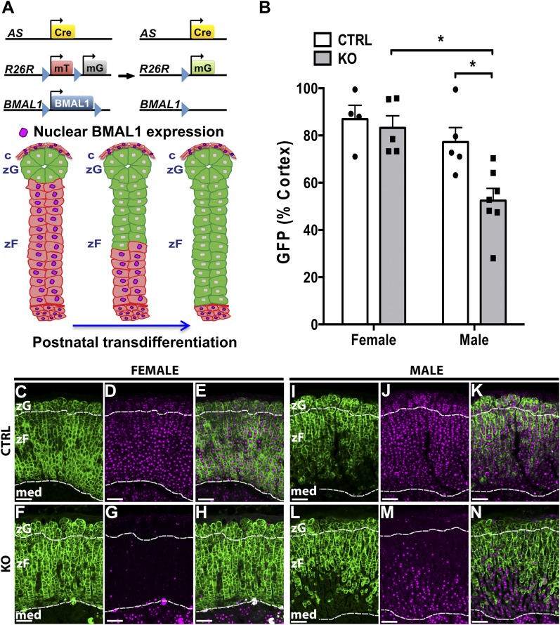
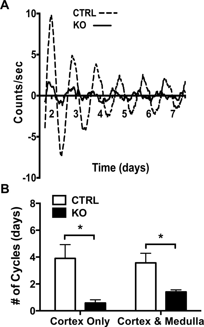
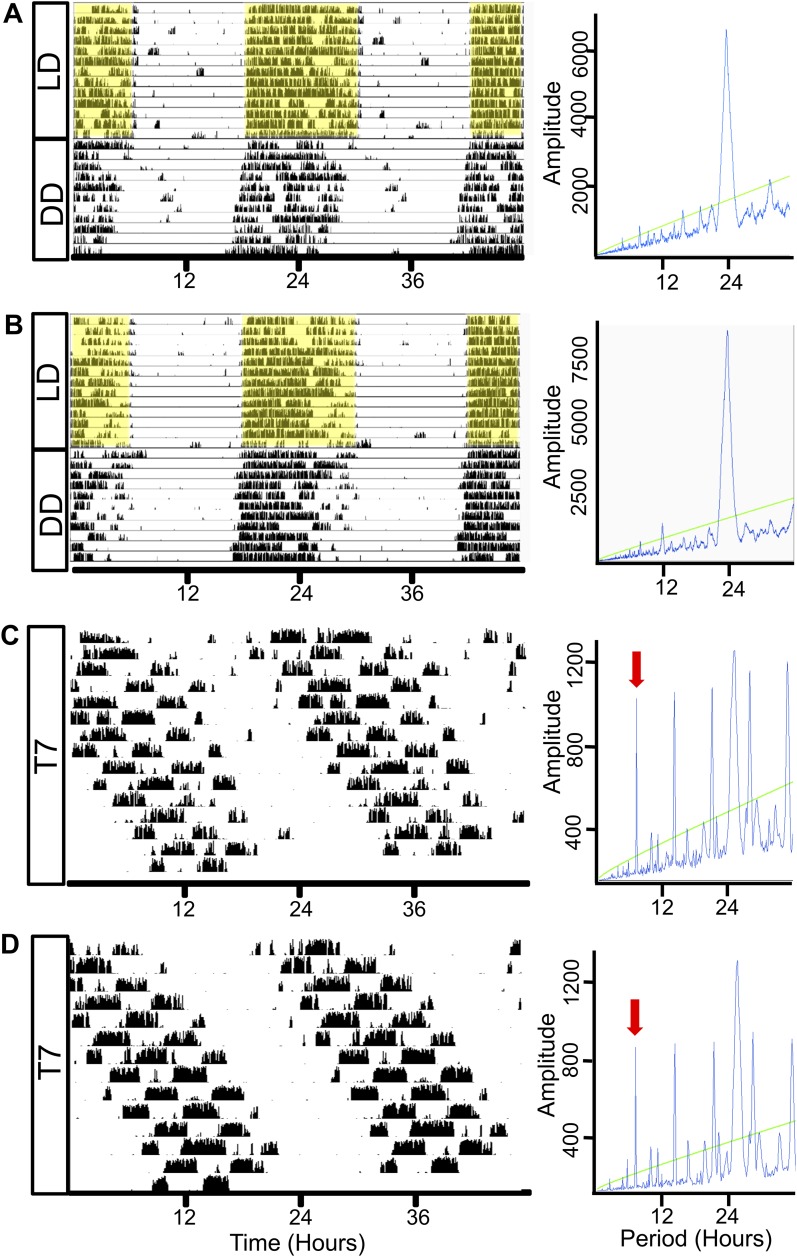
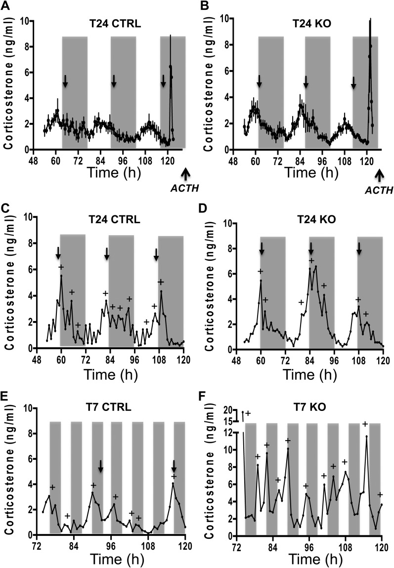
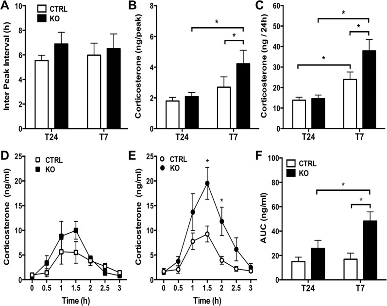
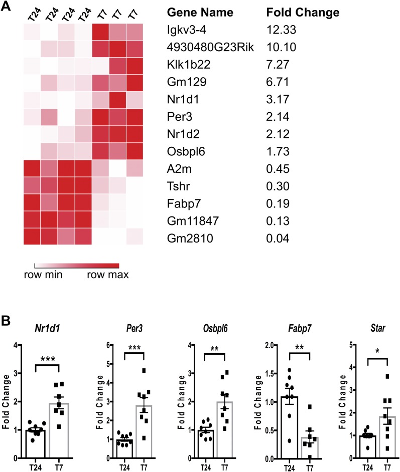
The glucocorticoid (GC) rhythm is entrained to light-dark (LD) cycles via a molecular clock in the suprachiasmatic nucleus (SCN) and is maintained by an adrenal clock synchronized by SCN-dependent signals. Targeted deletion of the core clock gene Bmal1 can disrupt adrenal clock function. The requirement of the adrenal clock to stabilize the circadian GC rhythm during exposure to aberrant LD cycles was determined using novel aldosterone synthase (AS)Cre/+::Bmal1Fl/Fl mice in which Bmal1 deletion occurred during postnatal adrenal transdifferentiation. To examine whether adrenal Bmal1 deletion results in loss of the adrenal clock, mice were crossed with mPER2::Luciferase (mPER2Luc/+) mice. Adrenals from ASCre/+::Bmal1+/+::PER2Luc/+ [control (CTRL)] mice show mPER2Luc rhythms ex vivo, whereas slices from ASCre/+::Bmal1Fl/Fl::PER2Luc/+ [knockout (KO)] mice show dampened rhythms. To monitor corticosterone rhythmicity, mice were implanted with subcutaneous microdialysis probes and sampled at 60-minute intervals for up to 3 days under 12:12-hour [τ (T) 24] LD or 3.5:3.5-hour (T7) LD cycles. Corticosterone rhythms were entrained to T24 LD in CTRL and KO mice. Under T7 LD, circadian corticosterone rhythms persisted in most CTRL mice but not KO mice. Hyperadrenocorticism also was observed in KO mice under T7 LD, reflected by increased corticosterone peak amplitude, total daily corticosterone, and responses to ACTH. Analysis of dysregulated adrenal genes in KO mice exposed to aberrant light identified candidates involved in cholesterol metabolism and trafficking, including steroidogenic acute regulatory protein, which could increase steroidogenesis. Our results show that the adrenal clock functions to buffer steroidogenic responses to aberrant light and stabilize circadian GC rhythmicity.


Figures

Figure 1.
Figure 1.
Analysis of postnatal transdifferentiation in male and female adrenal ASCre/+::Bmal1Fl/Fl KO mouse. (A) Adrenal clock KO (ASCre/+::Bmal1Fl/Fl::R26RmTom/mGFP/+) mice were generated by breeding ASCre/+ mice with the mTomato/mGFP Cre reporter and floxed Bmal1 mice. During postnatal transdifferentiation, zG cells migrated inward and differentiated into zF cells (19). Cre-dependent deletion of Bmal1 was predicted to occur in parallel with postnatal transdifferentiation reflected by GFP expression. (B) The extent of postnatal transdifferentiation (area of cortical GFP expression/total cortical area × 100) was not different between female ASCre/+::Bmal1+/+::R26RmTom/mGFP/+ CTRL (n = 4) and KO mice (n = 6) at 8 to 9 mo of age. However, male KO mice (n = 7) showed reduced postnatal transdifferentiation compared with female KO mice and male CTRL mice (n = 5). (C, F, I, L) In adrenals from female and male CTRL and KO mice, the zG was characterized by globular nests of cells underlying the capsule, and the adjacent zF consisted of parallel radial cords of cells. No clear differences in morphology were observed in KO mice. (C–E) In a female CTRL mouse (8.0 mo old), mGFP expression and nuclear BMAL1 labeling extended throughout the adrenal cortex from the zG through the zF; 95% of cortical area expressed mGFP. (F–H) In a female KO mouse (8.6 mo old), expression of mGFP was associated with loss of nuclear BMAL1 labeling and extended throughout the adrenal cortex; 94% of cortical area expressed mGFP. (I–K) In a male CTRL mouse (7.6 mo old), expression of GFP did not extend completely through the zF; 77% of cortical area expressed mGFP. (L–N) In a male KO mouse (8.6 mo old), mGFP expression and Bmal1 deletion did not extend completely throughout the adrenal cortex; only 56% of cortical area expressed mGFP. Magenta: BMAL1 immunofluorescence labeling. Borders between zG and zF and between cortex and medulla are denoted by dashed lines. Scale bar, 100 µm. med, medulla.
Figure 2.
Figure 2.
Adrenal Bmal1 deletion results in reduced adrenal mPER2Luc rhythms. (A) Adrenal slices from an ASCre/+::Bmal1+/+::PER2Luc CTRL mouse show mPER2Luc rhythms that persist for ∼7 d in vitro, whereas slices from an ASCre/+::Bmal1Fl/Fl::PER2Luc KO mouse show dampened rhythms. (B) Rhythms are diminished in slices of adrenal cortex only and cortex with medulla in KO mice, reflecting the loss of the endogenous cortical clock. Mean ± SEM, n = 5 to 7 mice, *P < 0.05.
Figure 3.
Figure 3.
Wheel-running activity under T24 LD, DD, and T7 LD in adrenal clock KO mice. Representative activity records from individual female (A) CTRL and (B) KO mice maintained under a T24 (12:12-h) LD cycle regimen for 10 to 14 d and then transferred to DD for 10 to 14 d. Both (A) CTRL (left panel) and (B) KO (left panel) mice demonstrate light entrainment in T24 LD as indicated by enhanced activity in the dark period. Shaded yellow regions indicate periods of darkness in T24 LD. Under DD, rhythms are free-running; periodograms (right panels) show a predominant circadian periodicity and measured periods (τ) are not different between CTRL (23.83 ± 0.02 h, n = 5) and adrenal clock KO mice (23.81 ± 0.05 h, n = 4). Activity records of (C) CTRL and (D) KO mice housed for 14 d in ultradian light cycles of T7 (3.5:3.5 h) LD. Both CTRL and KO mice are active during dark periods under T7 conditions; however, periodograms (right panels) show a lengthened circadian periodicity in CTRL (24.85 ± 0.09 h, n = 6) and KO (24.82 ± 0.15 h, n = 5) mice. Green line: cutoff for significant periodicity, P < 0.05. Red arrows: 7-h component of periodogram.
Figure 4.
Figure 4.
Circadian corticosterone rhythmicity under T24 LD and T7 LD in adrenal ASCre/+::Bmal1Fl/Fl KO mice. Seventy-two-hour profiles [mean ± SEM (n = 5 to 6)] of subcutaneous dialysate corticosterone in (A) CTRL and (B) KO mice and in individual female (C) CTRL and (D) KO mice under T24 LD. Peak corticosterone occurred daily at the onset of subjective night in CTRL and KO mice under T24 LD. Forty-eight-hour profiles of dialysate corticosterone in individual (E) CTRL and (F) KO mice under T7 LD. Circadian rhythmicity (defined by CircWave with peak phase denoted by black arrows) was observed in CTRL and KO mice under T24 LD and in CTRL mice under T7 LD; circadian rhythmicity was lost under T7 in most KO mice. Microdialysis samples were collected at 60-min intervals. Gray bars indicate the periods of darkness. Profiles in individual mice were analyzed to detect corticosterone peaks (denoted by +) using PULSAR.
Figure 5.
Figure 5.
Increased corticosterone responses under T7 LD in adrenal ASCre/+::Bmal1Fl/Fl KO mice. PULSAR analysis showed that increased (B) peak corticosterone amplitude, but not (A) peak frequency, occurs under T7 LD compared with T24 LD, resulting in increased (C) total daily corticosterone production. Both (B) peak amplitude and (C) total daily corticosterone increased in KO compared with CTRL mice under T7 LD. Mice were injected with ACTH (0.3 μg, subcutaneously) at the end of the sampling period. Increased corticosterone responsiveness to ACTH was observed in KO mice compared with CTRL mice on (E) T7 LD but not (D) T24 LD. (F) Total corticosterone in response to ACTH [area under the curve (AUC)] was increased in KO mice under T7LD compared with CTRL mice under T7LD and KO mice under T24 LD. Mean ± SEM, n = 5 to 6 mice, *P < 0.05.
Figure 6.
Figure 6.
Gene expression analysis of adrenals from female ASCre/Cre::Bmal1Fl/Fl KO mice under T24 LD and T7 LD. (A) RNA sequencing analysis revealed 13 genes significantly changed with a false discovery rate <0.05 and fold change >1.5 (upregulated) or <0.5 (downregulated) (n = 3 to 4). (B) Quantitative validation of genes relevant to the circadian clock and cholesterol metabolism using quantitative RT-PCR (n = 8). Mean ± SEM. *P < 0.05; **P < 0.01; ***P < 0.001.

References

    1. Mohawk JA, Green CB, Takahashi JS. Central and peripheral circadian clocks in mammals. Annu Rev Neurosci. 2012;35(1):445–462. - PMC - PubMed
    1. Menet JS, Rosbash M. When brain clocks lose track of time: cause or consequence of neuropsychiatric disorders. Curr Opin Neurobiol. 2011;21(6):849–857. - PMC - PubMed
    1. Zelinski EL, Deibel SH, McDonald RJ. The trouble with circadian clock dysfunction: multiple deleterious effects on the brain and body. Neurosci Biobehav Rev. 2014;40:80–101. - PubMed
    1. Reppert SM, Weaver DR. Coordination of circadian timing in mammals. Nature. 2002;418(6901):935–941. - PubMed
    1. Dibner C, Schibler U, Albrecht U. The mammalian circadian timing system: organization and coordination of central and peripheral clocks. Annu Rev Physiol. 2010;72(1):517–549. - PubMed

Publication types

MeSH terms