. 2018 Nov 30;46(21):11169-11183.
doi: 10.1093/nar/gky915.

Proteomic profiling and functional characterization of post-translational modifications of the fission yeast RNA exosome

Affiliations

Proteomic profiling and functional characterization of post-translational modifications of the fission yeast RNA exosome

Caroline Telekawa et al. Nucleic Acids Res. .

Abstract

The RNA exosome is a conserved multi-subunit complex essential for processing and degradation of several types of RNAs. Although many of the functions of the RNA exosome are well established, whether the activity of this complex is regulated remains unclear. Here we performed a proteomic analysis of the RNA exosome complex purified from Schizosaccharomyces pombe and identified 39 post-translational modifications (PTMs), including phosphorylation, methylation, and acetylation sites. Interestingly, most of the modifications were identified in Dis3, a catalytic subunit of the RNA exosome, as well as in the exosome-associated RNA helicase, Mtr4. Functional analysis of selected PTM sites using modification-deficient and -mimetic versions of exosome subunits revealed substitutions that affected cell growth and exosome functions. Notably, our results suggest that site-specific phosphorylation in the catalytic center of Dis3 and in the helical bundle domain of Mtr4 control their activity. Our findings support a view in which post-translational modifications fine-tune exosome activity and add a layer of regulation to RNA degradation.


Figures

Figure 1.
Figure 1.
Affinity purification of the S. pombe RNA exosome complex. (A) SYPRO RUBY-stained SDS-PAGE of affinity purified Dis3-TAP (lane 1) and Rrp4-TAP (lane 2). Subunits of the RNA exosome identified by mass spectrometry are indicated on the right. (B and C) Enrichment of RNA exosome subunits after affinity purification of Dis3-TAP (B) and Rrp4-TAP (C) as plotted by relative protein abundance (total peptide intensity) up the y-axis and percentage sequence coverage (amino acids) out the x-axis. Components of the RNA exosome complex are identified by green (B) and purple (C) squares. (D) MS determination of peptide number and sequence coverage for the indicated exosome subunits in Dis3-TAP and Rrp4-TAP purifications.
Figure 2.
Figure 2.
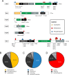
Repertoire of post-translational modifications (PTMs) identified on subunits of the fission yeast RNA exosome and Mtr4. (A) Schematic representation of the identified PTMs on the indicated exosome subunits and the Mtr4 helicase. Known domains in the indicated proteins: CR3, three cysteine residues motif; PIN, PilT N-terminal domain that catalyze endonuclease activity, CSD, cold-shock domains; RNB, RNase II catalytic domain that catalyses 3′-5′ exonuclease activity; S1, RNA-binding domain; PMC2NT, polycystin 2 N-terminal domain; EXO, 3′-5′ exonuclease domain; HRDC, helicase and RNase D C-terminal domain; ExoBD, exosome-binding domain; PH, ribonuclease PH-like domains; KH, K homology RNA-binding domain; PNPase, Polyribonucleotide nucleotidyltransferase domain; RecA, RecA-like domain. (B) Pie chart showing the distribution of PTMs detected in RNA exosome subunits and Mtr4 by MS. (C) Pie chart showing the distribution of the three types of phosphorylation sites identified in subunits of the RNA exosome and Mtr4. Phospho-peptides for which serine or threonine phosphorylation could not be distinguished are classified in light gray. (D) Pie chart showing the distribution of the two types of methylation sites identified in this study. Methylated-peptides for which arginine and lysine methylation could not be distinguished are classified in light gray.
Figure 3.
Figure 3.
Functional characterization of Dis3, Mtr4, and Rrp6 variants with substitutions at selected modified residues. (A–C) Doubling time calculated from growth curves of Pnmt-dis3 (A), Pnmt-mtr4 (B), and rrp6Δ (C) strains complemented with either the wild-type (WT) version of Dis3 (A), Mtr4 (B), and Rrp6 (C), or the indicated variants with substitutions at modified residues as determined by MS analysis. EV, empty vector control. In B, the 3KA version of Mtr4 corresponds to the multi-site substitution K910A-K911A-K912A. (D–F) RT-qPCR analysis of sme2 (meiRNA) expression using total RNA prepared from the strains described in panels A–C. RT-qPCR data were normalized to the housekeeping nda2 mRNA, and fold changes expressed relative to Pnmt-dis3 (D), Pnmt-mtr4 (E), and rrp6Δ (F) strains complemented with the wild-type version of Dis3, Mtr4, and Rrp6, respectively. Pnmt-dis3 and Pnmt-mtr4 strains were cultured in thiamine-supplemented medium to deplete endogenous Dis3 and Mtr4, respectively. The data and error bars represent the average and standard deviation from at least three independent experiments. P-values * ≤0.05, ** ≤0.01, *** ≤0.001, **** ≤0.0001; Student's t-test.
Figure 4.
Figure 4.
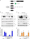
Phospho-mimetic versions of Dis3 and Mtr4 result in defective 5.8S rRNA synthesis. (A) Schematic of 7S pre-rRNA processing into mature 5.8S rRNA. Following endonucleolytic cleavage of the 27S rRNA precursor at site C2, the 7S pre-rRNA is trimmed 3′-5′ by the nuclear exosome assisted by the helicase activity of Mtr4 (green pacman). The resulting 6S intermediate is further processed by cytoplasmic ribonucleases (purple pacman) to generate the mature 5.8S rRNA. (B) Western blot (WB) and Northern blot (NB) analysis of wild-type (lane 1) and Pnmt-dis3 (lane 2–8) strains expressing the indicated phospho-deficient (lanes 5 and 7) and phospho-mimetic (lanes 6 and 8) versions of Dis3. EV, empty vector control. The position of the 7S pre-rRNA and 5.8S rRNA are indicated on the right. (C) Western blot (WB) and Northern blot (NB) analysis of wild-type (lane 1) and Pnmt-mtr4 (lane 2–6) strains expressing the indicated phospho-deficient (TASA; lane 5) and phospho-mimetic (TDSD; lane 6) versions of Mtr4. EV, empty vector control. The position of the 7S pre-rRNA and 5.8S rRNA are indicated on the right. The asterisk (*) shows the 5′-extended product specifically detected in the phospho-mimetic version of Mtr4. (B-C) The 5S rRNA and Tubulin were used as loading controls for northern and Western blots, respectively. (D and E) Quantification of 7S/5.8S ratios for the indicated versions of Dis3 (D) and Mtr4 (E). The calculated 7S/5.8S ratio was normalized to the 5S rRNA and expressed relative to Pnmt-dis3 (D) and Pnmt-mtr4 (E) strains complemented with the wild-type version of Dis3 and Mtr4, respectively. (B–E) Pnmt-dis3 and Pnmt-mtr4 strains were cultured in thiamine-supplemented medium to deplete endogenous Dis3 and Mtr4, respectively. The data and error bars represent the average and standard deviation from at least three independent experiments. P-values * ≤0.05, ** ≤0.01, *** ≤0.001; Student's t-test.
Figure 5.
Figure 5.
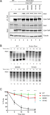
The presence of a negative charge that mimic the phosphorylated state of Ser-809 and Tyr-814 of Dis3 inhibits its catalytic activity. (A) Western blot analysis of total cell extracts (Input, bottom three panels) and affinity purifications (IP, top two panels) prepared from wild-type (lanes 1–2) and Pnmt-dis3 (lanes 3–8) strains that expressed either TAP-tagged (lanes 2–8) or untagged (lane 1) versions of Cls4 as well as the indicated versions of Dis3 (lanes 4–8). Pnmt-dis3 strains were cultured in thiamine-supplemented medium to deplete endogenous Dis3. (B) Analysis of RNA degradation kinetics of a 3′-phosphate AU-rich 49-nt RNA using affinity purified exosome complex prepared from extracts of Csl4-TAP strains that expressed wild-type (lanes 1–5), Endo/Exo-deficient (lanes 6–10), S809D (lanes 11–15), and Y814D (lanes 16–20) versions of Dis3. (C) Quantitative analysis of RNA decay assays as shown in panel B. The data and error bars represent the average and standard deviation from at least three independent experiments. ****P ≤ 0.0001, two-way ANOVA test.
Figure 6.
Figure 6.
Ser-809 and Tyr-814 of S. pombe Dis3 are evolutionarily conserved residues located near the exosome catalytic center. (A) Ribbon diagram representation of S. cerevisiae Dis3 (PDB: 4IFD) with a close-up view of the position of residues corresponding to Ser-809 (green) and Tyr-814 (magenta) in S. pombe Dis3. The position of the Asp-516 in S. pombe Dis3 (corresponding to Asp-551 in S. cerevisiae), which is critical for coordinating magnesium ions in the catalytic center of Dis3, is shown in blue. Single-stranded RNA is shown in orange. (B) Amino acid sequence alignment of Dis3 from fission and budding yeasts, Drosophila, mouse, and humans. Numbers on the right correspond to the position of the last amino acid shown for each sequence. Identical amino acids are shown in black outlined and similar amino acids are shown in gray outline. Ser-809 and Tyr-814 phosphorylation sites in S. pombe Dis3 are underlined.

Similar articles

Cited by

References

    1. Bentley D.L. Coupling mRNA processing with transcription in time and space. Nat. Rev. Genet. 2014; 15:163–175. - PMC - PubMed
    1. Bresson S., Tollervey D.. Surveillance-ready transcription: nuclear RNA decay as a default fate. Open Biol. 2018; 8:170270. - PMC - PubMed
    1. Andrulis E.D., Werner J., Nazarian A., Erdjument-Bromage H., Tempst P., Lis J.T.. The RNA processing exosome is linked to elongating RNA polymerase II in Drosophila. Nature. 2002; 420:837–841. - PubMed
    1. Chekanova J.A., Gregory B.D., Reverdatto S.V., Chen H., Kumar R., Hooker T., Yazaki J., Li P., Skiba N., Peng Q. et al. . Genome-wide high-resolution mapping of exosome substrates reveals hidden features in the Arabidopsis transcriptome. Cell. 2007; 131:1340–1353. - PubMed
    1. Lemay J.F., D’Amours A., Lemieux C., Lackner D.H., St-Sauver V.G., Bahler J., Bachand F.. The nuclear poly(A)-binding protein interacts with the exosome to promote synthesis of noncoding small nucleolar RNAs. Mol. Cell. 2010; 37:34–45. - PubMed

Publication types