Observational Study
. 2019 Apr;143(4):1380-1394.e9.
doi: 10.1016/j.jaci.2018.09.029. Epub 2018 Oct 13.

Mucosal bromodomain-containing protein 4 mediates aeroallergen-induced inflammation and remodeling

Affiliations
Observational Study

Mucosal bromodomain-containing protein 4 mediates aeroallergen-induced inflammation and remodeling

Bing Tian et al. J Allergy Clin Immunol. 2019 Apr.

Abstract

Background: Frequent exacerbations of allergic asthma lead to airway remodeling and a decrease in pulmonary function, producing morbidity. Cat dander is an aeroallergen associated with asthma risk.

Objective: We sought to elucidate the mechanism of cat dander-induced inflammation-remodeling.

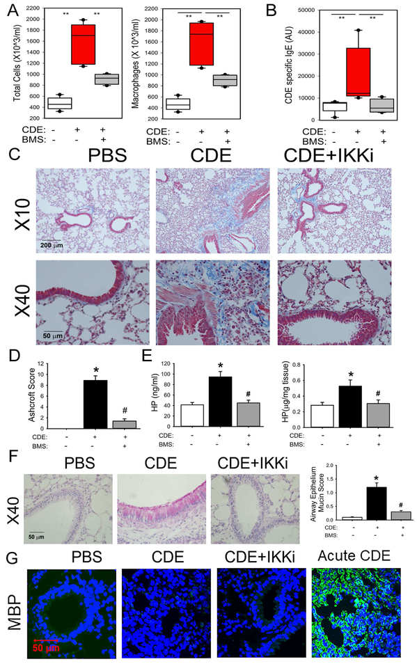
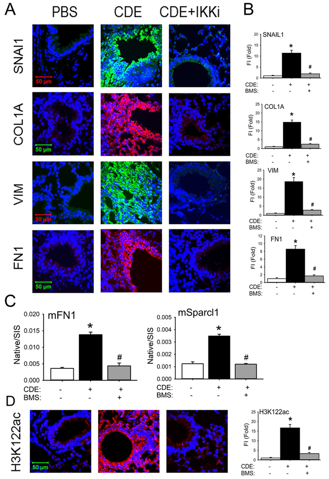
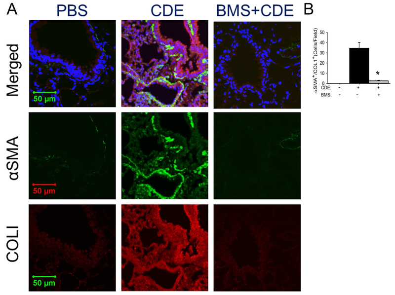
Methods: We identified remodeling in mucosal samples from allergic asthma by using quantitative RT-PCR. We developed a model of aeroallergen-induced experimental asthma using repetitive cat dander extract exposure. We measured airway inflammation using immunofluorescence, leukocyte recruitment, and quantitative RT-PCR. Airway remodeling was measured by using histology, collagen content, myofibroblast numbers, and selected reaction monitoring. Inducible nuclear factor κB (NF-κB)-BRD4 interaction was measured by using a proximity ligation assay in situ.

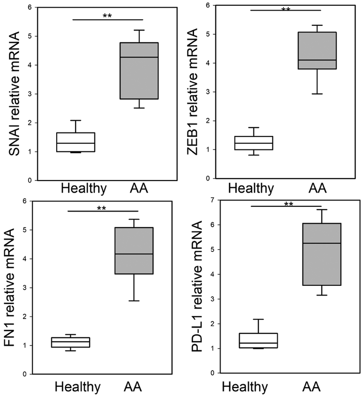
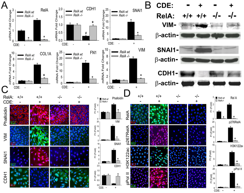
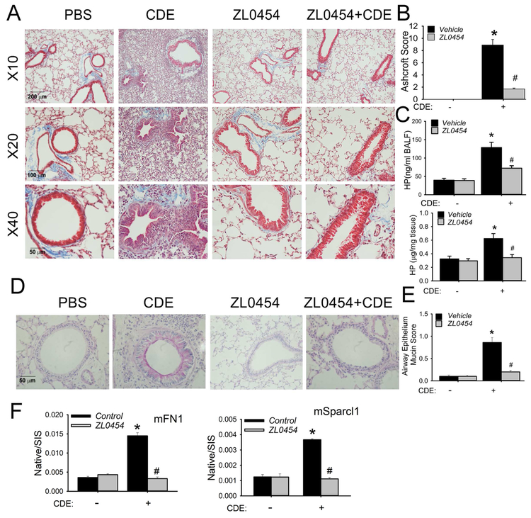
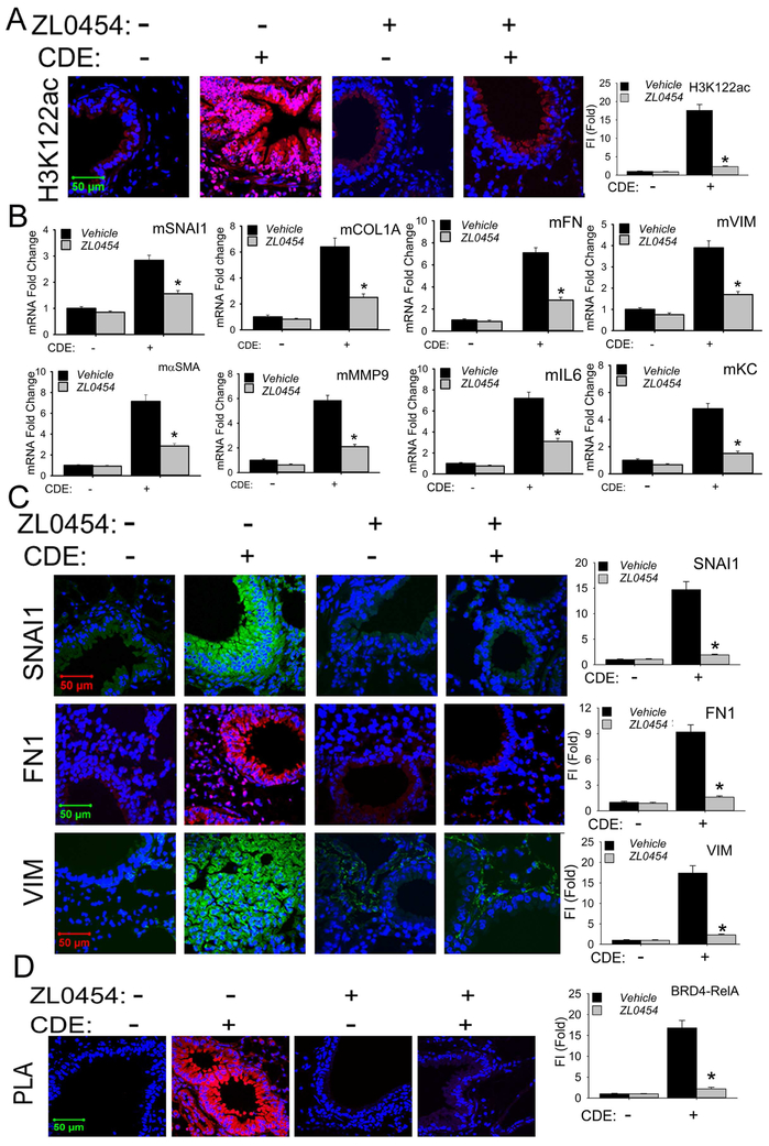
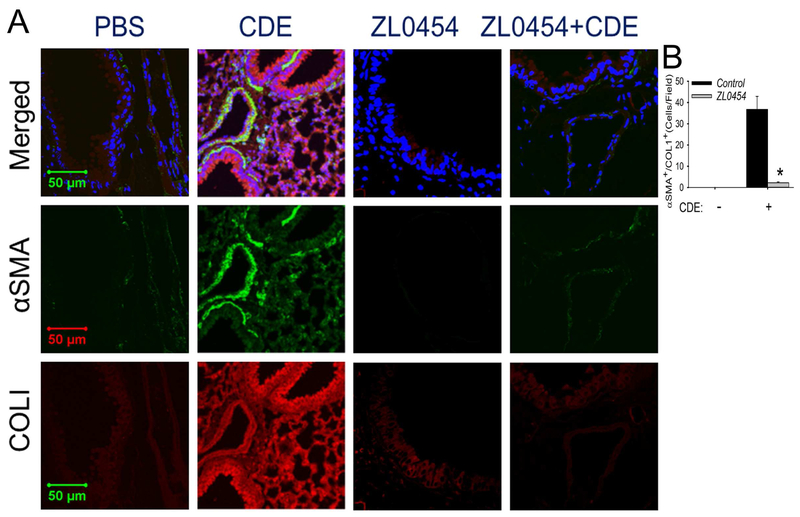
Results: Enhanced mesenchymal signatures are observed in bronchial biopsy specimens from patients with allergic asthma. Cat dander induces innate inflammation through NF-κB signaling, followed by production of a profibrogenic mesenchymal transition in primary human small airway epithelial cells. The IκB kinase-NF-κB signaling pathway is required for mucosal inflammation-coupled airway remodeling and myofibroblast expansion in the mouse model of aeroallergen exposure. Cat dander induces NF-κB/RelA to complex with and activate BRD4, resulting in modifying the chromatin environment of inflammatory and fibrogenic genes through its atypical histone acetyltransferase activity. A novel small-molecule BRD4 inhibitor (ZL0454) disrupts BRD4 binding to the NF-κB-RNA polymerase II complex and inhibits its histone acetyltransferase activity. ZL0454 prevents epithelial mesenchymal transition, myofibroblast expansion, IgE sensitization, and fibrosis in airways of naive mice exposed to cat dander.

Conclusions: NF-κB-inducible BRD4 activity mediates cat dander-induced inflammation and remodeling. Therapeutic modulation of the NF-κB-BRD4 pathway affects allergen-induced inflammation, epithelial cell-state changes, extracellular matrix production, and expansion of the subepithelial myofibroblast population.

Keywords: Bromodomain-containing protein 4; aeroallergen; airway remodeling; epigenetics; histone acetyltransferase activity; myofibroblast; nuclear factor κB.


Conflict of interest statement

Disclosure of potential conflict of interests: Drs. Brasier, Zhou, and Tian have disclosed intellectual property on the BRD4 inhibitors, but receive no license or royalty support.

Figures

Fig 1.
Fig 1.. EMT signature in mucosal samples in human severe asthma.
Q-RT-PCR for bronchial mucosal biopsies from normal or mild-moderate asthma for EMT regulators SNAI1, ZEB1, and FN1, or myofibroblast activation marker, PD-L1 mRNA expression. Shown as fold-change mRNA abundance normalized to PPIA (cyclophilin A). **, p<0.01, compared to healthy human samples (n=5 normals; 7 AAs), t-test.
Fig 2.
Fig 2.. NFκB/RelA mediates CDE-induced mesenchymal transition of airway epithelial cells.
A, RelA shRNA-expressing hSAECs treated with/without 2 μg/ml doxycycline (Dox), 5d. Afterwards, cells were treated with CDE (20 μg/mL) for 0 or 15 d prior to analysis by Q-RT-PCR. Fold changes in indicated mRNAs are shown. *p< 0.01 compared to control shRNA, #, p<0.01 compared to without CDE, n=3, t-test. B, Western immunoblots. Whole cell extracts from the same experiment were fractionated by SDS-PAGE and subjected to western immunoblot with indicated Abs, β-actin was probed as loading controls. Similar results were found in experiments repeated three times independently. C, Immunofluorescent confocal microscopy (IFCM) assays of WT and RelA-shRNA hSAECs. Cells were stained with either Alexa Fluor 568-conjugated phalloidin (upper panel, red color), or primary antibodies to VIM, SNAI1, and CDH1 Abs followed by secondary detection using Alexa 488-(green, for VIM and CDH1) or 568-(red, SNAI1) conjugated goat anti-rabbit IgG. Nuclei were counterstained with DAPI (blue). Images were acquired at 63X magnification. Right, quantifications of fluorescence intensities shown as fold changes compared to control hSAECs. * p<0.01, n = 5, t-test. FI: relative total fluorescence intensity. D, IFCM assays of total RelA, phospho-Ser276 RelA, H3K122 Ac, and phospho-Ser 2 CTD Pol II (pPol II). Secondary detection was Alexa 488-(green color, for RelA and H3K122ac) or 568-(red color, for p276 RelA and pPol II) conjugated goat anti-rabbit IgG. At the right are quantifications (X ± SD) of total fluorescence intensities, * p<0.01, n = 5, t-test.
Fig 3.
Fig 3.. Repetitive CDE (rCDE) exposure induces airway remodeling in mice.
C57BL6/J mice were pretreated with and without IKK inhibitor BMS345541 and given repetitive intranasal challenges of CDE. A, Total cells and macrophages count in the bronchoalveolar lavage fluid (BALF), expressed as total number of cells X 103/ml (left) and macrophages X 103/ml (right). **, p<0.01, n=5 mice per group. B, CDE specific serum IgE levels were quantitated. **, p<0.01, n=5, t-test. C, Masson Trichrome staining of lung sections from mice in the absence (left panel) or presence of CDE (middle), or those treated with rCDE and IKK inhibitor BMS345541 (right panel). The images were taken at magnifications of 10X and 40X respectively. D, Modified Ashcroft scoring for treatment groups. *, p<0.05, compared to without CDE; #, p<0.05, compared to CDE alone, t-test. E, Quantification of hydroxyproline. Left, hydroxyproline levels in BALF. Right, hydroxyproline content in total lung tissue. *, p<0.05, compared to without CDE; #, p<0.05 compared to CDE alone, t-test. F, PAS staining (pink) by treatment groups. At right is quantification of accumulated mucin in airway epithelial cells. *, p<0.05, compared to without CDE; #, p<0.05 compared to CDE alone, n=5 mice per group. G, IFCM for eosinophil Major Basic Protein (MBP, green color) with DAPI counterstain.
Fig 4.
Fig 4.. rCDE induces mucosal EMT in an IKK-NFκB-dependent manner.
A, IFCM of lung sections from control (PBS), CDE, or BMS+CDE treated mice. Sections were stained for the EMT markers SNAI1, VIM, COL1A, and FN1, Images are acquired at 63X magnification. B, quantitation of relative changes in fluorescence intensity for each treatment group. *, p<0.05, compared to without CDE; #, p<0.05 compared to CDE alone, n=5, t-test. C, Stable isotope dilution-Selected Reaction Monitoring (SID-SRM) of extracellular matrix proteins, mFN1 and mSparcl1 in BALF. Shown are mean ± SD of native to stable isotope standards (SIS) for n=5 animals in two technical replicates. *, p<0.05 compared to control; #, p<0.01 compared to CDE treatment only, t-test. D. IFCM of the BRD4 activation marker H3K122-Ac. Left panel, quantitation of relative changes in fluorescence intensity of H3K122-actin. *, p<0.05 compared to control; #, p<0.01 compared to CDE treatment only, t-test.
Fig 5.
Fig 5.. rCDE induces subepithelial myofibroblast expansion.
Confocal immunofluorescence microscopy of lung tissues in control (PBS), CDE, or BMS+CDE treated mice stained with rabbit anti-alpha smooth muscle actin (αSMA, green color) and mouse anti-COL1 (red color) and counterstained with DAPI (blue color). Merged images shown as top, 63X. Experiments were independently repeated twice with 5 animals in each treated group. Total 10 fields of each treatment were examined by 2 investigators who were blind to the treatment groups (n=10, *, p<0.01, t-test).
Fig 6.
Fig 6.. BRD4 inhibitor blocks rCDE-induced airway remodeling.
C57BL/6 mice were subjected to 15 treatments with PBS (in), rCDE (20 μg/dose, in), ZL0454 (10 mg/kg body weight, ip) or rCDE + ZL0454 for total 30 d and lungs were harvested 12 d after the last CDE challenge. A, Masson-Trichrome staining taken at 10X, 20X, and 40X magnification. B, Modified Ashcroft Score by treatment group. *, p<0.05, compared to without CDE; #, p<0.05, compared to CDE alone, t-test. C, Upper, hydroxyproline level in BALF. Lower, hydroxyproline content in lung tissue. *, P<0.05, compared to without CDE; #, p<0.05 compared to CDE alone. D, PAS staining (pink) showing mucin production. E, Quantification of cellular mucin. *, p<0.05, compared to without CDE; #, p<0.05 compared to CDE alone, n=5. F, SID-SRM of mFN1 and mSparcl1 in BALF. Shown are mean ± SD of native to stable isotope standards (SIS) for n=5 animals in two technical replicates. *, p<0.05 compared to control; #, p<0.01 compared to CDE treatment only, t-test.
Fig 7.
Fig 7.. BRD4 inhibitor blocks mucosal mesenchymal transition in vivo.
A, Confocal immunofluorescence microscopy of H3K122 Ac (red color) in mouse lungs treated with PBS, rCDE, ZL0454 or rCDE + ZL0454 respectively. Lung sections were counterstained in DAPI (blue color). X63 magnification. At the right is quantifications of relative fluorescence intensity, * p<0.01, n = 5. B, Q-RT-PCR for mRNA expression of mesenchymal and ECM genes from total RNA of mouse lungs treated with PBS, rCDE, ZL0454 or rCDE + ZL0454. * p<0.01, n = 5, t-test. C, Confocal immunofluoresence microscopy for SNAI1 (green color), FN1 (red color), and VIM (green color) in mouse lungs treated with PBS, rCDE, ZL0454 or rCDE + ZL0454. Lung sections were counterstained in DAPI (blue color). X63 magnification. At right are quantitation of relative fluorescence intensities of SNAI, FN1, and VIM. *, p<0.01, compared to CDE alone, n=5. D, PLA assay of RelA-BRD4 molecular interactions in lung sections from PBS, rCDE, ZL0454 or rCDE + ZL0454- treated mice. Foci of interactions are amplified as red foci; sections are counterstained with DAPI (blue color). X63 magnification. At the right is quantification of PLA assay. *, p<0.01, compared to CDE alone, n=5, t-test.
Fig 8.
Fig 8.. BRD4 mediates allergen-induced myofibroblast transition.
Confocal immunofluorescence microscopy of lung sections in PBS, rCDE, ZL0454 or rCDE + ZL0454- treated mice stained with both primary antibodies of rabbit anti-alpha smooth muscle actin (αSMA, green color) and mouse anti-COL1 Abs (red color) and counterstained with DAPI (blue color). Merged images shown at top. X 63 magnification. Experiments were independently repeated twice with 5 animals in each treated group. Total 10 fields of each treatment were examined by 2 investigators who were blind to the treatment groups (n=10, *, p<0.01, t-test).

References

    1. Broide DH. Immunologic and inflammatory mechanisms that drive asthma progression to remodeling. J Allergy Clin Immunol 2008; 121:560–70; quiz 71-2. - PMC - PubMed
    1. Pawankar R Allergic diseases and asthma: a global public health concern and a call to action. World Allergy Organ J 2014; 7:12. - PMC - PubMed
    1. Dahl R, Andersen PS, Chivato T, Valovirta E, de Monchy J. National prevalence of respiratory allergic disorders. Respir Med 2004; 98:398–403. - PubMed
    1. Prakash Ys, Halayko AJ, Gosens R, Panettieri RA Jr., Camoretti-Mercado B, Penn RB, et al. An Official American Thoracic Society Research Statement: Current Challenges Facing Research and Therapeutic Advances in Airway Remodeling. Am J Respir Crit Care Med 2017; 195:e4–e19. - PubMed
    1. Hosoki K, Aguilera-Aguirre L, Brasier AR, Kurosky A, Boldogh I, Sur S. Facilitation of Allergic Sensitization and Allergic Airway Inflammation by Pollen-Induced Innate Neutrophil Recruitment. Am J Respir Cell Mol Biol 2016; 54:81–90. - PMC - PubMed

Publication types

MeSH terms