

Complete Genome Sequence of the Model Halovirus PhiH1 (ΦH1)
- PMID: 30322017
- PMCID: PMC6210493
- DOI: 10.3390/genes9100493
Complete Genome Sequence of the Model Halovirus PhiH1 (ΦH1)
Abstract
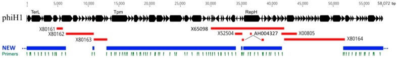
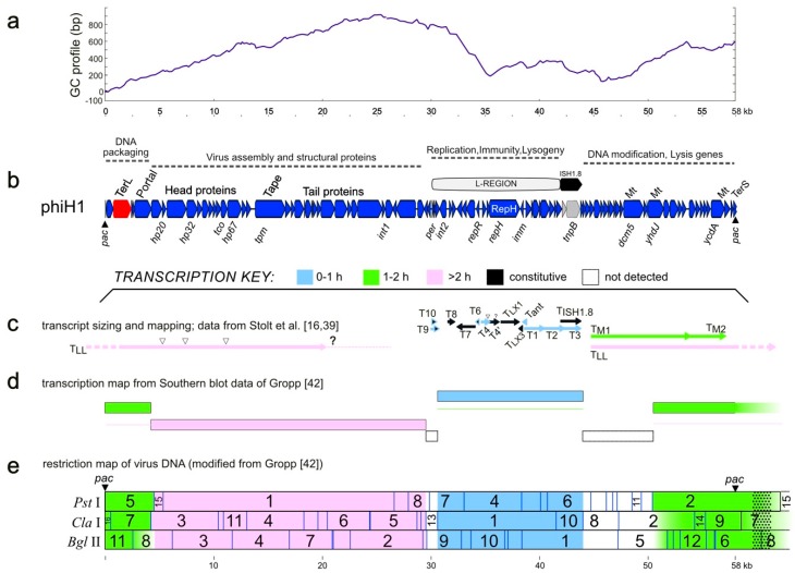
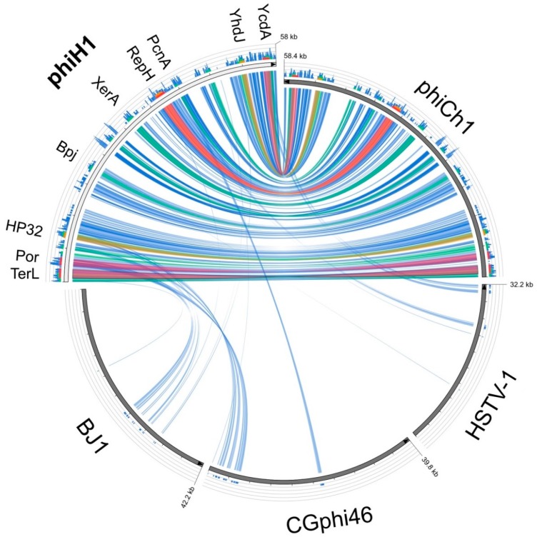
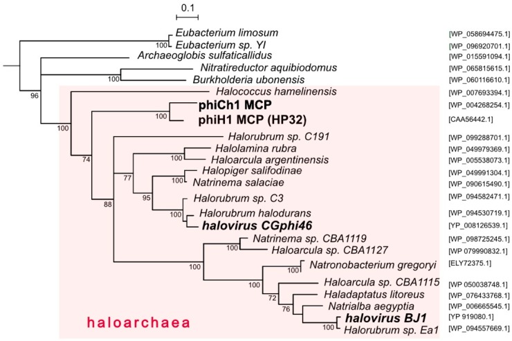
The halophilic myohalovirus Halobacterium virus phiH (ΦH) was first described in 1982 and was isolated from a spontaneously lysed culture of Halobacterium salinarum strain R1. Until 1994, it was used extensively as a model to study the molecular genetics of haloarchaea, but only parts of the viral genome were sequenced during this period. Using Sanger sequencing combined with high-coverage Illumina sequencing, the full genome sequence of the major variant (phiH1) of this halovirus has been determined. The dsDNA genome is 58,072 bp in length and carries 97 protein-coding genes. We have integrated this information with the previously described transcription mapping data. PhiH could be classified into Myoviridae Type1, Cluster 4 based on capsid assembly and structural proteins (VIRFAM). The closest relative was Natrialba virus phiCh1 (φCh1), which shared 63% nucleotide identity and displayed a high level of gene synteny. This close relationship was supported by phylogenetic tree reconstructions. The complete sequence of this historically important virus will allow its inclusion in studies of comparative genomics and virus diversity.
Keywords: Archaea; Halobacterium salinarum; genome inversion; haloarchaea; halobacteria; halophage; halovirus; virus.
Conflict of interest statement
The authors declare no conflict of interest.
Figures
Similar articles
-
Whole-genome comparison between the type strain of Halobacterium salinarum (DSM 3754T ) and the laboratory strains R1 and NRC-1.Microbiologyopen. 2020 Feb;9(2):e974. doi: 10.1002/mbo3.974. Epub 2019 Dec 3. Microbiologyopen. 2020. PMID: 31797576 Free PMC article.
-
The Novel Halovirus Hardycor1, and the Presence of Active (Induced) Proviruses in Four Haloarchaea.Genes (Basel). 2021 Jan 23;12(2):149. doi: 10.3390/genes12020149. Genes (Basel). 2021. PMID: 33498646 Free PMC article.
-
Halobacterium salinarum virus ChaoS9, a Novel Halovirus Related to PhiH1 and PhiCh1.Genes (Basel). 2019 Mar 1;10(3):194. doi: 10.3390/genes10030194. Genes (Basel). 2019. PMID: 30832293 Free PMC article.
-
The structural protein E of the archaeal virus phiCh1: evidence for processing in Natrialba magadii during virus maturation.Virology. 2000 Oct 25;276(2):376-87. doi: 10.1006/viro.2000.0565. Virology. 2000. PMID: 11040128
-
Natrialba magadii virus phiCh1: first complete nucleotide sequence and functional organization of a virus infecting a haloalkaliphilic archaeon.Mol Microbiol. 2002 Aug;45(3):851-63. doi: 10.1046/j.1365-2958.2002.03064.x. Mol Microbiol. 2002. PMID: 12139629
Cited by
-
Whole-genome comparison between the type strain of Halobacterium salinarum (DSM 3754T ) and the laboratory strains R1 and NRC-1.Microbiologyopen. 2020 Feb;9(2):e974. doi: 10.1002/mbo3.974. Epub 2019 Dec 3. Microbiologyopen. 2020. PMID: 31797576 Free PMC article.
-
Revisiting evolutionary trajectories and the organization of the Pleolipoviridae family.PLoS Genet. 2023 Oct 13;19(10):e1010998. doi: 10.1371/journal.pgen.1010998. eCollection 2023 Oct. PLoS Genet. 2023. PMID: 37831715 Free PMC article.
-
Isolation of a virus causing a chronic infection in the archaeal model organism Haloferax volcanii reveals antiviral activities of a provirus.Proc Natl Acad Sci U S A. 2022 Aug 30;119(35):e2205037119. doi: 10.1073/pnas.2205037119. Epub 2022 Aug 22. Proc Natl Acad Sci U S A. 2022. PMID: 35994644 Free PMC article.
-
Halovirus HF2 Intergenic Repeat Sequences Carry Promoters.Viruses. 2021 Nov 29;13(12):2388. doi: 10.3390/v13122388. Viruses. 2021. PMID: 34960657 Free PMC article.
-
The Novel Halovirus Hardycor1, and the Presence of Active (Induced) Proviruses in Four Haloarchaea.Genes (Basel). 2021 Jan 23;12(2):149. doi: 10.3390/genes12020149. Genes (Basel). 2021. PMID: 33498646 Free PMC article.
References
-
- Zillig W., Gropp F., Henschen A., Neumann H., Palm P., Reiter W.D., Rettenberger M., Schnabel H., Yeats S. Archaebacteria virus host systems. Syst. Appl. Microbiol. 1986;7:58–66. doi: 10.1016/S0723-2020(86)80124-2. - DOI
-
- Zillig W., Reiter W.-D., Palm P., Gropp F., Neumann H., Rettenberger M. Viruses of Archaebacteria. In: Calendar R., editor. The Bacteriophages. Plenum Publishing Corpn; New York, NY, USA: 1988.
-
- Schnabel H., Zillig W. Circular structure of the genome of phage ΦH in a lysogenic Halobacterium halobium. Mol. Gen. Genet. 1984;193:422–426. doi: 10.1007/BF00382078. - DOI
-
- Schnabel H., Schramm E., Schnabel R., Zillig W. Structural variability in the genome of phage ΦH of Halobacterium halobium. Mol. Gen. Genet. 1982;188:370–377. doi: 10.1007/BF00330036. - DOI
Grants and funding
LinkOut - more resources
Full Text Sources
