. 2018 Oct 12;8(10):824.
doi: 10.3390/nano8100824.

Copper Oxide Nanoparticles Cause a Dose-Dependent Toxicity via Inducing Reactive Oxygen Species in Drosophila

Affiliations

Copper Oxide Nanoparticles Cause a Dose-Dependent Toxicity via Inducing Reactive Oxygen Species in Drosophila

Eugene Baeg et al. Nanomaterials (Basel). .

Abstract

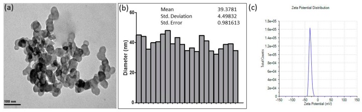
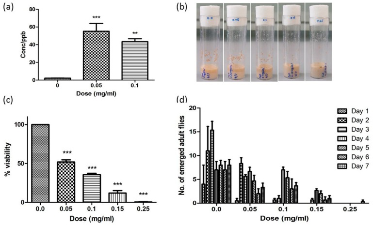
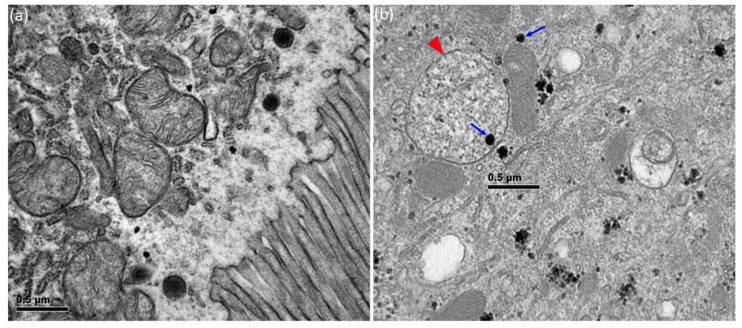
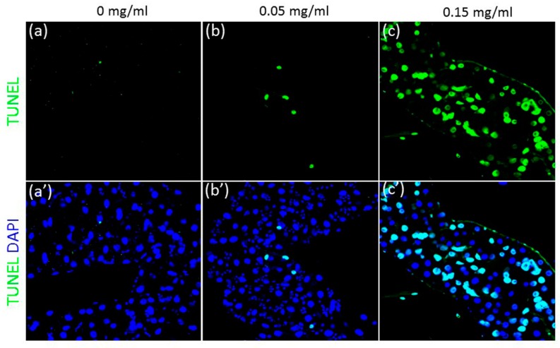
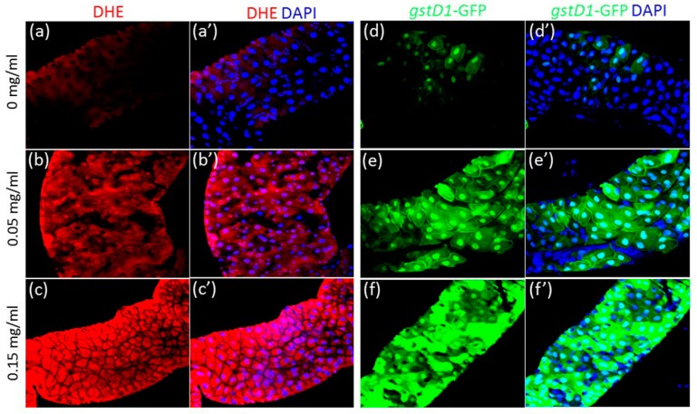
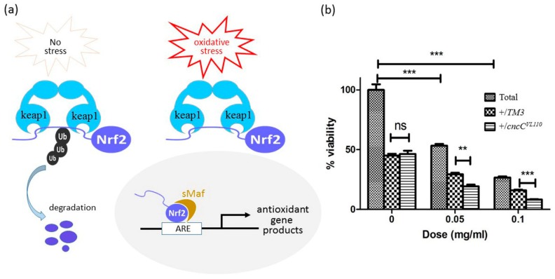
Copper oxide nanoparticles (CuONPs) have attracted considerable attention, because of their biocide potential and capability for optical imaging, however CuONPs were shown to be highly toxic in various experimental model systems. In this study, mechanism underlying CuONP-induced toxicity was investigated using Drosophila as an in vivo model. Upon oral route of administration, CuONPs accumulated in the body, and caused a dose-dependent decrease in egg-to-adult survivorship and a delay in development. In particular, transmission electron microscopy analysis revealed CuONPs were detected inside the intestinal epithelial cells and lumen. A drastic increase in apoptosis and reactive oxygen species was also observed in the gut exposed to CuONPs. Importantly, we found that inhibition of the transcription factor Nrf2 further enhances the toxicity caused by CuONPs. These observations suggest that CuONPs disrupt the gut homeostasis and that oxidative stress serves as one of the primary causes of CuONP-induced toxicity in Drosophila.

Keywords: Drosophila melanogaster; Nrf2; copper oxide nanoparticle; cytotoxicity; reactive oxygen species.


Conflict of interest statement

The authors declare no conflict of interest.

Figures

Figure 1
Figure 1
Characterization of Copper oxide nanoparticles (CuONPs). (a) Transmission electron microscopy (TEM) micrographs show spherical CuONPs with the size of approximately 40 nm; (b) The size distribution of CuONPs was determined by ImageJ software; (c) The zeta potential of CuONPs is −28.1 mV.
Figure 2
Figure 2
CuONP ingestion induces toxic effects in Drosophila. (a) CuONP-fed F1 progenies show an uptake and accumulation of CuONPs in the body. (b,c) A dose-dependent decline in the number of pupa and in egg-to-adult survivorship is observed upon CuONPs. (d) CuONP exposure causes a dose-dependent delay in development. Error bar = SEM, * p-value < 0.05; ** p-value < 0.01; *** p-value < 0.001.
Figure 3
Figure 3
Accumulation of CuONPs in the intestinal epithelial cells. (a) CuONPs with the size of 40–50 nm are not detected in the control intestinal epithelial cells; (b) CuONPs (arrows) are detected inside the cytoplasm of CuONP-exposed epithelia cells. Note that some of CuONPs are observed inside the vesicle (arrowhead).
Figure 4
Figure 4
CuONP exposure causes apoptosis of the gut epithelia cells. Flies were exposed to CuONPs at the concentration of (a,a’) 0 mg/mL, (b,b’) 0.05 mg/mL and (c,c’) 0.15 mg/mL. The third instar larval guts show a dose-dependent increase in the number of terminal deoxynucleotidyl transferase dUTP nick end labelling (TUNEL)-positive epithelial cells (green).
Figure 5
Figure 5
CuONPs induce excessive reactive oxygen species (ROS) production in the gut. Flies were exposed to CuONPs at the concentration of (a,a’,d,d’) 0 mg/mL, (b,b’,e,e’) 0.05 mg/mL and (c,c’,f,f’) 0.15 mg/mL. (a,a’) Dihydroethidium (DHE) staining that detects superoxide levels shows the basal levels of ROS in the control gut epithelial cells. (b,b’,c,c’) ROS levels become gradually increased upon CuONPs in a dose-dependent manner. Transgenic flies carrying the oxidative stress reporter gene GstD1-GFP were used to monitor the effects of CuONPs on intracellular ROS induction. (d,d’) The control gut shows weak GFP expression. (e,e’,f,f’) GFP expression is dose-dependently increased upon CuONPs.
Figure 6
Figure 6
ROS levels modulate the toxic effects caused by CuONPs. (a) Schematic diagram suggesting the role of Nrf2 in the regulation of antioxidant gene expression; (b) Introducing one mutant allele of CncC (the Drosophila homolog of Nrf2) further decreased the poor survivorship caused by CuONP exposure. Error bar = SEM, ** p-value < 0.01; *** p-value < 0.001.

References

    1. Pathakoti K., Manubolu M., Hwang H.-M. Chapter 48-Nanotechnology applications for environmental industry. In: Hussain C.M., editor. Handbook of Nanomaterials for Industrial Applications. Elsevier; Cambridge, MA, USA: 2018. pp. 894–907.
    1. Zhou M., Tian M., Li C. Copper-Based Nanomaterials for Cancer Imaging and Therapy. Bioconjugate Chem. 2016;27:1188–1199. doi: 10.1021/acs.bioconjchem.6b00156. - DOI - PubMed
    1. Hannig M., Kriener L., Hoth-Hannig W., Becker-Willinger C., Schmidt H. Influence of nanocomposite surface coating on biofilm formation in situ. J. Nanosci. Nanotechnol. 2007;7:4642–4648. - PubMed
    1. Monteiro D.R., Gorup L.F., Takamiya A.S., Ruvollo-Filho A.C., de Camargo E.R., Barbosa D.B. The growing importance of materials that prevent microbial adhesion: Antimicrobial effect of medical devices containing silver. Int. J. Antimicrob. Agents. 2009;34:103–110. doi: 10.1016/j.ijantimicag.2009.01.017. - DOI - PubMed
    1. Nations S., Long M., Wages M., Maul J.D., Theodorakis C.W., Cobb G.P. Subchronic and chronic developmental effects of copper oxide (CuO) nanoparticles on Xenopus laevis. Chemosphere. 2015;135:166–174. doi: 10.1016/j.chemosphere.2015.03.078. - DOI - PubMed

LinkOut - more resources