. 2018 Oct 13;7(10):142.
doi: 10.3390/antiox7100142.

Mitochondrial Arabidopsis thaliana TRXo Isoforms Bind an Iron⁻Sulfur Cluster and Reduce NFU Proteins In Vitro

Affiliations

Mitochondrial Arabidopsis thaliana TRXo Isoforms Bind an Iron⁻Sulfur Cluster and Reduce NFU Proteins In Vitro

Flavien Zannini et al. Antioxidants (Basel). .

Abstract

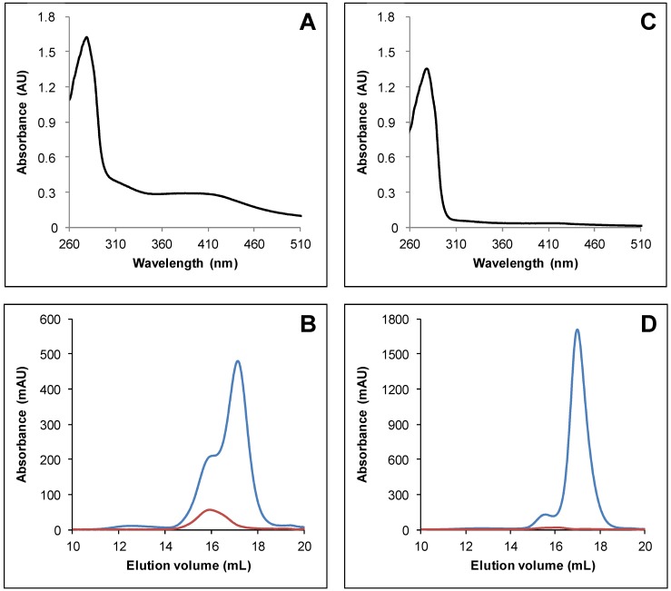
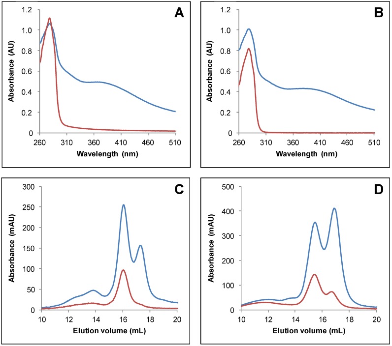
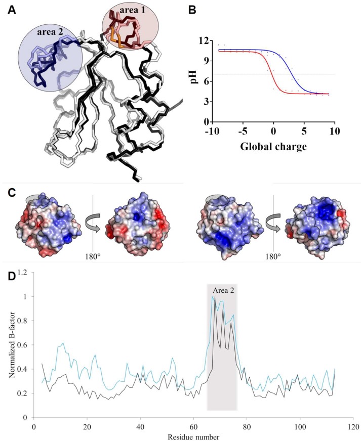
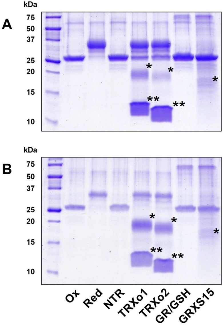
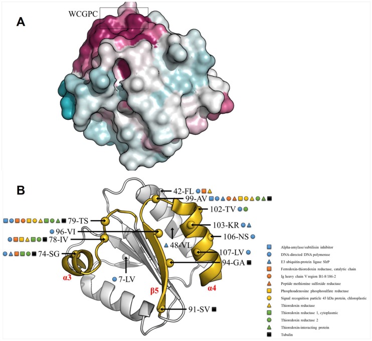
In plants, the mitochondrial thioredoxin (TRX) system generally comprises only one or two isoforms belonging to the TRX h or o classes, being less well developed compared to the numerous isoforms found in chloroplasts. Unlike most other plant species, Arabidopsis thaliana possesses two TRXo isoforms whose physiological functions remain unclear. Here, we performed a structure⁻function analysis to unravel the respective properties of the duplicated TRXo1 and TRXo2 isoforms. Surprisingly, when expressed in Escherichia coli, both recombinant proteins existed in an apo-monomeric form and in a homodimeric iron⁻sulfur (Fe-S) cluster-bridged form. In TRXo2, the [4Fe-4S] cluster is likely ligated in by the usual catalytic cysteines present in the conserved Trp-Cys-Gly-Pro-Cys signature. Solving the three-dimensional structure of both TRXo apo-forms pointed to marked differences in the surface charge distribution, notably in some area usually participating to protein⁻protein interactions with partners. However, we could not detect a difference in their capacity to reduce nitrogen-fixation-subunit-U (NFU)-like proteins, NFU4 or NFU5, two proteins participating in the maturation of certain mitochondrial Fe-S proteins and previously isolated as putative TRXo1 partners. Altogether, these results suggest that a novel regulation mechanism may prevail for mitochondrial TRXs o, possibly existing as a redox-inactive Fe-S cluster-bound form that could be rapidly converted in a redox-active form upon cluster degradation in specific physiological conditions.

Keywords: iron–sulfur cluster; mitochondria; redox regulation; thioredoxin.


Conflict of interest statement

The authors declare no conflict of interest.

Figures

Figure 1
Figure 1
Arabidopsis TRXo (thioredoxin-o) isoforms incorporate an Fe-S cluster after heterologous expression in E. coli. UV–visible absorption spectra of His-tagged recombinant TRXo2 (A) and TRXo1 (C). Spectra were recorded after an anaerobic purification in 30 mM Tris-HCl pH 8.0. Analytical gel filtration of His-tagged recombinant TRXo2 (B) and TRXo1 (D), purified in anaerobic conditions, was performed by loading 100 to 300 µg of protein onto a Superdex S200 10/300 column. The presence of the polypeptide and of the Fe-S cluster have been detected by the absorbance at 280 nm (blue line) and 420 nm (red line), respectively.
Figure 2
Figure 2
IscS-mediated in vitro Fe-S cluster reconstitution of Arabidopsis TRXo isoforms. UV–visible absorption spectra of TRXo2 (A) and TRXo1 (C) before (red line) and after (blue line) an anaerobic reconstitution performed in the presence of IscS in Tris NaCl buffer. Analytical gel filtration of reconstituted TRXo2 (B) and TRXo1 (D) was performed by loading 100 to 300 µg of protein (including 10 µM of EcIscS) onto a Superdex S200 10/300 column. The presence of the polypeptide and of the Fe-S cluster have been detected by the absorbance at 280 nm (blue line) and 420 nm (red line), respectively.
Figure 3
Figure 3
Monocysteinic variants of Arabidopsis TRXo2 isoform are mostly apo-monomers. UV–visible absorption spectra of His-tagged recombinant TRXo2 C37S (red line) and C40S (black line) (A) recorded after an anaerobic purification in 30 mM Tris-HCl pH 8.0. Analytical gel filtration of His-tagged recombinant TRXo2 C37S (B) and C40S (C) purified in anaerobic conditions, was performed by loading 100 to 300 µg of protein onto a Superdex S200 10/300 column. The presence of the polypeptide and of the Fe-S cluster have been detected by the absorbance at 280 nm (blue line) and 420 nm (red line), respectively.
Figure 4
Figure 4
Three-dimensional structure and sequence conservation of oxidized Arabidopsis TRXo isoforms. (A) Structure-based sequence alignment of Arabidopsis TRXo isoforms. Conserved residues are highlighted in black. (B) Three-dimensional structure of AtTRXo2 at 1.50 Å resolution. The X-ray structure of AtTRXo2 is shown as a ribbon representation with helices in red and strands in yellow. In addition, the side chains of Cys37 and Cys40 residues are shown as sticks. (C) Electron density around the Cys37-Cys40 disulfide bond. The maps shown are σA-weighted 2mFo-DFc maps contoured at 1.2σ (0.07 and 0.38 e/Å3 for AtTRXo1 and AtTRXo2, respectively). AtTRXo1 and AtTRXo2 are colored in white and black, respectively.
Figure 5
Figure 5
Structural features vs. charge and crystallographic B-factor distribution. (A) Structure superposition of the backbone trace of Arabidopsis TRXo isoforms. AtTRXo1 and AtTRXo2 are colored in white and black, respectively. The two divergent areas are circled in red and blue, respectively. (B) Global protein charge of AtTRXo1 (red) and AtTRXo2 (blue) as a function of pH as indicated by the PDB2PQR Server [54]. The dot line corresponds to pH 7.0. (C) Electrostatic potential mapped onto AtTRXo1 (left) and AtTRXo2 (right) structures at pH 8.1. The WCGPC signature residues are circled in black. (D) Flexibility of AtTRXo1 (black) and AtTRXo2 (blue) related to the crystallographic B-factor.
Figure 6
Figure 6
The disulfide bridge of mitochondrial NFUs (nitrogen-fixation-subunit-Us) is reduced by TRXs o but not GRXS15. The reduction of as-purified, oxidized forms of NFU4 (A) or NFU5 (B) was assessed after a 15 min incubation in the presence of the following reducing systems: NTR: NADPH + NTR; TRXo1: NADPH + NTR + TRXo1; TRXo2: NADPH + NTR + TRXo2; GR/GSH: NADPH + GR + GSH ; GRXS15: NADPH + GR + GSH + GRXS15. After alkylation with 2 kDa mPEG maleimide, proteins were separated on non-reducing SDS-PAGE (sodium dodecyl sulfate polyacrylamide gel electrophoresis). Reduced (Red) and oxidized (Ox) proteins served as controls. The stars indicate the alkylated (*) and non-alkylated (**) forms of the oxidoreductases in the respective regeneration systems when visible.
Figure 7
Figure 7
Thioredoxin interfacing residues. (A) Residue conservation among 500 thioredoxin orthologs of AtTRXo1 and AtTRXo2 using the ConSurf server [67] with the UniRef90 database (www.uniprot.org/uniref/). Residues are colored in white to purple, for least to most conserved residues. The conserved WCGPC motif is circled in black. (B) AtTRXo1 and AtTRXo2 sequences were blasted against the PDB to find protein–protein interactions involving thioredoxin homologs. 44 complexes were found using an E-Value Cutoff of 0.001. AtTRXo1 and AtTRXo2 non-conserved residues potentially involved in protein–protein interactions were mapped onto the X-ray structure of AtTRXo1. In each case the residue position, and the amino acids in one letter code for AtTRXo1 and AtTRXo2 are shown (ex: 78-IV; position 78, isoleucine and valine found in AtTRXo1 and AtTRXo2, respectively). The area comprising α3-helix, the loop between α3 and β4, β5-strand, and α4-helix is colored in yellow.

References

    1. Couturier J., Touraine B., Briat J.F., Gaymard F., Rouhier N. The iron–sulfur cluster assembly machineries in plants: Current knowledge and open questions. Front. Plant Sci. 2013;4:259. doi: 10.3389/fpls.2013.00259. - DOI - PMC - PubMed
    1. Braymer J.J., Lill R. Iron–sulfur cluster biogenesis and trafficking in mitochondria. J. Biol. Chem. 2017;292:12754–12763. doi: 10.1074/jbc.R117.787101. - DOI - PMC - PubMed
    1. Ciofi-Baffoni S., Nasta V., Banci L. Protein networks in the maturation of human iron–sulfur proteins. Metallomics. 2018;10:4972. doi: 10.1039/C7MT00269F. - DOI - PubMed
    1. Navrot N., Rouhier N., Gelhaye E., Jacquot J.P. Reactive oxygen species generation and antioxidant systems in plant mitochondria. Physiol. Plant. 2007;129:185–195. doi: 10.1111/j.1399-3054.2006.00777.x. - DOI
    1. Navrot N., Collin V., Gualberto J., Gelhaye E., Hirasawa M., Rey P., Knaff D.B., Issakidis E., Jacquot J.P., Rouhier N. Plant glutathione peroxidases are functional peroxiredoxins distributed in several subcellular compartments and regulated during biotic and abiotic stresses. Plant Physiol. 2006;142:1364–1379. doi: 10.1104/pp.106.089458. - DOI - PMC - PubMed

LinkOut - more resources