. 2018 Nov 6;115(9):1796-1807.
doi: 10.1016/j.bpj.2018.09.019. Epub 2018 Sep 28.

Microtubules Provide a Viscoelastic Resistance to Myocyte Motion

Affiliations

Microtubules Provide a Viscoelastic Resistance to Myocyte Motion

Matthew Alexander Caporizzo et al. Biophys J. .

Abstract

Background: Microtubules (MTs) buckle and bear load during myocyte contraction, a behavior enhanced by post-translational detyrosination. This buckling suggests a spring-like resistance against myocyte shortening, which could store energy and aid myocyte relaxation. Despite this visual suggestion of elastic behavior, the precise mechanical contribution of the cardiac MT network remains to be defined.

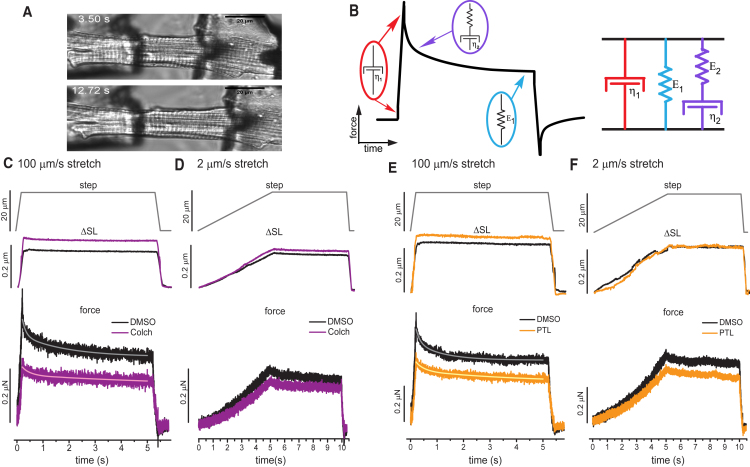
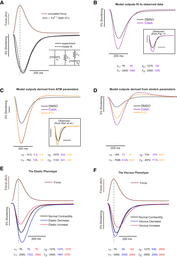
Methods: Here we experimentally and computationally probe the mechanical contribution of stable MTs and their influence on myocyte function. We use multiple approaches to interrogate viscoelasticity and cell shortening in primary murine myocytes in which either MTs are depolymerized or detyrosination is suppressed and use the results to inform a mathematical model of myocyte viscoelasticity.

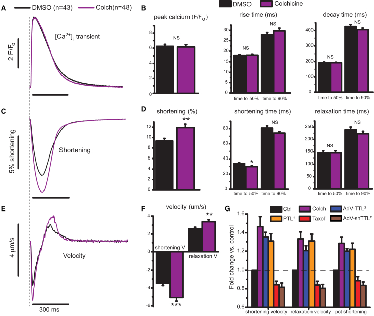
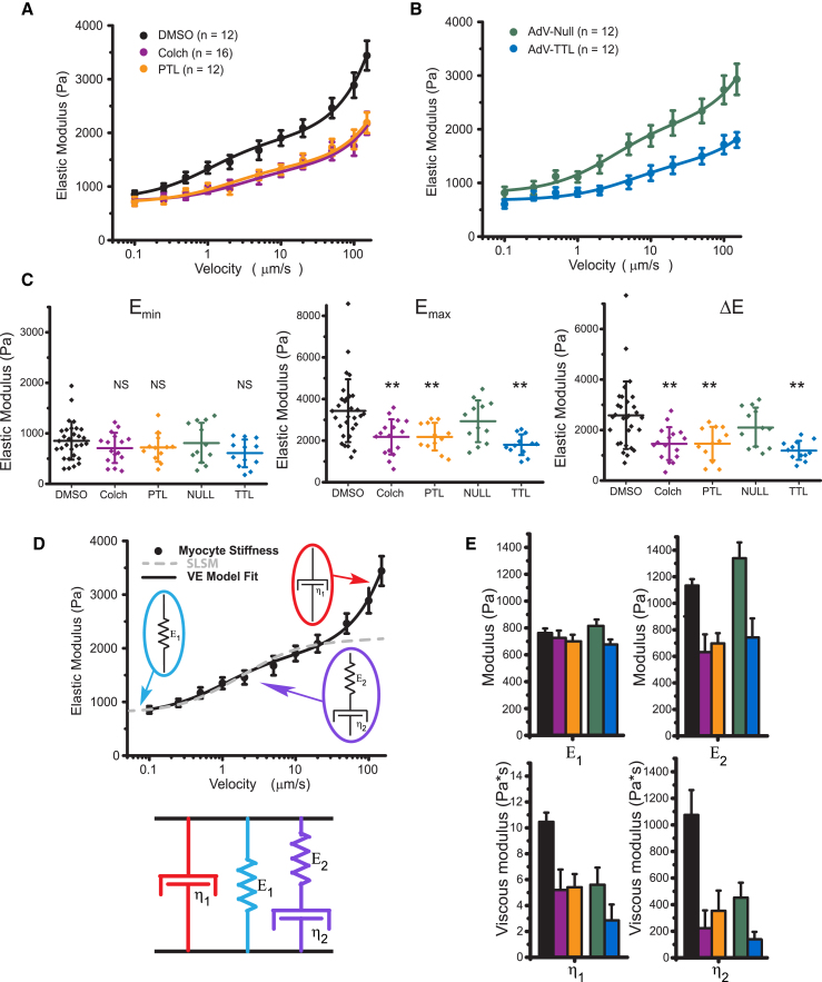
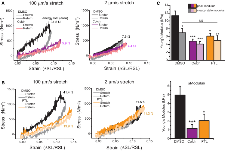
Results: MT ablation by colchicine concurrently enhances both the degree of shortening and speed of relaxation, a finding inconsistent with simple spring-like MT behavior and suggestive of a viscoelastic mechanism. Axial stretch and transverse indentation confirm that MTs increase myocyte viscoelasticity. Specifically, increasing the rate of strain amplifies the MT contribution to myocyte stiffness. Suppressing MT detyrosination with parthenolide or via overexpression of tubulin tyrosine ligase has mechanical consequences that closely resemble colchicine, suggesting that the mechanical impact of MTs relies on a detyrosination-dependent linkage with the myocyte cytoskeleton. Mathematical modeling affirms that alterations in cell shortening conferred by either MT destabilization or tyrosination can be attributed to internal changes in myocyte viscoelasticity.

Conclusions: The results suggest that the cardiac MT network regulates contractile amplitudes and kinetics by acting as a cytoskeletal shock-absorber, whereby MTs provide breakable cross-links between the sarcomeric and nonsarcomeric cytoskeleton that resist rapid length changes during both shortening and stretch.


Figures

Figure 1
Figure 1
MT depolymerization enhances myocyte contractility. (A) Normalized calcium transient (F/Fo) for control (black) and colchicine-treated (purple) cells. (B) Peak calcium, time to peak, and decay time determined from individual traces (N = 6 hearts, n = 43 dimethylsiloxane vehicle (DMSO) cells, n = 48 colchicine-treated cells). (C) Relative shortening (change in SL divided by resting SL) with time. (D) Peak shortening, shortening, and relaxation times from individual traces from identical cells analyzed in (B). (E) The average shortening velocity for DMSO- and colchicine-treated RCMs from (A) and (C). (F) Peak shortening and relaxation velocity determined for individual data points. Statistical significance was determined as *p<0.05, ∗∗p < 0.01 or ***p <0.001 compared to DMSO, Student’s t-test. (G) A comparison of changes in contractile velocities and fractional shortening upon various manipulations to depolymerize MTs or alter MT-dTyr. Each treatment condition is displayed normalized to its relative control (for example, DMSO treated for PTL or a null-encoding adenovirus (AdV-Null) for AdV-TTL). Colchicine data are from the current study. PTL and taxol data are replotted from (16). TTL overexpression and knock-down data are replotted from (2). Data in (Β), (D), (F), and (G) are means with standard error bars. To see this figure in color, go online.
Figure 2
Figure 2
Transverse indentation at variable rates to assess myocyte viscoelasticity. (A) Young’s modulus versus indentation velocity in freshly isolated RCMs treated with DMSO, colchicine, or PTL. Data points are means with standard error whiskers. (n = 12, 16, and 12 cells from three rats for DMSO, colchicine, and PTL, respectively). (B) Young’s modulus versus indentation velocity in RCMs 48 h post-transfection with AdV-Null, green, or AdV-TTL, blue. Data points are means with standard error whiskers. (n = 12 cells from three rats for both AdV-Null and AdV-TTL.) Solid lines in both (A) and (B) are best-fit curves to the VE model shown in (D). (C) The minimal Young’s modulus, Emin, is determined by the cell stiffness at the slowest (100 nm/s) indentation rate. The maximal Young’s modulus, Emax, is determined by the cell stiffness at the fastest (150 μm/s) indentation rate. The change in Young’s modulus over the velocity range measured, ΔE, is determined by EmaxEmin for each cell. Statistical significance was determined by one-way ANOVA with post hoc Tukey as p < 0.05, ∗∗p <0.01, or ∗∗∗p < 0.001 compared to DMSO or AdV-Null. Dot plots show mean line and whiskers as SD. (D) The VE model (a Maxwell element in parallel configuration with a Voigt element; below) for which the velocity-dependence of Young’s modulus can be fitted to extract the stiffness of each element (black trace). The dashed trace illustrates the velocity dependence of the Young’s modulus for a standard linear solid model fitted to the data and provides justification for the inclusion of the additional viscous element (η1). (E) VE parameters extracted from model fitted to data in (A) and (B). Bar plots show the estimated fitted value with whiskers showing the fit certainty. To see this figure in color, go online.
Figure 3
Figure 3
For a Figure360 author presentation of Fig. 3, see the figure legend at https://doi.org/10.1016/j.bpj.2018.9.019#mmc3 Axial stretch at variable rates after colchicine and PTL treatment of RCMs. (A) A transmitted light image of isolated adult RCM before and during tensile test. (B) The VE model (a Maxwell element in parallel configuration with a Voigt element) for which the time dependence of the stress-strain curve can be used to extract values for each parameter. The top row shows the imposed length change—a 20 μm length controller step over either a 200 ms (C and E) or 5 s (D and F) stretch, followed by a 5 s hold and 200 ms return. The middle row shows the average change in SL in response to stretch. The bottom row shows the average change in force in response to stretch. (A and B) 100 μm/s stretch and 2 μm/s stretch for DMSO and colchicine (N = 5 hearts, n = 17 cells DMSO, n = 18 cells colchicine). (C and D) 100 μm/s stretch and 2 μm/s stretch for DMSO and PTL. (N = 5 hearts, n = 14 cells DMSO, n = 17 cells colchicine). Solid traces overlaid on force-relaxation curves are best fits to double exponential decay function. To see this figure in color, go online.
Figure 4
Figure 4
Stress-strain curves calculated from axial stretch. (A) Stress-strain loops for DMSO- and colchicine-treated cells at 100 and 2 μm/s. (B) Stress-strain loops for DMSO- and PTL-treated cells at 100 and 2 μm/s. (C) Peak and steady-state modulus (top) and relaxation modulus (peak − steady state) (bottom) for individual cells treated with DMSO, PTL, or colchicine. Statistical significance was determined by one-way ANOVA with post hoc Tukey as p < 0.05, ∗∗p < 0.01, or ∗∗∗p < 0.001 compared to DMSO. To see this figure in color, go online.
Figure 5
Figure 5
VE model of cardiomyocyte contractility. (A) The stress-strain relationship defined by the VE model (bottom right inset) converts myofilament force (brown) to shortening (black dotted trace). By changing the stiffness of individual VE elements (values are shown in (B), bottom), experimentally observed shortening (black solid trace) can be modeled (black dashed trace) by cytoskeletal viscoelasticity. (B) Model fits (dashed traces) to observed DMSO and colchicine contractility data (boxed inset). Best-fit parameters are shown below the plot. (C) Modeled contractility from VE parameters derived from transverse indentation studies (Fig. 2). Previously observed data for PTL treatment are provided for comparison (inset). (D) Modeled contractility from parameters derived from tensile tests (Fig. 3). (E) Simulated changes in contractility for myocyte for which there is an isolated reduction (blue) or increase (red) in myocyte elasticity. (F) Simulated changes in contractility for which myocyte viscosity has been decreased (blue) or increased (red). To see this figure in color, go online.

References

    1. Fassett J.T., Xu X., Chen Y. Microtubule actin cross-linking factor 1 regulates cardiomyocyte microtubule distribution and adaptation to hemodynamic overload. PLoS One. 2013;8:e73887. - PMC - PubMed
    1. Robison P., Caporizzo M.A., Prosser B.L. Detyrosinated microtubules buckle and bear load in contracting cardiomyocytes. Science. 2016;352:aaf0659. - PMC - PubMed
    1. Sato H., Nagai T., Cooper G., IV Microtubule stabilization in pressure overload cardiac hypertrophy. J. Cell Biol. 1997;139:963–973. - PMC - PubMed
    1. Chen C.Y., Caporizzo M.A., Prosser B.L. Suppression of detyrosinated microtubules improves cardiomyocyte function in human heart failure. Nat. Med. 2018;24:1225–1233. - PMC - PubMed
    1. Janke C., Bulinski J.C. Post-translational regulation of the microtubule cytoskeleton: mechanisms and functions. Nat. Rev. Mol. Cell Biol. 2011;12:773–786. - PubMed

Publication types

LinkOut - more resources