. 2018 Oct 30;115(44):E10457-E10466.
doi: 10.1073/pnas.1812130115. Epub 2018 Oct 15.

ADP-ribosyl-binding and hydrolase activities of the alphavirus nsP3 macrodomain are critical for initiation of virus replication

Affiliations

ADP-ribosyl-binding and hydrolase activities of the alphavirus nsP3 macrodomain are critical for initiation of virus replication

Rachy Abraham et al. Proc Natl Acad Sci U S A. .

Abstract

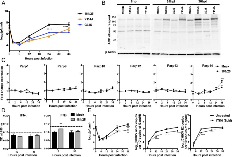
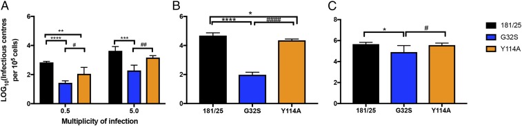
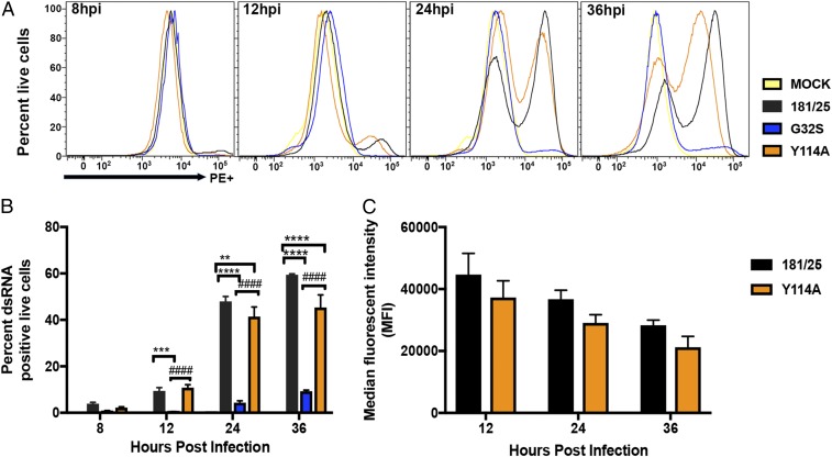
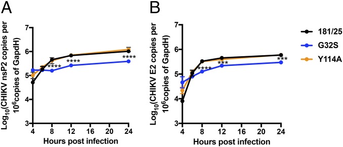
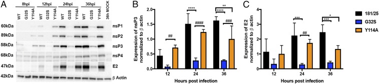
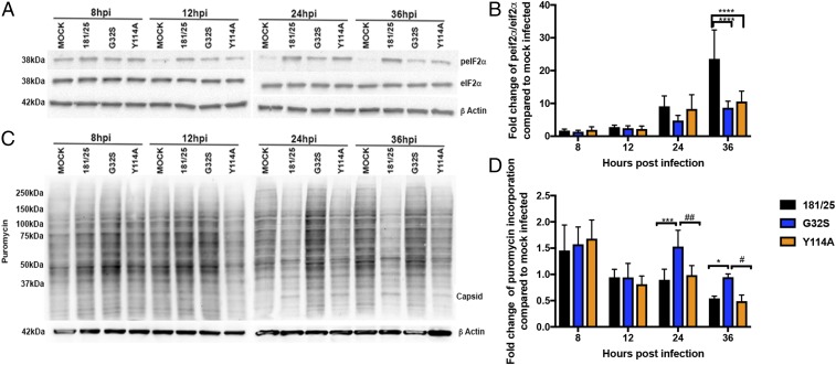
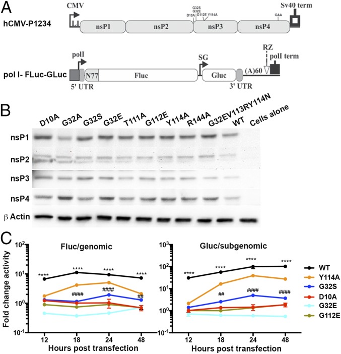
Alphaviruses are plus-strand RNA viruses that cause encephalitis, rash, and arthritis. The nonstructural protein (nsP) precursor polyprotein is translated from genomic RNA and processed into four nsPs. nsP3 has a highly conserved macrodomain (MD) that binds ADP-ribose (ADPr), which can be conjugated to protein as a posttranslational modification involving transfer of ADPr from NAD+ by poly ADPr polymerases (PARPs). The nsP3MD also removes ADPr from mono ADP-ribosylated (MARylated) substrates. To determine which aspects of alphavirus replication require nsP3MD ADPr-binding and/or hydrolysis function, we studied NSC34 neuronal cells infected with chikungunya virus (CHIKV). Infection induced ADP-ribosylation of cellular proteins without increasing PARP expression, and inhibition of MARylation decreased virus replication. CHIKV with a G32S mutation that reduced ADPr-binding and hydrolase activities was less efficient than WT CHIKV in establishing infection and in producing nsPs, dsRNA, viral RNA, and infectious virus. CHIKV with a Y114A mutation that increased ADPr binding but reduced hydrolase activity, established infection like WT CHIKV, rapidly induced nsP translation, and shut off host protein synthesis with reduced amplification of dsRNA. To assess replicase function independent of virus infection, a transreplicase system was used. Mutant nsP3MDs D10A, G32E, and G112E with no binding or hydrolase activity had no replicase activity, G32S had little, and Y114A was intermediate to WT. Therefore, ADP ribosylation of proteins and nsP3MD ADPr binding are necessary for initiation of alphavirus replication, while hydrolase activity facilitates amplification of replication complexes. These observations are consistent with observed nsP3MD conservation and limited tolerance for mutation.

Keywords: ADP ribosylation; PARP; alphavirus; macrodomain; replication complexes.


Conflict of interest statement

The authors declare no conflict of interest.

Figures

Fig. 1.
Fig. 1.
CHIKV infection facilitates virus replication in NSC34 cells by increasing ADP ribosylation of cellular proteins without inducing PARP gene transcription. (A) NSC34 cells were infected with CHIKV 181/25 (WT) and nsP3MD mutants G32S and Y114A at an MOI of 10. Virus production was measured by plaque formation in Vero cells. Each value represents the average from three independent experiments; error bars indicate SDs. ***P < 0.001, ****P < 0.0001 (181/25 vs. nsP3MD mutants G32S and Y114A). (Adapted with permission from ref. .) (B) NSC34 cells were infected with CHIKV 181/25 and nsP3MD mutants G32S and Y114A at an MOI of 5, and cell lysates were immunoblotted for ADPr. Antibody against β-actin was used for loading controls. A representative image from three independent experiments is shown. (C) NSC34 cells were infected with CHIKV 181/25 at an MOI of 5, and PARP mRNA expression was measured by qRT-PCR. Mock-infected cells (solid line) were compared with CHIKV-infected cells (dashed line). CT values were normalized to Gapdh, and fold change was calculated relative to uninfected 0-h (ΔΔCT) data. Each value represents the average ± SD from three independent experiments. ***P < 0.001 (mock vs. infected). (D) NSC34 cells were infected with CHIKV 181/25 (MOI = 5), and supernatant fluids were assayed for IFNα and IFNβ by enzyme immunoassay. Dashed lines indicate the lower limit of detection for the assays. (E) NSC34 cells were infected with CHIKV 181/25 (MOI = 1) and were treated or not treated with the pan mono-ADPr inhibitor ITK6 (5 μM) at the time of infection. Supernatant fluids were assayed for virus production by plaque formation in Vero cells (Left), and intracellular RNAs were assayed by qRT-PCR for viral genomic (nsP2; Middle) and genomic+sg E2 RNA and were compared with standard curves of CHIKV RNAs (Right). Each value represents the average ± SD from three independent experiments. ***P < 0.001, ****P < 0.0001.
Fig. 2.
Fig. 2.
nsP3MD mutations affecting ADP ribosyl binding and hydrolase activities differentially affect the initiation of infection. (A) Infectious center assays for NSC34 cells infected at MOIs of 0.5 and 5 with CHIKV 181/25 (WT) and nsP3MD mutants G32S or Y114A. Infected cells were trypsinized, viable cells were counted, and serially diluted cells were plated on Vero cells to identify virus-producing cells by plaque assay. (B and C) Infectious center assays for NSC34 (B) and BHK21 (C) cells after electroporation of 10 μg of viral RNA transcribed in vitro from full-length clones of 181/25 and nsP3MD G32S and Y114A mutants into 105 cells that were plated onto subconfluent monolayers of BHK21 cells to identify virus-producing cells by plaque formation. The data are presented as log10 infectious centers per 105 cells. Each value represents the average ± SD from three independent experiments; *P < 0.05; **P < 0.01; ***P < 0.001; ****P < 0.0001 (WT vs. G32S/Y114A); #P < 0.05; ##P < 0.01, ####P < 0.0001 (Y114A vs. G32S).
Fig. 3.
Fig. 3.
Formation of replication complexes containing dsRNA. NSC34 cells infected at an MOI of 5 with CHIKV 181/25 (WT) virus and nsP3MD mutants G32S or Y114A were gated for live cells, fixed, permeabilized, stained for dsRNA, and analyzed by flow cytometry. (A) Representative histograms from three independent experiments. (B) Quantification of the percent of live cells positive for dsRNA. (C) The median fluorescent intensities for WT and Y114A-infected cells. The data are presented as the means ± SD from three independent experiments; **P < 0.01; ***P < 0.001; ****P < 0.0001 (WT vs. G32S/Y114A); ####P < 0.0001 (Y114A vs. G32S). hpi, hours postinfection.
Fig. 4.
Fig. 4.
Synthesis of viral RNAs. Levels of genomic (A) and genomic plus sg (B) RNA from NSC34 cells infected with CHIKV 181/25 (WT) and nsP3MD mutants G32S or Y114A at an MOI of 5 were measured by qRT-PCR. Total cellular RNA was collected, and cDNA was produced and was quantified by qPCR compared with a standard curve of CHIKV cDNA fragments corresponding to genomic or sgRNA. The data represent the means ± SD from three independent experiments. ***P < 0.001; ****P < 0.0001 (WT/Y114A vs. G32S).
Fig. 5.
Fig. 5.
Effect of nsP3MD mutation on viral protein synthesis. NSC34 cells were infected with CHIKV 181/25 (WT) or nsP3MD mutants G32S or Y114A at an MOI of 5, and cell lysates were probed by immunoblot for production of viral proteins using rabbit polyclonal antibodies to nsP1, nsP2, nsP3, nsP4, and E2. Antibody against β-actin was used for loading controls. (A) Representative image. (B and C) Quantification of expression of nsP3 (B) and E2 (C) relative to actin by densitometry on blots from three independent experiments. Data indicate the mean ± SD. *P < 0.05, **P < 0.01, ***P < 0.001, ****P < 0.0001 (WT vs. mutant); ##P < 0.01, ###P < 0.001, ####P < 0.0001 (Y114A vs. G32S). hpi, hours postinfection.
Fig. 6.
Fig. 6.
Effect of nsP3MD mutation on phosphorylation of eIf2α and host protein translation. NSC34 cells were infected with CHIKV 181/25 (WT) or nsP3MD mutants G32S or Y114A at an MOI of 5. (A and B) Cell lysates were probed by immunoblot for levels of total and phosphorylated eIF2α. Antibody against β-actin was used for loading controls. (A) Representative image. (B) Blots from three independent experiments were quantitated by densitometry, and the ratios of phosphorylated eIF2α to total eIF2α were calculated. Data are expressed as the mean fold change over mock-infected cells ± SD. (C and D) Host protein translation was analyzed by the incorporation of puromycin. At the time points indicated, medium was replaced with medium containing puromycin (5 μg/mL) and was incubated for 10 min. Cell lysates were probed by immunoblot with antibody to puromycin to detect newly synthesized protein. Antibody against β-actin was used for loading controls. (C) Representative image. (D) Blots from three independent experiments were quantitated by densitometry, and puromycin incorporation was compared with mock-infected cells. Data are presented as the mean fold change over mock ± SD. *P < 0.05, ***P < 0.001, ****P < 0.0001 (WT vs. mutant); #P < 0.05, ##P < 0.01 (Y114A vs. G32S).
Fig. 7.
Fig. 7.
Analysis of nsP function in the absence of virus infection using a transreplicase system. (A) The transreplicase system includes an hCMV-P1234 plasmid with and without nsP3MD mutations, an identical inactivated nsP4 polymerase hCMV-P1234GAA plasmid, and a pol I 5′ UTR–Fluc–sg promoter–Gluc–3′ UTR plasmid. (B) NSC34 cells were transfected with 1 μg of each plasmid and were incubated at 37 °C for 24 h. Viral protein production was analyzed by immunoblotting with antibodies to CHIKV nsP1, nsP2, nsP3, nsP4, and E2. Antibody against β-actin was used for loading controls. The image is representative of three independent experiments. (C) Fluc reporter activity for genomic RNA synthesis and Gluc reporter activity for subgenomic RNA synthesis. Luciferase activities were read on a luminometer and were normalized to the total protein content in the lysate. The reporter activities generated by the inactive replicase P1234GAA were subtracted from reporter activities generated by the active replicase P1234 and are reported as fold change. The data are presented as the means ± SD from three independent experiments. ****P < 0.0001, WT vs. G32S; ##P < 0.01, ####P < 0.0001, Y114A vs. G32S.

References

    1. Gubler DJ. The global emergence/resurgence of arboviral diseases as public health problems. Arch Med Res. 2002;33:330–342. - PubMed
    1. Weaver SC, Lecuit M. Chikungunya virus and the global spread of a mosquito-borne disease. N Engl J Med. 2015;372:1231–1239. - PubMed
    1. Ronca SE, Dineley KT, Paessler S. Neurological sequelae resulting from encephalitic alphavirus infection. Front Microbiol. 2016;7:959. - PMC - PubMed
    1. Chandak NH, et al. Neurological complications of chikungunya virus infection. Neurol India. 2009;57:177–180. - PubMed
    1. Strauss JH, Strauss EG. The alphaviruses: Gene expression, replication, and evolution. Microbiol Rev. 1994;58:491–562. - PMC - PubMed

Publication types