. 2018 Oct 30;115(44):11292-11297.
doi: 10.1073/pnas.1811974115. Epub 2018 Oct 15.

Herpesvirus trigger accelerates neuroinflammation in a nonhuman primate model of multiple sclerosis

Affiliations

Herpesvirus trigger accelerates neuroinflammation in a nonhuman primate model of multiple sclerosis

Emily C Leibovitch et al. Proc Natl Acad Sci U S A. .

Abstract

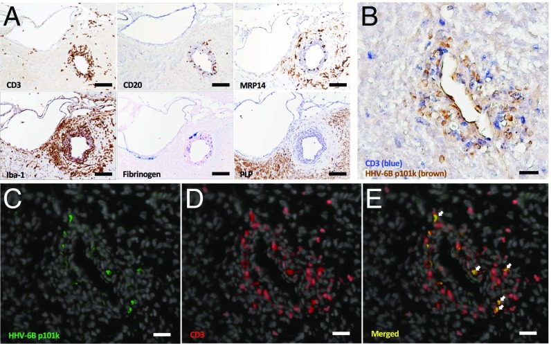
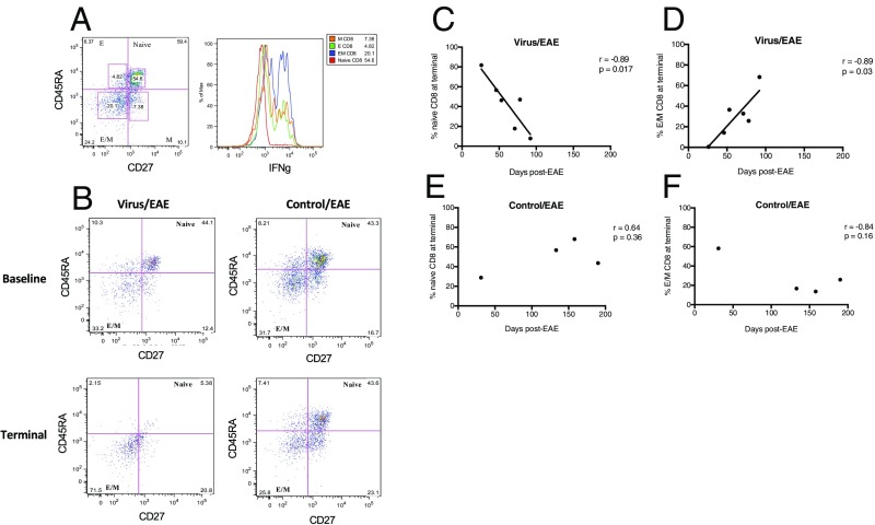
Pathogens, particularly human herpesviruses (HHVs), are implicated as triggers of disease onset/progression in multiple sclerosis (MS) and other neuroinflammatory disorders. However, the time between viral acquisition in childhood and disease onset in adulthood complicates the study of this association. Using nonhuman primates, we demonstrate that intranasal inoculations with HHV-6A and HHV-6B accelerate an MS-like neuroinflammatory disease, experimental autoimmune encephalomyelitis (EAE). Although animals inoculated intranasally with HHV-6 (virus/EAE marmosets) were asymptomatic, they exhibited significantly accelerated clinical EAE compared with control animals. Expansion of a proinflammatory CD8 subset correlated with post-EAE survival in virus/EAE marmosets, suggesting that a peripheral (viral?) antigen-driven expansion may have occurred post-EAE induction. HHV-6 viral antigen in virus/EAE marmosets was markedly elevated and concentrated in brain lesions, similar to previously reported localizations of HHV-6 in MS brain lesions. Collectively, we demonstrate that asymptomatic intranasal viral acquisition accelerates subsequent neuroinflammation in a nonhuman primate model of MS.

Keywords: EAE; human herpesvirus 6; marmoset; multiple sclerosis; viral trigger.


Conflict of interest statement

The authors declare no conflict of interest.

Figures

Fig. 1.
Fig. 1.
HHV-6 viral antigen occasionally detected in the brain after intranasal inoculations. Two of nine examined brain regions from HHV-6B–inoculated marmoset M26 contained detectable HHV-6 late antigen p101k (red; arrows), with cytoplasmic localization. (A) Olfactory bulb. (B) Hippocampus. (Scale bar: 50 µm; Inset scale bar: 5 µm.)
Fig. 2.
Fig. 2.
Significantly accelerated clinical EAE in HHV-6 intranasally inoculated marmosets. (A) Reduced time to symptom onset in virus/EAE marmosets compared with SupT1 control/EAE marmosets (unpaired t test, P = 0.04). (B) Shortened survival post-EAE induction in virus/EAE marmosets compared with SupT1 control/EAE marmosets (unpaired t test, P = 0.03).
Fig. 3.
Fig. 3.
Characterization of EAE brain lesions. PD (A) and T1 (B) in vivo images from HHV-6B/EAE marmoset M28. Two white matter lesions are numbered. (C) Final PD and T1 lesion volumes by marmoset for all groups. (D and E) Virus/EAE marmosets survived for less time after the onset of PD (unpaired t test, P = 0.04) (D) and T1 (unpaired t test, P = 0.03) (E) brain lesions. (F) HHV-6B/EAE marmosets had the greatest T1 lesion burden.
Fig. 4.
Fig. 4.
HHV-6 viral antigen is up-regulated in virus/EAE marmoset brains. Arrows indicate positivity for HHV-6 late antigen p101k (red) in inflamed brain regions from representative virus/EAE marmoset M28. (A and B) Temporal white matter around putamen. (C) Leptomeninges around hippocampal fissure. (Scale bar: 50 µm.)
Fig. 5.
Fig. 5.
Characterization of HHV-6–positive brain lesions from a virus/EAE marmoset. (A) Serial sections of an HHV-6–positive perivascular EAE lesion demonstrate CD3 T and CD20 B cell infiltrates, MRP14+ macrophages/monocytes, Iba-1+ microglia, blood-brain barrier leakiness evidenced by fibrinogen deposition, and demyelination evidenced by the absence of myelin PLP. (Scale bar: 100 µm.) (B) HHV-6 antigen (brown) colocalizes with CD3 T cells (blue). (C) HHV-6–infected cells in green (Alexa 488). (D) CD3 T cells in red (Alexa 594), with DAPI counterstain (gray). (E) Merged image with arrows showing colocalization of HHV-6 viral antigen with CD3 T cells. (Scale bar: BE, 50 µm.) The lesion pictured in A is located in the gray matter (lateral dorsal thalamic nucleus), whereas the lesion pictured in BE is located in the temporal white matter. Lesions are from HHV-6B/EAE marmoset M28.
Fig. 6.
Fig. 6.
Increase in IFN-γ–producing effector/memory (E/M) CD8 T cells significantly correlates with disease duration in virus-inoculated marmosets. (A) Representative flow cytometry plot of CD8+ CD45RA/CD27 T cell subsets, with histogram showing highest IFN-γ production by E/M subset. E, effector; M, memory. (B) Representative flow cytometry plots showing a decrease in naïve CD8+ T cells at terminal EAE time points in virus/EAE, but not control/EAE, marmosets. (C and D) Contraction of naïve CD8 (C) and expansion of E/M CD8 (D) T cells correlate with EAE disease duration in virus-inoculated marmosets. (E and F) In the control/EAE marmosets, no such correlation was observed for either the naïve CD8 (E) or the E/M CD8 (F) subset.

Comment in

  • Reply to Zahednasab et al.: HHV-6 and marmoset EAE.
    Leibovitch EC, Jacobson S. Leibovitch EC, et al. Proc Natl Acad Sci U S A. 2018 Dec 26;115(52):E12127. doi: 10.1073/pnas.1818755115. Epub 2018 Dec 11. Proc Natl Acad Sci U S A. 2018. PMID: 30538192 Free PMC article. No abstract available.
  • Role of HHV-6 subtypes in accelerating EAE progression.
    Zahednasab H, Keyvani H, Karampour S, Harirchian MH. Zahednasab H, et al. Proc Natl Acad Sci U S A. 2018 Dec 26;115(52):E12126. doi: 10.1073/pnas.1817967115. Epub 2018 Dec 11. Proc Natl Acad Sci U S A. 2018. PMID: 30538193 Free PMC article. No abstract available.

References

    1. Compston A, Lassman H, McDonald I. McAlpine’s Multiple Sclerosis, 4th Ed Churchill Livingstone/Elsevier; London: 2006. The story of multiple sclerosis.
    1. Virtanen JO, Jacobson S. Viruses and multiple sclerosis. CNS Neurol Disord Drug Targets. 2012;11:528–544. - PMC - PubMed
    1. Merelli E, et al. Human herpes virus 6 and human herpes virus 8 DNA sequences in brains of multiple sclerosis patients, normal adults and children. J Neurol. 1997;244:450–454. - PubMed
    1. Leibovitch EC, Jacobson S. Evidence linking HHV-6 with multiple sclerosis: An update. Curr Opin Virol. 2014;9:127–133. - PMC - PubMed
    1. Opsahl ML, Kennedy PG. Early and late HHV-6 gene transcripts in multiple sclerosis lesions and normal appearing white matter. Brain. 2005;128:516–527. - PMC - PubMed