. 2018 Dec 14;293(50):19377-19386.
doi: 10.1074/jbc.RA118.005211. Epub 2018 Oct 15.

Processive incorporation of multiple selenocysteine residues is driven by a novel feature of the selenocysteine insertion sequence

Affiliations

Processive incorporation of multiple selenocysteine residues is driven by a novel feature of the selenocysteine insertion sequence

Sumangala P Shetty et al. J Biol Chem. .

Abstract

RNA stem loop structures have been frequently shown to regulate essential cellular processes. The selenocysteine insertion sequence (SECIS) element, found in the 3' UTRs of all selenoprotein mRNAs, is an example of such a structure, as it is required for the incorporation of the 21st amino acid, selenocysteine (Sec). Selenoprotein synthesis poses a mechanistic challenge because Sec is incorporated during translation in response to a stop codon (UGA). Although it is known that a SECIS-binding protein (SBP2) is required for Sec insertion, the mechanism of action remains elusive. Additional complexity is present in the synthesis of selenoprotein P (SELENOP), which is the only selenoprotein that contains multiple UGA codons and possesses two SECIS elements in its 3' UTR. Thus, full-length SELENOP synthesis requires processive Sec incorporation. Using zebrafish Selenop, in vitro translation assays, and 75Se labeling in HEK293 cells, we found here that processive Sec incorporation is an intrinsic property of the SECIS elements. Specifically, we identified critical features of SECIS elements that are required for processive Sec incorporation. A screen of the human SECIS elements revealed that most of these elements support processive Sec incorporation in vitro; however, we also found that the processivity of Sec incorporation into Selenop in cells is tightly regulated. We propose a model for processive Sec incorporation that involves differential recruitment of SECIS-binding proteins.

Keywords: 21st amino acid; RNA structure; mRNA; ribonucleoprotein (RNP); selenium; selenocysteine; selenoprotein; stem loop structure.


Conflict of interest statement

The authors declare that they have no conflicts of interest with the contents of this article. The content is solely the responsibility of the authors and does not necessarily represent the official views of the National Institutes of Health

Figures
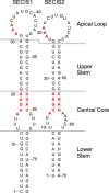
Figure 1.
Figure 1.
Schematic of SECIS elements. Red represents the known conserved motifs of SECIS elements. Each SECIS element is divided into unique sections that include the lower stem, internal loop, upper stem, and apical loop.
Figure 2.
Figure 2.
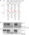
SECIS-1 versus SECIS-2 in in vitro translation and transfected cells. A, schematics of the constructs used in the study. The WT ZSelenop coding region is represented by the white boxes, in which the vertical lines stand for each selenocysteine codon. The ZSelenop coding region was ligated on the 3′ end with either full-length 3′ UTR, SECIS-1, or SECIS-2. In the SECIS2 U59C mutant, the first selenocysteine codon was mutated to a cysteine codon. B, left panel, capped WT ZSelenop and mutant mRNAs were translated in rabbit reticulocyte lysate (RRL) supplemented with 8 pmol of recombinant CT-SBP2 and analyzed using 75Se labeling. Arrows indicate full-length ZSelenop and early termination products. Radiolabeled proteins were resolved by SDS-PAGE and detected by PhosphorImager analysis. Right panel, 75Se-labeled medium from HEK293 cells transiently transfected with the constructs shown in A, resolved by SDS-PAGE and detected by PhosphorImager analysis. The arrows indicate full-length (FL) and early termination products.
Figure 3.
Figure 3.
Analysis of SECIS-1/SECIS-2 chimeras. A, top panel, schematic of SECIS-1 chimeras wherein either the lower stem, internal loop, upper stem, or apical loop of SECIS-1 is replaced with its equivalent from SECIS-2. SECIS 1 elements are shown in green and SECIS 2 in coral pink. Center panel, capped ZSelenop and chimera mRNAs were translated in RRL supplemented with 8 pmol of recombinant CT-SBP2 and analyzed using 75Se labeling. Arrows indicate full-length (FL) ZSelenop and early termination products (Term). Radiolabeled proteins were resolved by SDS-PAGE and detected by PhosphorImager analysis. Bottom panel, 75Se-labeled medium from transient cell lines expressing the constructs in the top panel, resolved by SDS-PAGE and detected by PhosphorImager analysis. Arrows indicate full-length and early termination products. B, top panel, schematic of SECIS-2 chimeras wherein either the lower stem, internal loop, upper stem, or apical loop of SECIS-2 is replaced with its equivalent from SECIS-1. SECIS 1 elements are shown in green and SECIS 2 in coral pink. Center panel, capped ZSelenop and chimera mRNAs were translated in RRL supplemented with 8 pmol of recombinant CT-SBP2 and analyzed using 75Se labeling. Arrows indicate full-length ZSelenop and early termination products. Radiolabeled proteins were resolved by SDS-PAGE and detected by PhosphorImager analysis. Bottom panel, 75Se-labeled medium from transient cell lines expressing the constructs in the top panel, resolved by SDS-PAGE and detected by PhosphorImager analysis. The arrows indicate full-length and early termination products. C, top panel, schematic of the processive SECIS-2 chimera containing the upper stem and apical loop of SECIS 1. SECIS 1 elements are shown in green and SECIS 2 in coral pink. Center panel, capped ZSelenop and chimera mRNAs were translated in RRL supplemented with 16 pmol of recombinant CT-SBP2 and analyzed using 75Se labeling. Arrows indicate full-length ZSelenop and early termination products. Radiolabeled proteins were resolved by SDS-PAGE and detected by PhosphorImager analysis. Bottom panel, 75Se-labeled medium from transient cell lines expressing the constructs in the top panel, resolved by SDS-PAGE and detected by PhosphorImager analysis. The arrows indicate full-length and early termination products.
Figure 4.
Figure 4.
Analysis of the SECIS-1 mutants. A, schematic of the SELENOP SECIS-1, SECIS-2, and SECIS-1 mutants. B, capped ZSelenop and mutant mRNAs were translated in RRL supplemented with 8 pmol of recombinant CT-SBP2 and analyzed using 75Se labeling. Arrows indicate full-length (FL) ZSelenop and early termination products (Term). Radiolabeled proteins were resolved by SDS-PAGE and detected by PhosphorImager analysis. C, 75Se-labeled medium from transient cell lines expressing the constructs in A, resolved by SDS-PAGE and detected by PhosphorImager analysis. The arrows indicate full-length and early termination products.
Figure 5.
Figure 5.
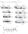
Screen for human SECIS elements for processivity. A, schematic of the ZSelenop coding region ligated to one of the 26 human SECIS elements. B, left panels, capped ZSelenop mRNAs ligated to different human SECIS elements were translated in RRL supplemented with 8 pmol of recombinant CT-SBP2 and analyzed using 75Se labeling. ZSelenop_FL3′ UTR, ZSECIS-1, and ZSECIS-2 were used as controls. Radiolabeled proteins were resolved by SDS-PAGE and detected by PhosphorImager analysis. Right panel, 75Se-labeled medium from transient cell lines expressing constructs with the ZSelenop coding region ligated to any one of the 26 human SECIS elements, resolved by SDS-PAGE and detected by PhosphorImager analysis. Arrows indicate full-length (FL) ZSelenop and early termination products (Term). C, qRT-PCR analysis of mRNA data obtained from the samples in B that either produced no product or very weak products. Equal amounts of total RNA were used for qRT-PCR, and the -fold increase in coding region deletion mutant mRNA relative to ZSelenop was determined by the comparative CT method. Data are plotted as the average ± S.D. for three independent experiments. p < 0.05 compared with the native Zselenop value.
Figure 6.
Figure 6.
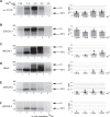
Effect of magnesium on processivity. A–F, left panel, in vitro RRL translation of capped ZSelenop mRNA with either zebrafish 3′ UTR or SECIS elements from zebrafish or human origin over a range of Mg2+ concentrations (millimolar). Samples were supplemented with 8 pmol of recombinant CT-SBP2 and analyzed using 75Se labeling. Radiolabeled proteins were resolved by SDS-PAGE and detected by PhosphorImager analysis. Arrows indicate full-length ZSelenop (FL) and early termination products (Term). Right panel, plots representing the ratio of full-length peptides to total peptides over a range of Mg2+ concentrations (millimolar). This was calculated as the ratio of full-length ZSelenop band intensity (normalized for selenium atoms) divided by the sum of full-length band intensity (normalized for selenium atoms) and early termination product band intensity. Quantification was performed with Imagequant. Data are plotted as the average ± S.D. for three independent experiments. p < 0.05 compared with the 1.25 mm Mg2+ reaction value.

References

    1. Berry M. J., Banu L., Chen Y. Y., Mandel S. J., Kieffer J. D., Harney J. W., and Larsen P. R. (1991) Recognition of UGA as a selenocysteine codon in type I deiodinase requires sequences in the 3′ untranslated region. Nature 353, 273–276. 10.1038/353273a0 - DOI - PubMed
    1. Fagegaltier D., Hubert N., Yamada K., Mizutani T., Carbon P., and Krol A. (2000) Characterization of mSelB, a novel mammalian elongation factor for selenoprotein translation. EMBO J. 19, 4796–4805. 10.1093/emboj/19.17.4796 - DOI - PMC - PubMed
    1. Tujebajeva R. M., Copeland P. R., Xu X. M., Carlson B. A., Harney J. W., Driscoll D. M., Hatfield D. L., and Berry M. J. (2000) Decoding apparatus for eukaryotic selenocysteine insertion. EMBO Rep. 1, 158–163. 10.1093/embo-reports/kvd033 - DOI - PMC - PubMed
    1. Lee B. J., Worland P. J., Davis J. N., Stadtman T. C., and Hatfield D. L. (1989) Identification of a selenocysteyl-tRNA(Ser) in mammalian cells that recognizes the nonsense codon, UGA. J. Biol. Chem. 264, 9724–9727. - PubMed
    1. Copeland P. R., Fletcher J. E., Carlson B. A., Hatfield D. L., and Driscoll D. M. (2000) A novel RNA binding protein, SBP2, is required for the translation of mammalian selenoprotein mRNAs. EMBO J. 19, 306–314. 10.1093/emboj/19.2.306 - DOI - PMC - PubMed

Publication types

LinkOut - more resources