. 2019 Aug;26(8):1396-1410.
doi: 10.1038/s41418-018-0216-2. Epub 2018 Oct 15.

Conditional deletion of platelet derived growth factor receptor alpha (Pdgfra) in urorectal mesenchyme causes mesenchyme apoptosis and urorectal developmental anomalies in mice

Affiliations

Conditional deletion of platelet derived growth factor receptor alpha (Pdgfra) in urorectal mesenchyme causes mesenchyme apoptosis and urorectal developmental anomalies in mice

Chen Qian et al. Cell Death Differ. 2019 Aug.

Abstract

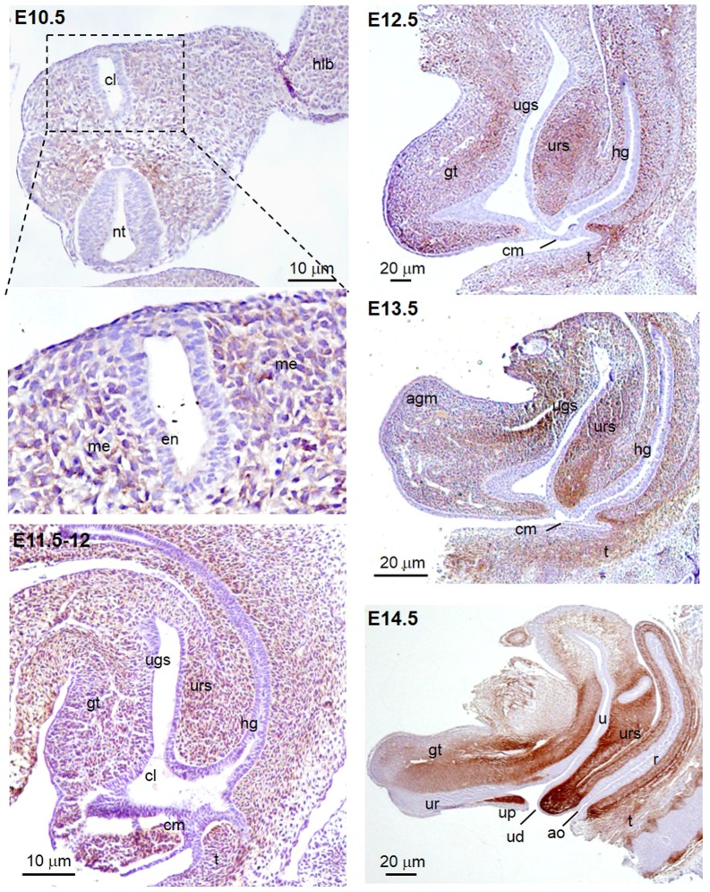
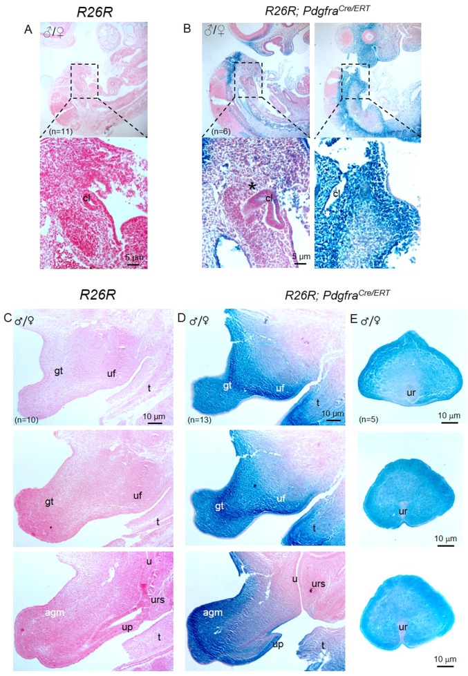
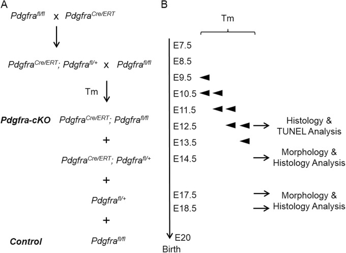
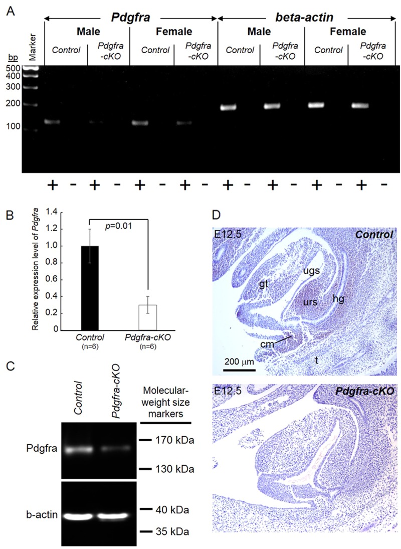
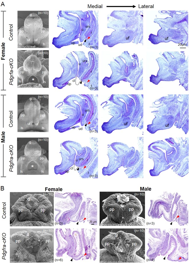
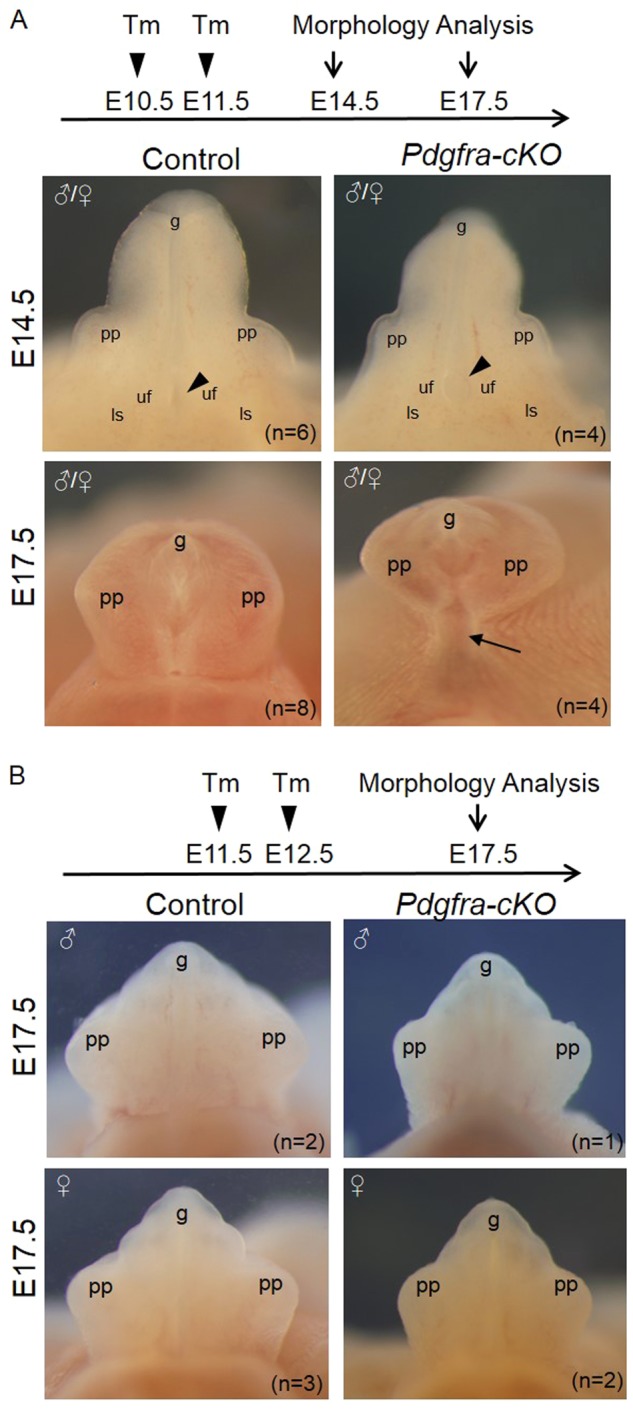
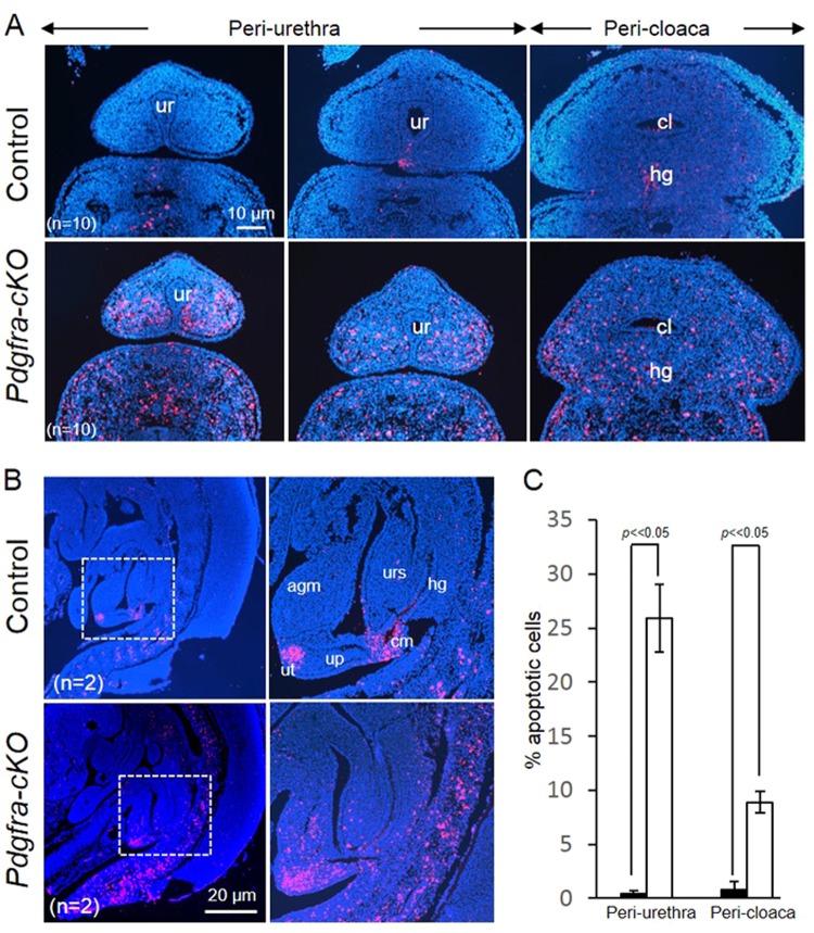
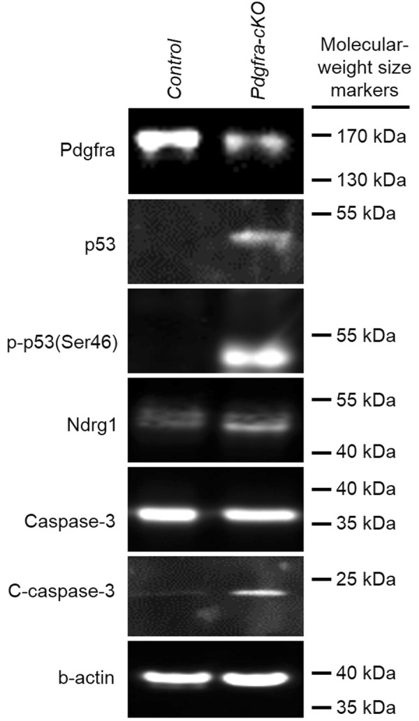
In mammals, urorectal development starts at early embryonic stage, defective urorectal development results in anorectal malformations, which are common congenital developmental defects of the anus and the urethra in newborns. The etiology and embryology of the defects are still largely unknown. Platelet-derived growth factor receptor alpha (Pdgfra) is a cell surface receptor tyrosine kinase, upon binding to its ligands (Pdgfa-d), mediates intracellular signaling and regulates embryonic development. The expression of Pdgfra is tightly regulated in the developing urorectal mesenchyme, and its dysregulation is associated with urorectal defects in animals with urorectal defects. Knockout of Pdgfra induces early embryo lethality which precludes investigation of Pdgfra in urorectal development. To address the temporal requirement of Pdgfra in urorectal development, we conditionally deleted Pdgfra in Pdgfra-expressing tissues using a tamoxifen inducible Cre-loxP approach in mice, examined the urorectal development in Pdgfra conditional knockout (Pdgfra-cKO) embryos. We showed that conditional deletion of Pdgfra in Pdgfra-expressing tissues at E10-E11 caused cloaca septation defect, anteriorly displaced anus, defective urogenital folds development and abnormal urethra tubularization in both male and female mice. Furthermore, we showed that Pdgfra was required for the survival of urorectal mesenchyme, deletion of Pdgfra caused apoptosis in the peri-cloacal, the peri-urethra and the urorectal septum mesenchyme of Pdgfra-cKO mutants, associated with an induction of p53, Ndrg1 and activation of caspase-3 in Pdgfra-cKO embryos. In conclusion, Pdgfra is required for the development and survival of the urorectal mesenchyme in embryo, dysregulated Pdgfra signaling induced urorectal defects in mice resembling human congenital diseases of anorectal malformations and hypospadias. Perturbation of PDGFRA signaling may contribute to anorectal malformations and hypospadias in human.


Conflict of interest statement

The authors declare that they have no conflict of interest.

Figures

Fig. 1
Fig. 1
Pdgfra was expressed in the peri-cloaca and genital tubercle mesenchyme of mouse embryos from E10.5 to E14.5. Immuno-histochemistry for Pdgfra was performed on transverse section of E10.5 and mid-sagittal sections of E11.5–E14.5 mouse embryos. Immuno-reactivity for Pdgfra (brown) was localized at the mesenchyme surrounding the cloaca of the budding genital tubercle at E10.5. From E11.5 to E13.5, Pdgfra expression was detected at the genital tubercle, urorectal septum and the hindgut mesenchyme. At E14.5, a more restricted expression of Pdgfra was localized at the mesenchyme adjacent to the urethra and the ureter, at the urorectal septum and at the urethral plate mesenchyme. cl cloaca, hlb hindlimb bud, nt neural tube gt genital tubercle, ugs urogenital sinus, urs urorectal septum, hg hindgut, cm cloaca membrane, t tail, ur urethra, up urethral plate, ud urethral duct, ao anal opening, u ureter, r rectum
Fig. 2
Fig. 2
PdgfraCre/ERT drives tamoxifen induced Cre recombination to the peri-cloaca and genital tubercle mesenchyme in mice. Tamoxifen was given to pregnant mice at E9.5 and E10.5, embryos were collected at E11.5 (a, b) or at E14.5 (c, d) for X-gal staining and sectioning. Strong X-gal staining was only observed in R26R; PdgfraCre/ERT embryos but not in R26R embryos. Region highlighted was shown underneath. cl cloaca, gt genital tubercle, u ureter, urs urorectal septum, up urethral plate, uf urogenital fold, agm apical genital mesenchyme, t tail. n = number of embryos analyzed
Fig. 3
Fig. 3
Conditional deletion of Pdgfra in Pdgfra-expressing tissues in mouse embryos. a Crossing scheme of PdgfraCre/ERT mice with Pdgfrafl/fl to generate conditional knockout (Pdgfra-cKO, PdgfraCre/ERT; Pdgfrafl/fl) and control (Ctrl, Pdgfrafl/fl) embryos. b Tamoxifen (Tm) was given to pregnant females at various embryo days by intragastric gavage, and embryos were collected for analysis
Fig. 4
Fig. 4
Conditional deletion of Pdgfra in mouse embryos. Tamoxifen was given to pregnant mice at E9.5 and E10.5, E14.5 embryos were collected to isolate total RNAs of the urorectum for RT-PCR analysis (a) and qPCR analysis (b). Expression of beta-actin RNA was used as internal control. “+” means with RT; “−” means without RT (negative control). Relative expression levels of Pdgfra (mean ± S.E.M.) in control and mutant urorectum were shown and the relative expression level of Pdgfra in controls were taken arbitrarily as one. Western blot analysis was performed on E14.5 urorectum to detect the expression of Pdgfra and beta-actin (b-actin) was used as loading controls (c). E12.5 embryos were collected for immuno-histochemistry (d). Pdgfra expression (brown) was detected in the urorectal mesenchyme of E12.5 control embryos. In contrast, no Pdgfra expression was detected in the urorectal mesenchyme of E12.5 Pdgfra-cKO. gt genital tubercle, ugs urogenital sinus, urs urorectal septum, hg hindgut, cm cloaca membrane. n = number of embryos analyzed
Fig. 5
Fig. 5
Conditional deletion of Pdgfra in the mesenchyme of the developing external genitalia causes defective external genitalia development. Tamoxifen was given to pregnant mice at E9.5 and E10.5, embryos were collected at E14.5 (a) and E18.5 (b) for scanning electron microscopy and histology analysis. Medial to lateral sagittal sections of E14.5 control and Pdgfra conditional knockout (Pdgfra-cKOs) embryos revealed that the urorectal septum (urs) and the urogenital fold (uf) were not developed normally in female and male Pdgfra-cKOs. The urs was shorter and not reaching the level of the cloaca membrane in Pdgfra-cKOs (dotted lines indicated the expected normal locations of the urs). Prepuce and urethra were not developed normally in E18.5 male and female Pdgfra-cKOs. Anal openings were anteriorly displaced in male and female Pdgfra-cKOs. Red arrowhead indicates the location of the tail base. Black arrowhead indicates the anal opening. pp prepuce, uf urogenital fold, ls labioscrotal fold, urs urorectal septum, r rectum, u urethra, cl cloaca, up urethral plate, ud urethral duct, t tail, g glans. n = number of embryos analyzed
Fig. 6
Fig. 6
Conditional deletion of Pdgfra in the mesenchyme of the developing external genitalia at later embryonic stages resulted in milder defective external genitalia development. Tamoxifen was given to pregnant mice at E10.5 and E11.5 (a) or E11.5 and E12.5 (b), embryos were collected at E14.5 and/or E17.5 for morphology analysis. Black arrowhead indicates the location of the urethral opening. Black arrow indicates the proximal untubularized urethra. pp prepuce, uf urogenital fold, ls labioscrotal fold, g glans. n = number of embryos analyzed
Fig. 7
Fig. 7
Elevated cell death of the urorectal mesenchyme of Pdgfra-conditional knockout embryos. Transverse (a) and mid-sagittal (b) sections of E12.5 mutant and control embryos were analyzed using TUNEL analysis. c Percentages of apoptotic cells in the peri-urethra and peri-cloaca mesenchyme of wild-type (filled bar) and Pdgfra-cKO embryos (un-filled bars) were shown. Apoptotic cells (red) were abundantly localized at the mesenchyme surrounding the cloaca and the urethra (a); at the urorectal septum, apical genital mesenchyme and the urethral plate of the mutant embryos. In contrast, apoptotic cells were localized only at the tip of the urethra (future urethra orifice) and at the cloaca membrane. Regions highlighted with dotted line were magnified and shown on the right. ur urethra, cl cloaca, agm apical genital mesenchyme, up urethral plate, ut urethral tip, cm cloaca membrane urs urorectal septum. n = number of embryos analyzed
Fig. 8
Fig. 8
Deletion of Pdgfra induced phosphorylation of p53 and activation of Caspase 3 in the genital tubercle. Tamoxifen was given to pregnant mice at E9.5 and E10.5, embryos were collected at E12.5 to isolate protein of the urorectum. Proteins of 6 Pdgfra-cKO and control urorectum were pooled and separated by polyacrylamide gel electrophoresis. Expressions of Pdgfra, Caspase 3, activated Caspase 3 (C-Caspase 3), p53, phosphorylated p53 (p-p53(Ser46)), Ndrg1 and beta-actin (b-actin) were analyzed by western blotting

References

    1. Perriton CL, Powles N, Chiang C, Maconochie MK, Cohn MJ. Sonic hedgehog signaling from the urethral epithelium controls external genital development. Dev Biol. 2002;247:26–46. doi: 10.1006/dbio.2002.0668. - DOI - PubMed
    1. Cohn MJ. Development of the external genitalia: conserved and divergent mechanisms of appendage patterning. Dev Dyn. 2011;240:1108–15. doi: 10.1002/dvdy.22631. - DOI - PMC - PubMed
    1. Springer A, van den Heijkant M, Baumann S. Worldwide prevalence of hypospadias. J Pediatr Urol. 2016;12:152 e151–157. doi: 10.1016/j.jpurol.2016.08.003. - DOI - PubMed
    1. Kohler N, Lipton A. Platelets as a source of fibroblast growth-promoting activity. Exp Cell Res. 1974;87:297–301. doi: 10.1016/0014-4827(74)90484-4. - DOI - PubMed
    1. Ross R, Glomset J, Kariya B, Harker L. A platelet-dependent serum factor that stimulates the proliferation of arterial smooth muscle cells in vitro. Proc Natl Acad Sci USA. 1974;71:1207–10. doi: 10.1073/pnas.71.4.1207. - DOI - PMC - PubMed

Publication types

Substances