. 2018 Nov;19(11):1248-1256.
doi: 10.1038/s41590-018-0227-7. Epub 2018 Oct 15.

Characterization of a potent and highly unusual minimally enhancing antibody directed against dengue virus

Affiliations

Characterization of a potent and highly unusual minimally enhancing antibody directed against dengue virus

Max Renner et al. Nat Immunol. 2018 Nov.

Abstract

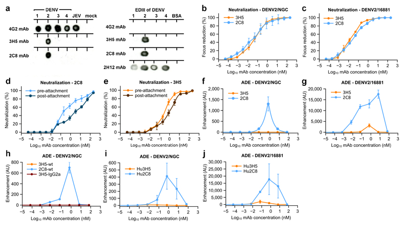
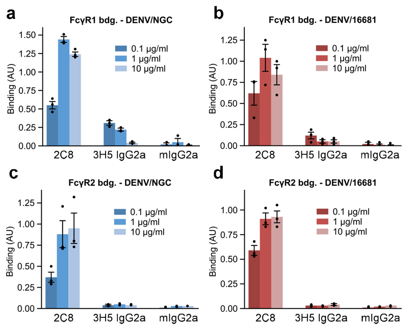
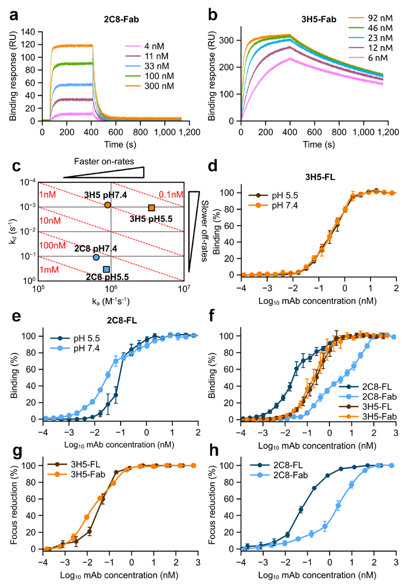
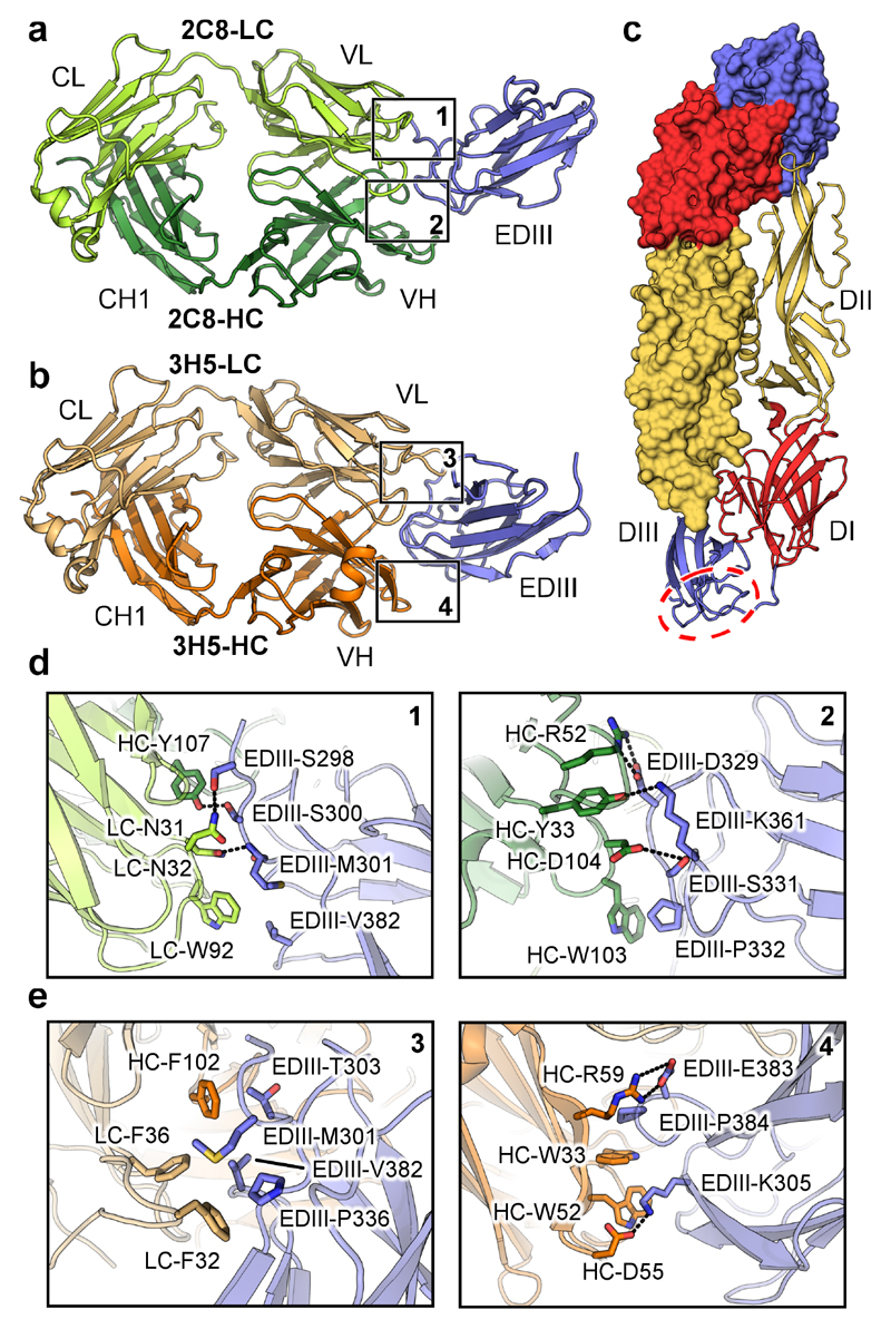
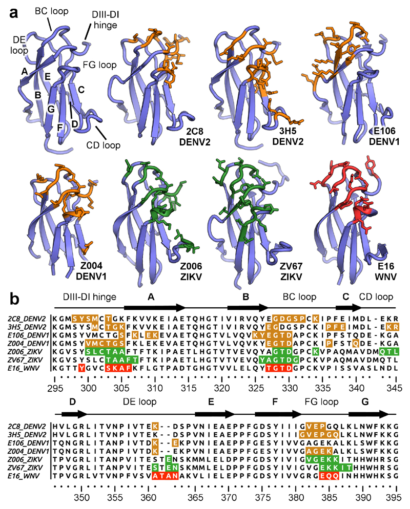
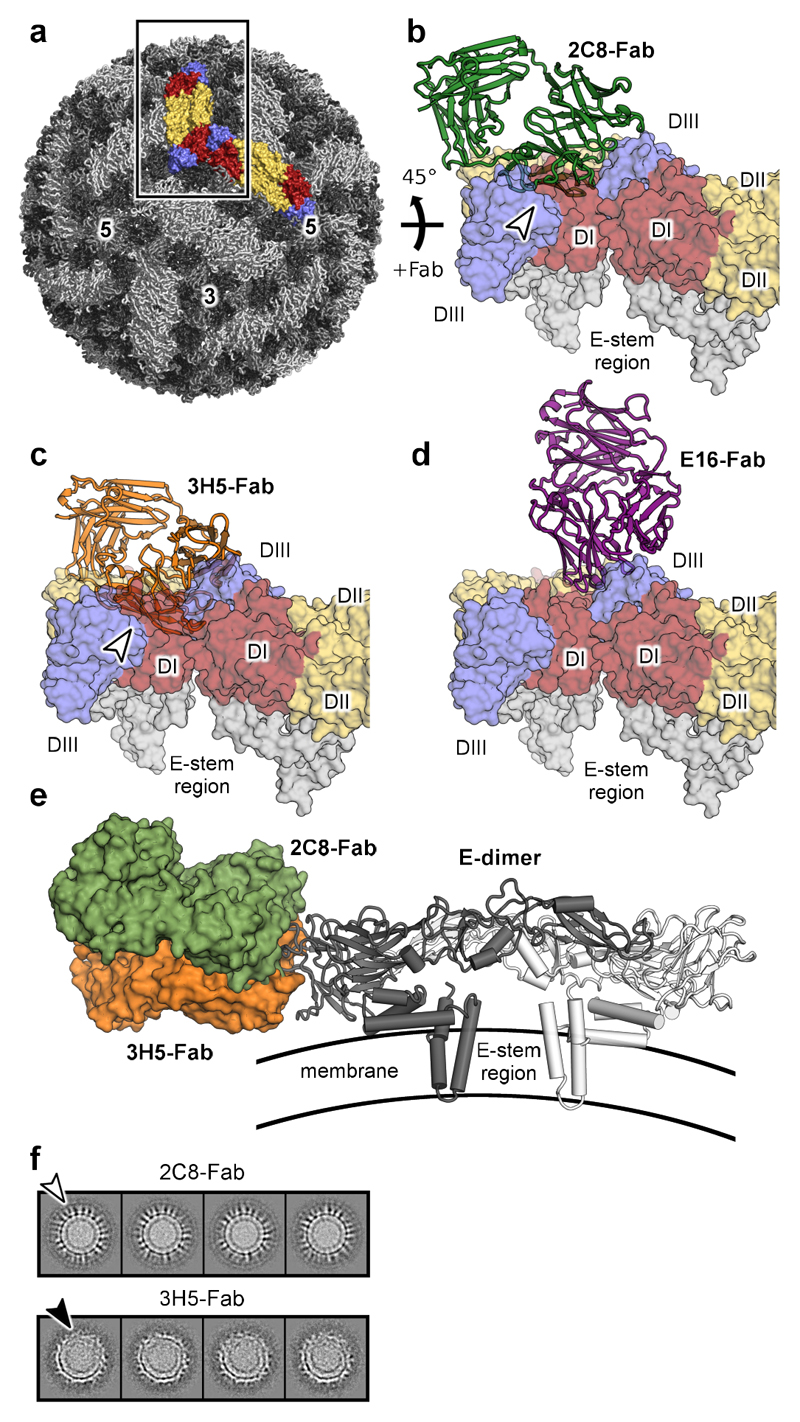
Dengue virus is a major pathogen, and severe infections can lead to life-threatening dengue hemorrhagic fever. Dengue virus exists as four serotypes, and dengue hemorrhagic fever is often associated with secondary heterologous infections. Antibody-dependent enhancement (ADE) may drive higher viral loads in these secondary infections and is purported to result from antibodies that recognize dengue virus but fail to fully neutralize it. Here we characterize two antibodies, 2C8 and 3H5, that bind to the envelope protein. Antibody 3H5 is highly unusual as it not only is potently neutralizing but also promotes little if any ADE, whereas antibody 2C8 has strong capacity to promote ADE. We show that 3H5 shows resilient binding in endosomal pH conditions and neutralizes at low occupancy. Immunocomplexes of 3H5 and dengue virus do not efficiently interact with Fcγ receptors, which we propose is due to the binding mode of 3H5 and constitutes the primary mechanism of how ADE is avoided.


Conflict of interest statement

Competing interests

The authors declare no competing interests.

Figures

Figure 1
Figure 1. Neutralization and enhancement characteristics of the anti-DENV2 antibodies 3H5 and 2C8.
a, Dot blot showing specificity of 3H5 and 2C8. Virus or recombinant EDIII of the 4 DENV serotypes were probed with the indicated antibodies. The dot blot was repeated three times independently with similar results. Antibodies 4G2 and 2H12 were included as controls. 4G2 is a cross-reactive anti-dengue and anti-Japanese encephalitis virus (JEV) mAb directed against EDII. 2H12 is a cross-reactive, EDIII-specific mAb. b and c, Neutralization activities of 3H5 (orange) and 2C8 (blue) with DENV2 strains NGC and 16681 as indicated. d and e, Pre- and post-attachment neutralization of DENV2 by 2C8 and 3H5 as indicated. Pre-attachment neutralization was determined with virus preincubated with the corresponding antibody prior to transfer to Vero cells. For post-attachment neutralization virus was incubated with the Vero cells before addition of antibody. f-j, Enhancement properties of 3H5, 2C8, 3H5 switched to IgG2a isotype (3H5-IgG2a), and humanized 3H5 and 2C8 (Hu3H5 and Hu2C8) as indicated. For all neutralization assays and antibody-dependent enhancement assays three technical replicates were carried out and the data are shown as mean ± s.e.m.
Figure 2
Figure 2. Binding of DENV immunecomplexes to Fcγ receptors.
a-d, ELISA binding assays of 2C8-DENV2 or 3H5-DENV2 immune complexes to Fcγ-receptor 1 (FcγR1) or Fcγ-receptor 2a (FcγR2a). Data is shown for DENV2 strains NGC and 16681 as indicated. Antibody/virus complexes were pre-incubated and added to plates coated with FcγR1 or FcγR2a. Mouse IgG2a (mIgG2a) was included as a negative control. Three technical replicates of all ELISA experiments were performed and data are shown as mean ± s.e.m.
Figure 3
Figure 3. Binding properties of 2C8 and 3H5.
a and b, Representative SPR profiles of 2C8 and 3H5 Fabs, respectively, binding to recombinant EDIII. Varying concentrations of Fab were flowed over immobilized EDIII of DENV2. c, On/off rate map showing binding affinities and rate constants for 2C8 (blue) and 3H5 (orange) at pH 7.4 (circles) and pH 5.5 (squares). Log-scale plot of kd (y-axis) against ka (x-axis). In this plot, affinity (kd/ka) increases from bottom left to top right, resulting in iso-affinity diagonals (red dotted lines). d and e, ELISA binding assays of full-length 2C8 and full-length 3H5 (2C8-FL and 3H5-FL) measured at pH 7.4 and pH 5.5 as indicated. (f) ELISA binding assay of full-length 3H5 (brown) and 2C8 (light blue) and corresponding Fabs (orange and dark blue, respectively) to DENV2 virions. (g and h) Neutralization activities of Fabs and full-length antibodies (FL) of 2C8 (g) and 3H5 (h). Three technical replicates of all neutralization and ELISA assays were carried out and the data are shown as mean ± s.e.m.
Figure 4
Figure 4. Crystal structures of 2C8 and 3H5 EDIII complexes.
a, 2C8 Fab (heavy chain in dark green, light chain in light green) in complex with EDIII (blue). Light chain (LC) and heavy chain (HC) as well as constant light (CL), variable light (VL), constant heavy (CH1), and variable heavy (VH) domains are labelled. b, 3H5 Fab (heavy chain in orange, light chain in light orange) in complex with EDIII (blue). c, Overview of the E-protein architecture. A head-to-tail dimer is shown with one monomer rendered as surface and colored by domain (DI in red, DII in yellow and DIII in blue). The lateral ridge region of DIII is indicated by a red dotted circle. d and e, Key contacts between heavy chain (HC) and light chain (LC) residues of 2C8 and 3H5 with EDIII. The locations of the highlighted interaction surfaces on the full structures is indicated by numbered boxes in a and b. Polar contacts are indicated by dotted lines. Domains are colored as above.
Figure 5
Figure 5. Epitope recognition by 3H5 and 2C8.
a, EDIII epitopes recognized by lateral ridge antibodies directed against different flaviviruses. The antibody designations and targeted viruses are indicated. The general architecture of EDIII is shown in the top left and important regions are labelled. Residues engaged by antibodies on dengue virus EDIIIs are colored in orange, ZIKV EDIIIs in green and WNV EDIIIs in red. All contacted residues are rendered as sticks. b, Sequence alignment of DENV, ZIKV, and WNV EDIIIs with highlighted antibody epitopes (coloring as above, antibodies and viruses indicated on the left of sequences). EDIII regions are labelled above the sequences.
Figure 6
Figure 6. Fab binding in the context of the mature virion.
a, Structure of mature DENV2 at 3.5 Å resolution (pdbID: 3J27). Two neighboring E-protein dimers on the viral surface are highlighted and rendered as surfaces (DI colored in red, DII in yellow, and DIII in blue). b-d, Docking of 2C8, 3H5, and E16 Fabs onto the virion. Two neighboring E dimers (as indicated by the box in A and rotated by 45°) on the mature virus are shown as surfaces with domains labelled. Clashes with antibodies are indicated with white arrows. e, Comparison of 2C8-Fab and 3H5-Fab docked onto a E dimer. 2C8 (green) and 3H5 (orange) Fabs were docked onto pdbID 3J27 by aligning the EDIII potion of the structures. The Fabs are shown as surfaces and the E-dimer is displayed in cartoon representation. A side view is of the E dimer on the viral surface is shown. The approximate location of the viral membrane is shown schematically. f,2D-class averages from cryo-EM of DENV-2 bound to the indicated Fabs. The white arrow indicates density corresponding to 2C8-Fabs protruding from the viral surface. The black arrow indicates deformed regions of the virion with disordered appearance.

Comment in

References

    1. Bhatt S, et al. The global distribution and burden of dengue. Nature. 2013;496:504–507. - PMC - PubMed
    1. Halstead SB. Neutralization and antibody-dependent enhancement of dengue viruses. Adv Virus Res. 2003;60:421–467. - PubMed
    1. Halstead SB, O’Rourke EJ. Antibody-enhanced dengue virus infection in primate leukocytes. Nature. 1977;265:739–741. - PubMed
    1. Guzman MG, Alvarez M, Halstead SB. Secondary infection as a risk factor for dengue hemorrhagic fever/dengue shock syndrome: an historical perspective and role of antibody-dependent enhancement of infection. Arch Virol. 2013;158:1445–1459. - PubMed
    1. Pierson TC, et al. The stoichiometry of antibody-mediated neutralization and enhancement of West Nile virus infection. Cell Host Microbe. 2007;1:135–145. - PMC - PubMed

Publication types

MeSH terms