. 2018 Oct 10:7:75.
doi: 10.1038/s41377-018-0070-5. eCollection 2018.

All-optical forward-viewing photoacoustic probe for high-resolution 3D endoscopy

Affiliations

All-optical forward-viewing photoacoustic probe for high-resolution 3D endoscopy

Rehman Ansari et al. Light Sci Appl. .

Abstract

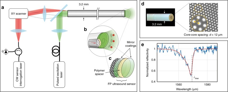
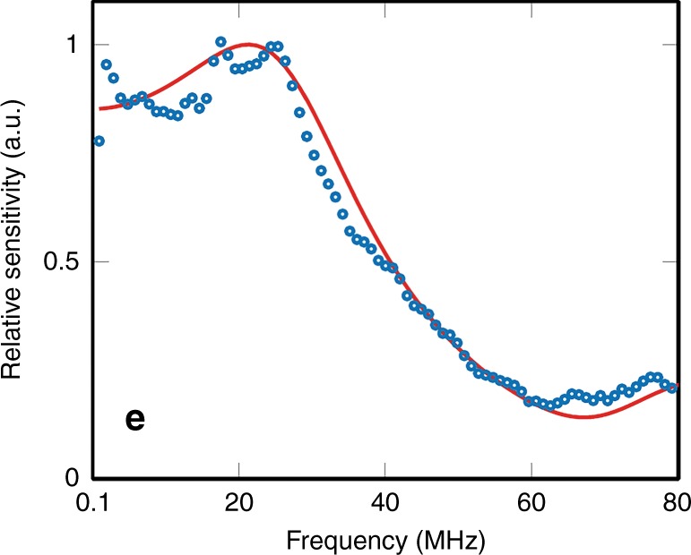
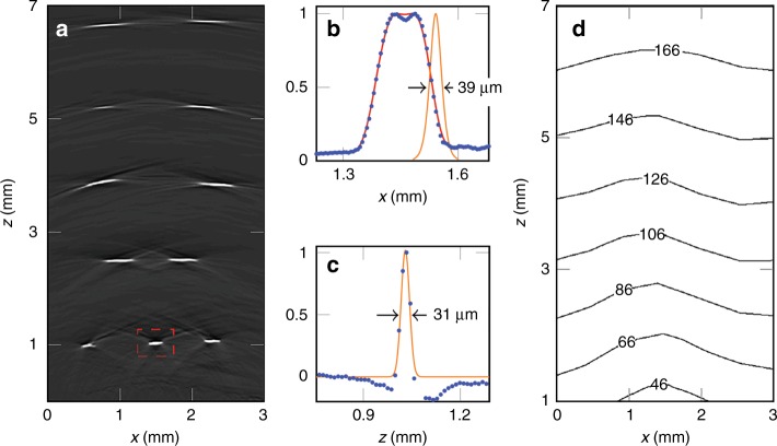
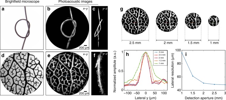
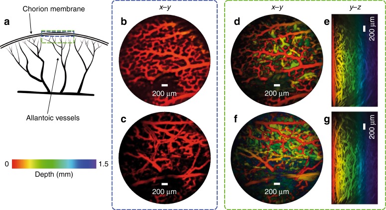
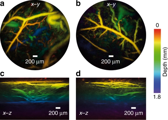
A miniature forward-viewing endoscopic probe that provides high-resolution 3D photoacoustic images is demonstrated. The probe is of outer diameter 3.2 mm and comprised of a transparent Fabry-Pérot (FP) polymer-film ultrasound sensor that is located at the distal end of a rigid optical fiber bundle. Excitation laser pulses are coupled simultaneously into all cores of the bundle and are transmitted through the FP sensor to provide wide-field tissue illumination at the distal end. The resulting photoacoustic waves are mapped in 2D by sequentially scanning the input end of the bundle with an interrogation laser beam in order to individually address different points on the FP sensor. In this way, the sensor acts as a high-density ultrasound array that is comprised of 50,000 individual elements, each of which is 12 µm in diameter, within the 3.2 mm diameter footprint of the probe. The fine spatial sampling that this affords, along with the wide bandwidth (f -3dB = 34 MHz) of the sensor, enables a high-resolution photoacoustic image to be reconstructed. The measured on-axis lateral resolution of the probe was depth-dependent and ranged from 45-170 µm for depths between 1 and 7 mm, and the vertical resolution was 31 µm over the same depth range. The system was evaluated by acquiring 3D images of absorbing phantoms and the microvascular anatomies of a duck embryo and mouse skin. Excellent image fidelity was demonstrated. It is anticipated that this type of probe could find application as a tool for guiding laparoscopic procedures, fetal surgery and other minimally invasive interventions that require a millimeter-scale forward-viewing 3D photoacoustic imaging probe.


Conflict of interest statement

The authors declare that they have no conflict of interest.

Figures

Fig. 1
Fig. 1. All-optical forward-viewing photoacoustic endoscopy probe.
a A schematic representation of the probe. b A magnified visualization of the distal end shows individual fiber-optic cores in the coherent fiber bundle and the Fabry-Pérot (FP) ultrasound sensor. c The FP sensor is deposited on the distal end of the fiber bundle and is comprised of two dielectric mirror coatings that are sandwiched between a Parylene C spacer layer (15 µm thick)—see also Supplementary Fig. S1. d A bright-field microscope image of the tip of the fiber bundle (prior to deposition of the FP sensor), which shows a subset of the 50,000 fiber cores. Suppl. Fig. S2 shows an image of the entire bundle, which illustrates all 50,000 cores and was obtained by scanning the interrogation beam over the proximal end of the bundle with the FP sensor in situ. e The interferometer transfer function (ITF) from a single core position. Blue: the measured ITF. Red: an asymmetric Lorentzian fit to the measured ITF
Fig. 2
Fig. 2. Frequency response of the FP sensor from a single fiber-optic core.
Blue dots: measured response, red line: modeled frequency response
Fig. 3
Fig. 3. Spatial resolution of the probe.
a x–z cross-section extracted from the reconstructed 3D photoacoustic image of a multi-layer ribbon phantom showing the individual absorbing ribbons over the probe field of view; the apparent curvature of the features in the top three rows is not an image artifact but reflects the true geometry of the phantom. b, c Lateral and vertical profiles (blue filled circles), respectively, through the highlighted feature in a; The lateral resolution is given by the FWHM of a Gaussian function that is fitted to the derivative of the falling edge (orange line in b). The vertical resolution is given by the FWHM of a Gaussian function that is fitted (orange line in c) to the vertical profile. d A contour plot that shows the variation in the lateral spatial resolution over the x–z plane. Incident fluence: 18 mJ/cm2
Fig. 4
Fig. 4. High-resolution 3D photoacoustic images of phantoms.
a A bright-field microscope image (3 mm across) of a 100 µm diameter synthetic hair knot. b, c Reconstructed photoacoustic images of the synthetic hair knot shown as x–y and y–z MIPs, respectively. d A bright-field microscope image of a leaf skeleton phantom, e, f Reconstructed photoacoustic images of the leaf phantom shown as x–y and y–z MIPs, respectively. g Reconstructed photoacoustic images of the leaf phantom for four detection aperture diameters: 2.5, 2, 1.5, and 1 mm. h Profiles of a feature (indicated by horizontal red dotted lines) from the leaf phantom images in g for each detection aperture. i A plot of the lateral spatial resolution (evaluated by taking the FWHM width of a Gaussian function that is fitted to the profiles in h) as a function of the detection aperture. Incident fluence: 18 mJ/cm2
Fig. 5
Fig. 5. Photoacoustic images of an ex vivo duck embryo.
a A schematic diagram of an avian embryonic vasculature. b, c x–y MIPs for depth range z = 0–200 µm of two regions of the same embryo, which show the microvascular anatomy of the chorioallantoic membrane. d, f x–y MIPs for the depth range z = 0–1.5 mm for the same two regions as in b and c, respectively. e, g y–z MIPs. Excitation laser wavelength: 590 nm, PRF: 30 Hz, and incident fluence: 15 mJ/cm2
Fig. 6
Fig. 6. Photoacoustic images of mouse abdominal skin microvasculature.
a, b x–y MIPs for the depth range z = 0–2 mm of two regions, c, d Corresponding vertical x–z MIPs of the same regions as in a, b. Excitation laser wavelength: 590 nm, PRF: 30 Hz, and incident fluence: 15 mJ/cm2

References

    1. Beard P. Biomedical photoacoustic imaging. Interface Focus. 2011;1:602–631. doi: 10.1098/rsfs.2011.0028. - DOI - PMC - PubMed
    1. Wang LV, Yao J. A practical guide to photoacoustic tomography in the life sciences. Nat. Methods. 2016;13:627–638. doi: 10.1038/nmeth.3925. - DOI - PMC - PubMed
    1. Oraevsky Alexander, Karabutov Alexander. Biomedical Photonics Handbook. 2003. Optoacoustic Tomography.
    1. Ntziachristos V, Razansky D. Molecular imaging by means of multispectral optoacoustic tomography (MSOT) Chem. Rev. 2010;110:2783–2794. doi: 10.1021/cr9002566. - DOI - PubMed
    1. Valluru KS, Wilson KE, Willmann JK. Photoacoustic imaging in oncology: translational preclinical and early clinical experience. Radiology. 2016;280:332–349. doi: 10.1148/radiol.16151414. - DOI - PMC - PubMed