. 2018 Sep 1;8(9):1725-1738.
eCollection 2018.

Targeting PDK4 inhibits breast cancer metabolism

Affiliations

Targeting PDK4 inhibits breast cancer metabolism

Maheedhara R Guda et al. Am J Cancer Res. .

Abstract

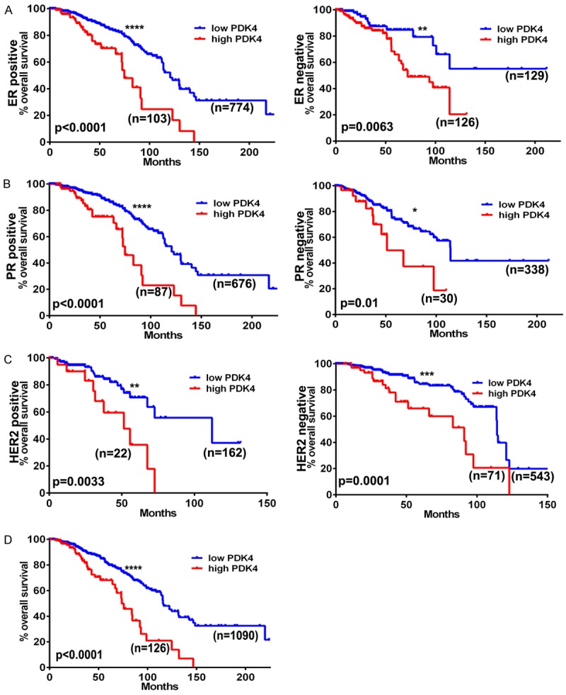
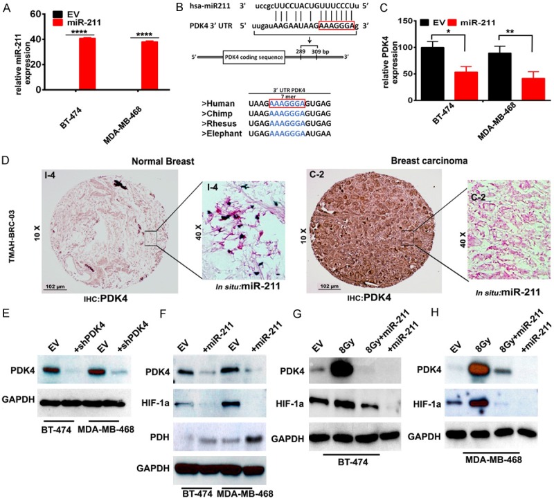
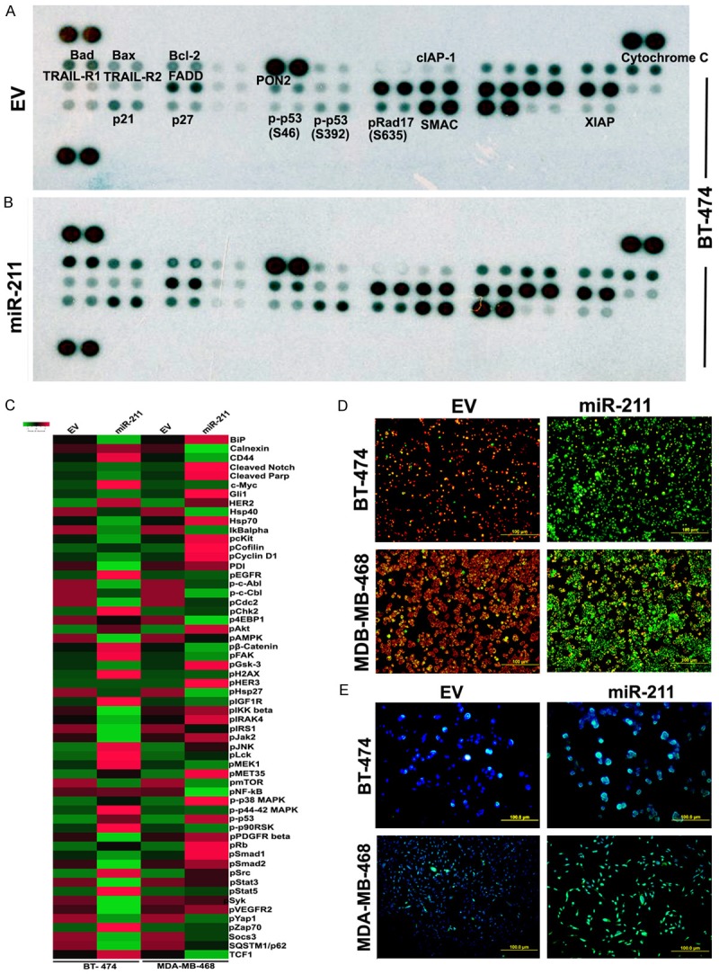
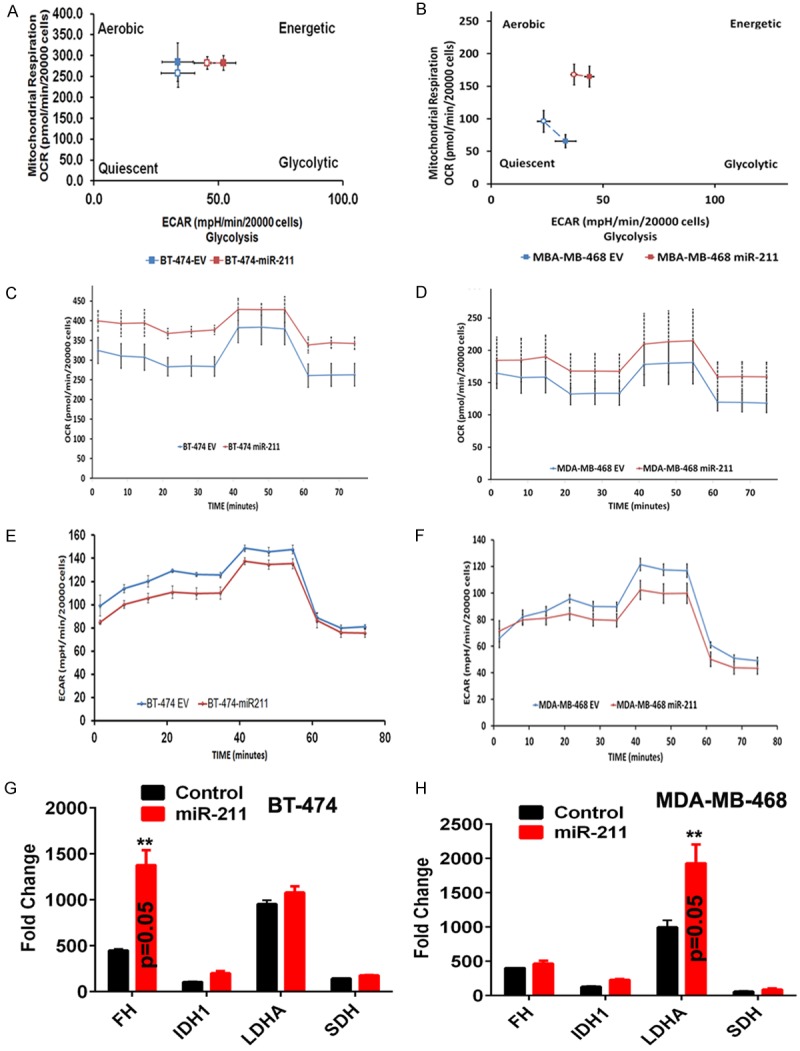
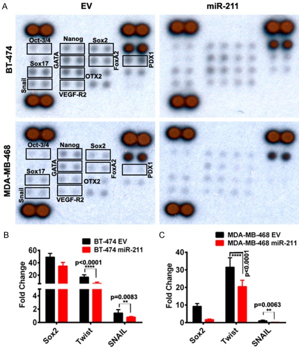
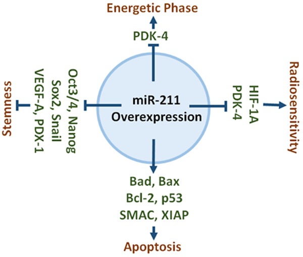
Dysregulated metabolism in the form of aerobic glycolysis occurs in many cancers including breast carcinoma. Here, we report PDK4 (pyruvate dehydrogenase kinase 4) as key enzyme implicated in the control of glucose metabolism and mitochondrial respiration is relatively highly expressed in breast cancers, and its expression correlates with poor patient outcomes. Silencing of PDK4 and ectopic expression of miR-211 attenuates PDK4 expression in breast cancer cells. Interestingly, low miR-211 expression is significantly associated with shorter overall survival and reveals an inverse correlation between expression of miR-211 and PDK4. We have found that depletion of PDK4 by miR-211 shows an oxidative phosphorylation-dominant phenotype consisting of the reduction of glucose with increased expression of PDH and key enzymes of the TCA cycle. miR-211 expression causes alteration of mitochondrial membrane potential and induces mitochondrial apoptosis as observed via IPAD assay. Further, by inhibiting PDK4 expression, miR-211 promotes a phenotype shift towards a pro-glycolytic state evidenced by decreased extracellular acidification rate (ECAR); increased oxygen consumption rate (OCR); and increased spare respiratory capacity in breast cancer cell lines. Taken together this data establishes a molecular connection between PDK4 and miR-211 and suggests that targeting miR-211 to inhibit PDK4 could represent a novel therapeutic strategy in breast cancers.

Keywords: PDK4; Warburg effect; altered metabolism; apoptosis; miR-211.


Conflict of interest statement

None.

Figures

Figure 1
Figure 1
PDK4 status correlates with survivorship in various breast cancer genotypes. Kaplan-Meier curves generated from the cancer genome atlas database showing patient populations dichotomized by PDK4 expression (low = blue, high = red) graphing survival time given (A) estrogen receptor, (B) progesterone receptor, or (C) HER2 receptor status, and (D) overall survivorship.
Figure 2
Figure 2
miR-211 is down regulated in breast cancer. (A) RT-PCR analysis to detect miR-211 endogenous levels in BT-474 and MDA-MB-468 cell lines. (B) Schematic representation of miR-211 sequence alignment with 3’UTR of human PDK4. The nucleotides in red box are putative positions of interaction. The region of miR-211 binding to PDK4 is conserved across the species. (C) RT-PCR analysis to detect mRNA levels of PDK4 in empty vector and miR-211 over expressing plasmid transfected BT-474 and MDA-MB-468 cells. (D) Negative IHC of normal breast tissue for PDK4 protein alongside inset in situ hybridization showing expression of miR-211 RNA (left panel); IHC of breast carcinoma demonstrating PDK4 positivity and inset image showing relative absence of miR-211 RNA via in situ hybridization (right panel). (E) Western blot analysis of PDK4 in PDK4 silenced (shPDK4) cells (F) miR-211 transfected cells probed for PDK4, HIF1α and PDH proteins using specific antibodies (EV = Empty vector). Total protein lysates obtained after 8 Gy radiation, miR-211 transfection, and 8 Gy radiation + miR-211 transfection from (G) BT-474 and (H) MDA-MB-468 cells were collected after 72 h. Immuno blot analysis of PDK4 and HIF1α levels were shown in Panel (G and H) respectively. GAPDH was used as a loading control.
Figure 3
Figure 3
miR-211 promote mitochondrial apoptosis of breast cancer cells in vitro. Representative expression levels of various apoptosis -related proteins in BT-474 cells transfected with EV (A and B) miR-211. Illustrated images showed increased levels of several apoptotic proteins after mir-211 silencing. (C) BT-474 and (D) MDA-MB-468 cells were transfected with EV and miR-211 were subjected to IPAD assay, which measure simultaneous expression or activation of 70 proteins involving various activated signaling pathways in tumors. The differential expression is presented as heatmaps using a free software available online (www.shinyheatmap.com). (D) Both BT-474 and MBA-MD-468 cells transfected with miR-211 and EV were treated with JC-1 dye to record the alteration in mitochondrial membrane potential (Red = live cells; Green = dead cells). (E) Cells were stained for apoptosis using TUNEL assay.
Figure 4
Figure 4
miR-211 suppresses the glycolytic pathway; modulate cellular bioenergetics. Both the EV- and miR-211 transfected cells were plated in Seahorse XFp 8 well plates. For all the experiments that involve Seahorse XFp, cell medium is replaced with bicarbonate free medium, one hour before use. Cell energy phenotype (A and B) assay was performed to measure the metabolic phenotype in EV and miR-211 transfected cells. Mitostress test (C and D) and Glycolytic stress test (E and F) were performed to assess the spare respiratory capacities, oxygen consumption rates (OXPHOS) and extracellular acidification rates (glycolysis) in EV and miR-211 transfected BT-474 and MDA-MB-468 cells. All the experiments were performed in duplicates. (G and H) RT-PCR analysis.
Figure 5
Figure 5
miR-211 overexpression reduced breast cancer stemness in vitro. (A) Around ~200 mg of total protein was extracted from EV and miR-211 transfected breast cancer cell. The experiment was conducted in accordance with the manufacturer’s instructions. Human pluripotent stem cell antibody array (#ARY010) was used to measure the change in the expression profiles of stemness related markers. Representative pictographs showed decreased levels of stem cell markers in miR-211 treatments. (B and C) Validation of differentially expressed stem cells markers in (A) using RT-PCR analysis.
Figure 6
Figure 6
Schematic representation of role of miR-211 in different functions. Overexpression of miR-211 increase the expression of apoptotic genes, reduce the expression of stemness markers, and decrease the expression of metabolic markers.

References

    1. Global Burden of Disease Cancer Collaboration. Fitzmaurice C, Allen C, Barber RM, Barregard L, Bhutta ZA, Brenner H, Dicker DJ, Chimed-Orchir O, Dandona R, Dandona L, Fleming T, Forouzanfar MH, Hancock J, Hay RJ, Hunter-Merrill R, Huynh C, Hosgood HD, Johnson CO, Jonas JB, Khubchandani J, Kumar GA, Kutz M, Lan Q, Larson HJ, Liang X, Lim SS, Lopez AD, MacIntyre MF, Marczak L, Marquez N, Mokdad AH, Pinho C, Pourmalek F, Salomon JA, Sanabria JR, Sandar L, Sartorius B, Schwartz SM, Shackelford KA, Shibuya K, Stanaway J, Steiner C, Sun J, Takahashi K, Vollset SE, Vos T, Wagner JA, Wang H, Westerman R, Zeeb H, Zoeckler L, Abd-Allah F, Ahmed MB, Alabed S, Alam NK, Aldhahri SF, Alem G, Alemayohu MA, Ali R, Al-Raddadi R, Amare A, Amoako Y, Artaman A, Asayesh H, Atnafu N, Awasthi A, Saleem HB, Barac A, Bedi N, Bensenor I, Berhane A, Bernabé E, Betsu B, Binagwaho A, Boneya D, Campos-Nonato I, Castañeda-Orjuela C, Catalá-López F, Chiang P, Chibueze C, Chitheer A, Choi JY, Cowie B, Damtew S, das Neves J, Dey S, Dharmaratne S, Dhillon P, Ding E, Driscoll T, Ekwueme D, Endries AY, Farvid M, Farzadfar F, Fernandes J, Fischer F, G/Hiwot TT, Gebru A, Gopalani S, Hailu A, Horino M, Horita N, Husseini A, Huybrechts I, Inoue M, Islami F, Jakovljevic M, James S, Javanbakht M, Jee SH, Kasaeian A, Kedir MS, Khader YS, Khang YH, Kim D, Leigh J, Linn S, Lunevicius R, El Razek HMA, Malekzadeh R, Malta DC, Marcenes W, Markos D, Melaku YA, Meles KG, Mendoza W, Mengiste DT, Meretoja TJ, Miller TR, Mohammad KA, Mohammadi A, Mohammed S, Moradi-Lakeh M, Nagel G, Nand D, Le Nguyen Q, Nolte S, Ogbo FA, Oladimeji KE, Oren E, Pa M, Park EK, Pereira DM, Plass D, Qorbani M, Radfar A, Rafay A, Rahman M, Rana SM, Søreide K, Satpathy M, Sawhney M, Sepanlou SG, Shaikh MA, She J, Shiue I, Shore HR, Shrime MG, So S, Soneji S, Stathopoulou V, Stroumpoulis K, Sufiyan MB, Sykes BL, Tabarés-Seisdedos R, Tadese F, Tedla BA, Tessema GA, Thakur JS, Tran BX, Ukwaja KN, Uzochukwu BSC, Vlassov VV, Weiderpass E, Wubshet Terefe M, Yebyo HG, Yimam HH, Yonemoto N, Younis MZ, Yu C, Zaidi Z, Zaki MES, Zenebe ZM, Murray CJL, Naghavi M. Global, regional, and national cancer incidence, mortality, years of life lost, years lived with disability, and disability-adjusted life-years for 32 cancer groups, 1990 to 2015: a systematic analysis for the global burden of disease study. JAMA Oncol. 2017;3:524–48. - PMC - PubMed
    1. Gajria D, Chandarlapaty S. HER2-amplified breast cancer: mechanisms of trastuzumab resistance and novel targeted therapies. Expert Rev Anticancer Ther. 2011;11:263–75. - PMC - PubMed
    1. Li L, Kang L, Zhao W, Feng Y, Liu W, Wang T, Mai H, Huang J, Chen S, Liang Y, Han J, Xu X, Ye Q. miR-30a-5p suppresses breast tumor growth and metastasis through inhibition of LDHA-mediated warburg effect. Cancer Lett. 2017;400:89–98. - PubMed
    1. Miao Y, Zhang LF, Guo R, Liang S, Zhang M, Shi S, Shang-Guan CF, Liu MF, Li B. (18)F-FDG PET/CT for monitoring the response of breast cancer to miR-143-based therapeutics by targeting tumor glycolysis. Mol Ther Nucleic Acids. 2016;5:e357. - PMC - PubMed
    1. Velpula KK, Bhasin A, Asuthkar S, Tsung AJ. Combined targeting of PDK1 and EGFR triggers regression of glioblastoma by reversing the warburg effect. Cancer Res. 2013;73:7277–89. - PubMed

LinkOut - more resources