. 2019 Feb;92(1094):20180389.
doi: 10.1259/bjr.20180389. Epub 2018 Nov 1.

Current and future applications of 3D printing in congenital cardiology and cardiac surgery

Affiliations

Current and future applications of 3D printing in congenital cardiology and cardiac surgery

Elena Giulia Milano et al. Br J Radiol. 2019 Feb.

Abstract

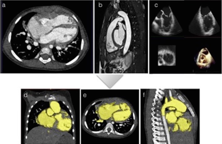
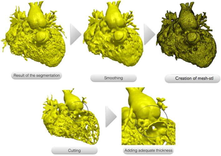
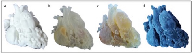
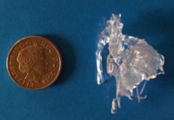
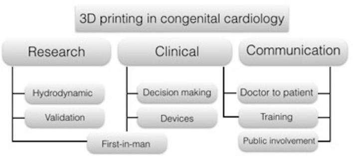
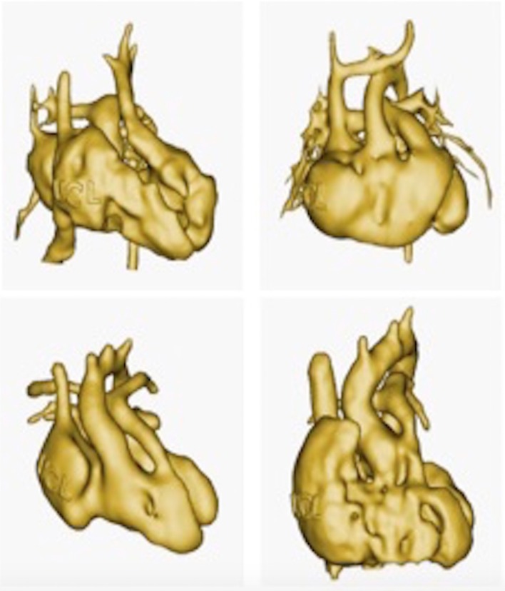
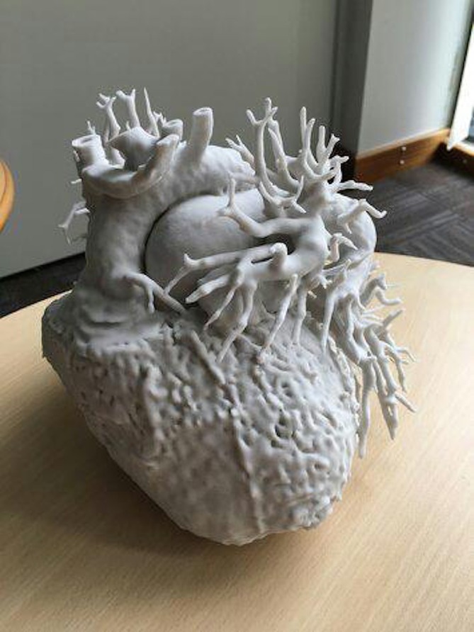
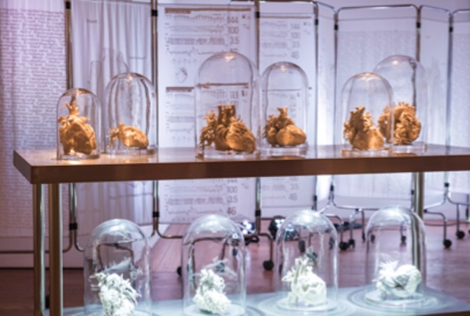
Three-dimensional (3D) printing technology in congenital cardiology and cardiac surgery has experienced a rapid development over the last decade. In presence of complex cardiac and extra-cardiac anatomies, the creation of a physical, patient-specific model is attractive to most clinicians. However, at the present time, there is still a lack of strong scientific evidence of the benefit of 3D models in clinical practice and only qualitative evaluation of the models has been used to investigate their clinical use. 3D models can be printed in rigid or flexible materials, and the original size can be augmented depending on the application the models are needed for. The most common applications of 3D models at present include procedural planning of complex surgical or interventional cases, in vitro simulation for research purposes, training and communication with patients and families. The aim of this pictorial review is to describe the basic principles of this technology and present its current and future applications.


Figures

Figure 1.
Figure 1.
All imaging modalities can be used input images for the creation of 3D models. (a) Non-gated cardiac CT images; (b) Balance SSFP with ECG gating and respiratory navigation can be used to acquire 3D images of the heart in cardiac MRI. (c) 3D echocardiography provides excellent visualization of the atrio-ventricular valves. Panels (d), (e) and (f) are examples of the segmentation process with a commercially available software (ScanIP, Synopsis, US) using tresholding and manual editing from multiplanar non-gated CT images of a patient with apical VSD. This step allows the operator to identify and separate the different cardiac and extracardiac structures: the blood pool usually present a higher signal intensity comparing to the myocardiaum and vascular walls and this automated software identifies the endocardial contours creating a colour mask (showed in yellow in panel d,e,f) that need to be carefully checked for accuracy by the operator. Only the volume included in the segmentation process, and highlighted in yellow, will be used for the creation of the model.
Figure 2.
Figure 2.
The 3D images resulting from the segmentation process usually present a dyshomogenous surface due to the presence of multiple trabeculations, mostly inside the right ventricle. This file is modified though a smoothing tool using a commercial available software (ScanIP, Synopsis, US) and converted to a surface mesh file called “.stl”. The surface can be cut exposing the region of interest for the surgeon. In our case, the right and left ventricle free walls have been removed to allow the visualization of the multiple apical VSD. Finally a 1.2 mm thickness is added to the surface to make it suitable for printing.
Figure 3.
Figure 3.
Depending on the application of the model, different materials can be used. In this figure, the same heart of a 1-year-old patient with double outlet right ventricle was printed in (a) Nylon; (b) Transparent resin (c) Compliant rubber-like material and (d) Compliant rubber-like material black
Figure 4.
Figure 4.
Foetal ultrasound images were used as input to create a 1:1 3D model of a foetal heart. Foetal heart models can be printed also in bigger scale to better appreciate small cardiac structures. In this image, a foetal heart model is compared to 1-penny coin (diameter 20.3 mm)
Figure 5.
Figure 5.
Application of 3D models in congenital cardiology and cardiac surgery. Biomedical engineers use the physical model to create experimental set ups and test hydrodynamic conditions in patient specific settings; these setups are used also to validate computation models. Physical models are used for clinical purposes in surgical planning and decision-making of complex procedures, as well as to test new application of devices to patient specific anatomy. Medical students and trainee can benefit from the use of three-dimensional models during cardiac morphology courses as well as cardiac surgeon from practicing complex procedures. Comparing to medical images, physical models are much easier to understand for patients and parents.
Figure 6.
Figure 6.
Patient specific model was used to test a range of devices potentially suitable for the patient case, checking geometrical anchoring and suitability to patient anatomy.
Figure 7.
Figure 7.
3D model from MRI images of a 16-year-old patient with congenitally corrected transposition of the great arteries, ventricular septal defect and pulmonary stenosis who underwent multiple surgeries with interposition of a conduit between the right sided left ventricle and the main pulmonary artery. The 3D model was used to plan further intervention on the conduit.
Figure 8.
Figure 8.
A 14-year-old female patient who was referred for cardiac CT after echocardiography showing prominent coronary artery flow, suggesting presence of fistula or anomalous artery connection. CT images confirmed the diagnosis of anomalous origin of the circumflex coronary artery from right pulmonary artery (a and b). 3D model (c) was manufactured (d) for better understanding of coronary anatomy and dimensions.
Figure 9.
Figure 9.
Complex congenital case: double outlet right ventricle with transposition of the great arteries and non-committed VSD, repaired with LV to aorta baffle presenting with symptoms of left ventricular outflow tract (LVOT) obstruction. Model manufactured on clinical request for assessment of LVOT anatomy. The yellow arrow shows the LV to aorta intracardiac baffle. (a,b) Two views from CT data, (c) 3D reconstruction, (d) 3D-printed model.
Figure 10.
Figure 10.
Complex congenital case: A 11-year-old patient truncus arteriosus presenting with conduit and pulmonary stenosis. The model was used to assess the relationship between the aorta and the pulmonary arteries in order to plan the surgery. (a) CT images showing the position of the branch pulmonary arteries and the aorta. (b) Segmentation process to create the 3D model (highlighted in red); (c) 3D reconstruction used as input file to create the 3D model; (d) 3D model printed in white resin.
Figure 11.
Figure 11.
Patient-specific model of right ventricular outflow tract is connected to a hydrodynamic circuit to simulate realistic flow and pressure conditions to be assessed with MRI acquisitions. These in vitro setups are used to simulate hydrodynamic conditions and study the complex physiology of patients with CHD, typically performing parametric studies. Mock circulatory systems incorporating patient-specific models can also be designed to be MRI compatible and thus allow for the acquisition of detailed visual information, such as 4D MRI flow sequences.
Figure 12.
Figure 12.
In this example, a cardiac surgery fellow is practicing on the creation of an intracardiac baffle from the VSD to the aorta in a patient with DORV using a 3D model printed in compliant rubber-like material.
Figure 13.
Figure 13.
The use of 3D model is particularly valuable for education in congenital cardiology and cardiac surgery. Patient anatomies are often unique and it is difficult to capture the wide range of anatomical variations that occur in the context of the same cardiac malformation. In this example, four patient specific virtual 3D models of double outlet right ventricle are imaged. (From 3D library at http://www.ucl.ac.uk/cardiac-engineering/research/library-of-3d-anatomies),
Figure 14.
Figure 14.
Example of a 3D model of an adult patient with transposition of the great arteries who underwent Mustard procedure. The model was printed with a off-white plaster powder on a Projet 660 printer, and was then used during a workshop with the patient.
Figure 15.
Figure 15.
Making the Invisible Visible II, Sofie Layton, 2018 (detail). Installation including 3D printed hearts of paediatric and adult patients with congenital heart disease, and with soundscape by Jules Maxwell. As part of “The Heart of the Matter” exhibition (www.insidetheheart.org)

References

    1. Garekar S, Bharati A, Chokhandre M, Mali S, Trivedi B, Changela VP, et al. . Clinical application and multidisciplinary assessment of three dimensional printing in double outlet right ventricle with remote ventricular septal defect. World J Pediatr Congenit Heart Surg 2016; 7: 344–50. doi: 10.1177/2150135116645604 - DOI - PubMed
    1. Bramlet M, Olivieri L, Farooqi K, Ripley B, Coakley M. Impact of three-dimensional printing on the study and treatment of congenital heart disease. Circ Res 2017; 120: 904–7. doi: 10.1161/CIRCRESAHA.116.310546 - DOI - PMC - PubMed
    1. Farooqi KM, Nielsen JC, Uppu SC, Srivastava S, Parness IA, Sanz J, et al. . Use of 3-dimensional printing to demonstrate complex intracardiac relationships in double-outlet right ventricle for surgical planning. Circ Cardiovasc Imaging 2015; 8. doi: 10.1161/CIRCIMAGING.114.003043 - DOI - PubMed
    1. Valverde I, Gomez-Ciriza G, Hussain T, Suarez-Mejias C, Velasco-Forte MN, Byrne N, et al. . Three-dimensional printed models for surgical planning of complex congenital heart defects: an international multicentre study. Eur J Cardiothorac Surg 2017; 52: 1139–48. doi: 10.1093/ejcts/ezx208 - DOI - PubMed
    1. Schievano S, Migliavacca F, Coats L, Khambadkone S, Carminati M, Wilson N, et al. . Percutaneous pulmonary valve implantation based on rapid prototyping of right ventricular outflow tract and pulmonary trunk from MR data. Radiology 2007; 242: 490–7. doi: 10.1148/radiol.2422051994 - DOI - PubMed